
Issue published October 15, 2026 Previous issue

- Volume 136, Issue 20
On the cover:
ISG15+ Müller glia expansion in diabetic retinopathy
Niu et al report a diabetes-induced ISG15+ Müller glial subtype, triggered by microglial immune signaling, that weakens antioxidant defenses and contributes to early retinal vascular damage in murine diabetic retinopathy. The cover image shows murine retinal vasculature isolated using trypsin digestion followed by PAS staining, with acellular capillaries visible.
Niu et al report a diabetes-induced ISG15+ Müller glial subtype, triggered by microglial immune signaling, that weakens antioxidant defenses and contributes to early retinal vascular damage in murine diabetic retinopathy. The cover image shows murine retinal vasculature isolated using trypsin digestion followed by PAS staining, with acellular capillaries visible.
Go to section:
-
Review Series
×
Abstract
Cell-cycle dysregulation is a central driver of breast cancer progression and a major therapeutic vulnerability, particularly in hormone receptor–positive/human epidermal growth factor receptor 2–negative (HR+/HER2–) disease. The clinical success of cyclin-dependent kinase (CDK) 4/6 inhibitors combined with endocrine therapy has transformed treatment paradigms and established cell-cycle targeting as a cornerstone of care. However, important challenges remain, including hematologic toxicity, incomplete and largely cytostatic responses, and the near-universal development of resistance. These limitations have highlighted the need for next-generation CDK-directed strategies capable of improving selectivity, extending disease control, and overcoming adaptive escape pathways. This Review examines the evolving landscape of CDK-targeted therapeutics in breast cancer, with particular emphasis on emerging CDK4-selective inhibitors, CDK2-directed approaches, and broader multi-CDK strategies. We discuss the biological rationale underpinning these agents, the mechanisms of resistance that have informed their development, and the clinical opportunities they may create across both advanced and early-stage settings. By refining target specificity and addressing resistance-associated cell-cycle rewiring, next-generation CDK inhibitors may reduce toxicity, enhance treatment durability, and enable more effective combination and sequencing strategies. Collectively, these advances promise to move breast cancer treatment beyond current CDK4/6 inhibition toward more precise, adaptable, and durable cell-cycle–directed therapy.
Authors
Daria Seriak, Jun Yong Kim, Geoffrey I. Shapiro, Antonio Giordano
×Abstract
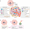
One in eight women will be diagnosed with breast cancer in their lifetime. Most breast cancers express estrogen receptor α (ERα) and depend on its signaling, which promotes tumor growth, survival, and metastatic spread through hormone-dependent transcriptional reprogramming. Selective estrogen receptor degraders/downregulators (SERDs), such as fulvestrant (faslodex), which was FDA approved in 2002, are second-line endocrine therapies for the treatment of advanced metastatic disease. Tumors containing activating hotspot mutations in ESR1 (the gene encoding ERα) retain some sensitivity to fulvestrant but require higher doses to achieve inhibition of the mutant receptor. Substantial efforts to develop next-generation SERDs with improved pharmacology over fulvestrant have led to the FDA approval of the first-in-class orally available SERD elacestrant (Orserdu), followed by imlunestrant (Inluriyo), and the anticipated approval of several more therapies. Excitingly, recent studies show that in addition to their previously described antitumoral mechanisms, SERDs also act by increasing antitumor immunity. In this Review, we detail the current SERD clinical landscape and our emerging understanding of the interplay among SERDs, breast cancer cells, and the tumor immune microenvironment.
Authors
Kristen S. Young, Sean W. Fanning
×Abstract
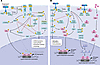
Somatic alterations in the phosphoinositide 3-kinase (PI3K) pathway are frequent oncogenic drivers in cancer and are particularly relevant in estrogen receptor–positive breast cancer, where PI3K signaling integrates hormonal, growth factor, and metabolic inputs. Therapeutic targeting of PI3K has been limited by toxicity, pathway crosstalk, and adaptive resistance. Recent advances in structural and functional characterization of PIK3CA mutations have enabled the development of more selective inhibitors, including PI3K isoform–selective and emerging allosteric PI3Ka mutant–targeted compounds. These approaches aim to improve the therapeutic index while maintaining pathway inhibition. In parallel, combination strategies with endocrine therapy and CDK4/6 inhibitors have demonstrated clinical benefit in selected patient populations. However, responses are often transient owing to compensatory signaling and pathway reactivation. Additional metabolic and transcriptional adaptations further contribute to sustaining tumor cell survival. Overall, PI3K-targeted therapy is evolving along two complementary paths: increased mutant-selective inhibition to improve therapeutic index and broader pathway-level strategies designed to overcome adaptive resistance and achieve more durable responses. Ongoing efforts focus on optimizing combination strategies, enhancing development of predictive biomarkers, and improving therapeutic sequencing to achieve durable responses while minimizing toxicity.
Authors
Fabiana Napolitano, Nisha Unni, Carlos L. Arteaga

-
Reviews
×
Abstract
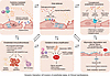
Sepsis is a systemic vascular disorder in which endothelial activation, dysfunction, and structural injury contribute to shock, microcirculatory failure, immunothrombosis, and multiorgan dysfunction. Sepsis-associated endotheliopathy is not a uniform process but a dynamic trajectory that evolves from adaptive endothelial activation toward functional disruption, structural injury, and organ-specific vascular failure. In this Review, we propose a phenotype- and trajectory-oriented framework that integrates clinical manifestations, molecular mechanisms, and translational assessment of endothelial dysfunction in sepsis. We focus on four major clinically recognizable modes of endothelial dysfunction that often overlap and evolve over the course of sepsis. We further discuss biology-anchored biomarker panels, bedside physiological surrogates, and serial trajectory-based assessment as complementary approaches for identifying endothelial states, refining risk stratification, and guiding future precision strategies. Finally, we describe implications for biomarker-enriched clinical trials, microvascular-informed resuscitation, and endotype-guided endothelial cell–targeted therapies. Viewing sepsis through the lens of dynamic endotheliopathy provides a framework linking vascular biology to clinically interpretable trajectories and may facilitate the development of personalized approaches to endothelial cell–directed care.
Authors
Song Qin, Ninan Dai, Fengyun Xu, Cindy Zhuang, Judith Hellman
×Abstract
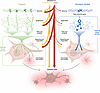
Most neurodegenerative diseases reflect patient-specific combinations of polygenic risk variants interacting with aging and environmental factors, rather than a single causative mutation. Phase III clinical trial failure rates for neurodegenerative indications exceed 95%, reflecting in part the limitations of existing preclinical systems to fully model the human genetic context of disease. Patient-derived induced pluripotent stem cells (iPSCs) have the potential to address this shortcoming by faithfully recapitulating human cell biology and preserving each patient’s complete genetic architecture. This Review examines iPSC-based models of neurodegeneration across three themes. First, we discuss mechanisms revealed by these models, from convergent pathways uncovered by familial mutations to early evidence that sporadic disease biology is accessible at sufficient cohort scale. Second, we evaluate culture systems from 2-dimensional monocultures to organoids, examining how increasing complexity expands addressable biological questions while introducing trade-offs. Third, we address the aging problem: reprogramming erases epigenetic signatures of donor aging, removing the cellular context in which late-onset sporadic neurodegeneration emerges. We discuss strategies approximating aging in iPSC-derived neurons and highlight direct conversion strategies that preserve native epigenetic age, enabling access to age-dependent phenotypes. We propose a framework matching model systems to biological questions, improving mechanistic understanding and therapeutic development.
Authors
Shijie Wang, Lisa Elia, Weiyi Liu, Steven Finkbeiner
-
Commentaries
×
Abstract
Lactation insufficiency in postpartum women has been associated with obesity and insulin resistance, among other risk factors. Challenges related to translating findings from animal models of lactation and the limited ability to study pregnant and lactating women in clinical trials have impeded more detailed mechanistic understanding of this relationship. In this issue of the JCI, Meng et al. combined studies in human milk samples and in vitro mammary epithelial cell models from humans and rodents to provide compelling evidence that mitochondrial dysfunction mediates the link between insulin resistance and lactation insufficiency. Notably, they observed that chronic exposure to high levels of insulin led to mitochondrial fragmentation and loss of volume in mouse mammary epithelial cells. This framework linking mitochondria, insulin signaling, and lactation should be extended to in vivo models and primary human tissues to better define the mechanisms supporting successful lactation.
Authors
Laura L. Hernandez
×Abstract
Efferocytosis, or the clearance of apoptotic cells by macrophages, has been implicated in tissue resolution that promotes disease regression in atherosclerosis and other diseases of chronic inflammation. The mechanisms by which efferocytosis leads to pro-resolving macrophage reprogramming have not been fully defined. In this issue of the JCI, Avrampou and colleagues found that efferocytosis activates DNA-methyltransferase-3A (DNMT3A) to promote base excision repair and suppress oxidized DNA. Moreover, they provide evidence that DNMT3A enabled tissue repair in the context of plaque regression in a mouse model of atherosclerosis. Human studies have linked DNMT3A mutation–mediated clonal hematopoiesis (DNMT3A-CH) with atherosclerosis. The findings of Avrampou et al. linking DNMT3A function to efferocytosis-mediated tissue repair suggest that this pathway could be modulated therapeutically to minimize cardiovascular events in patients.
Authors
Aarti Asnani
×Abstract
Trauma-related neuropsychiatric disorders are commonly treated with exposure-based therapy, an approach based on fear extinction, in which fear memory and expression are suppressed by repeated exposure to the inciting fear stimulus. Fear extinction–focused research often investigates mechanisms of fear inhibition, but positive affect may also contribute to facilitating extinction. In this issue of the JCI, Lin et al. used activity-dependent neuronal labeling in a model of fear extinction to tag extinction-activated medial prefrontal cortex (mPFC) ensembles, identifying functionally distinct projections to the zona incerta (ZI) and supramamillary nucleus (SuM). While mPFC-ZI projections governed extinction retrieval, mPFC-SuM projections encoded positive affect and reinforced fear extinction persistence. Additionally, Lin et al. discovered that SIRT1-expressing mPFC-SuM extinction-recruited projections control extinction relapse vulnerability, showing that SIRT1 overexpression can be leveraged to reduce fear relapse. Overall, this study shows that key cortico-hypothalamic projections manage extinction retrieval and positive affect to promote fear extinction.
Authors
Quan-Son Eric Le, Luis E. Rosas-Vidal
×Abstract
Friedreich’s ataxia (FRDA) is a triplet repeat expansion disorder with multisystem, progressive symptoms caused by reduced frataxin protein levels and compromised mitochondrial function. Restoration of frataxin protein is a focus of clinical trials involving adeno-associated viral vectors, but substantial barriers exist because of the immune response they elicit. In this issue of the JCI, Sivakumar et al. harnessed CRISPR/Cas9 editing of endogenous hematopoietic stem and progenitor cells (HSPCs) to excise the frataxin gene (FXN) repeat expansion ex vivo, followed by autologous transplantation. A single, systemic infusion of corrected HSPCs resulted in long-term engraftment and sustained multiorgan benefit in a humanized mouse model of FRDA. This improvement in brain, cardiac, and pancreatic symptoms, mediated by myeloid progeny of the engrafted stem cells, provides strong support for the potential effectiveness of this approach in humans and bypasses the need to correct the FXN deficiency in the affected tissue and cells directly.
Authors
Lucy R. Osborne

-
Research Letters
×
Abstract
Authors
Diwakar Turaga, Chang-Ru Tsai, Yuka Morikawa, Hanna J. Tadros, Yi Zhao, Lalita Wadhwa, Iki Adachi, Xiao Li, James F. Martin
×Abstract
Authors
Chenjian Gu, Yili Fang, Yixuan Wang, Eric Tycksen, Gayathri Kondepati, Chuang Li, Kendrah O. Kidd, Jun Liu, Fumihiko Urano, Maria Lindahl, Anthony J. Bleyer, Srikanth Singamaneni, Zhao Sun, Ying Maggie Chen
×Abstract
Authors
Diana B. Voss, Emily J. Shields, Andrey Poleshko, Li Li, Jun Li, Lizhou Wang, Mariacristina Calcagno, Rajan Jain, Cheryl L. Smith, Jonathan A. Epstein.


-
Research Articles
×
Abstract
Transport of nucleoside chemotherapeutic drugs into tumor cells is primarily accomplished through equilibrative nucleoside transporter 1 (ENT1), which is considered constitutively active. Drugs are redistributed via ENT1 across lipid bilayers via facilitated diffusion. Here we discover that ENT1 is not constitutively active, but rather requires activation of acid sphingomyelinase (ASMase) by gemcitabine, generating ceramide-rich platforms (CRPs) on external plasma membranes of endothelial and tumor cells into which ENT1 inserts, dimerizing therein to functionalize transmembrane gemcitabine transport. Whereas sarcoma cells synthesize minimal ASMase, they take up gemcitabine poorly in vitro and in murine xenografts. A strategy designed to augment gemcitabine-induced ASMase secretion into the extravascular space by ASMase-rich neo-angiogenic cells, which then targets tumor cell plasma membranes, yields “bystander” CRPs on sarcoma cells and ENT1 insertion therein, conferring markedly enhanced gemcitabine uptake and xenograft response. Engaging this biology in a prospective phase II clinical trial in advanced sarcoma yielded robust volumetric changes in evaluated tumors that developed early and were often durable.
Authors
Aditya Ganju, Shyam Rao, Mark A. Dickson, Robert A. Lefkowitz, Chris Thompson, Jin Cheng, Katia Manova, Adriana Haimovitz-Friedman, Gary Schwartz, Zhigang Zhang, Zvi Fuks, William D. Tap, Richard Kolesnick
×Abstract
Microglia play essential yet poorly understood roles in brain development, including axon guidance, regulation of neurogenesis, and pruning of neuronal projections. Congenital hydrocephalus (CH), characterized by enlarged ventricles filled with cerebrospinal fluid (CSF), is a leading cause of pediatric brain surgery, but its molecular mechanisms remain unclear. We have identified what we believe to be novel, recurrent, damaging missense variants in the SH3-binding domain of the adaptor protein growth factor receptor-bound protein 2 (GRB2) in unrelated patients with CH. GRB2 is significantly coexpressed with 1 of its known upstream RTK partners, CSF1R, in the developing human brain, particularly in a microglial subtype associated with regulation of neural stem cells. IP validated GRB2-CSF1R binding in mouse microglial cells and a human monocyte cell line. Cx3cr1-Grb2fl/fl mice engineered with conditional deletion of Grb2 in microglia exhibit congenital absence of microglia and early postnatal severe communicating (nonobstructive) hydrocephalus, mimicking the condition of patients with the GRB2 mutation. The severe ventriculomegaly of Cx3cr1-Grb2fl/fl mice is associated with both depletion of cerebral cortical neurons and impairment of glia-lymphatic-mediated CSF flow. Together, these findings implicate a role of GRB2 in microglia that could be essential for brain development and CSF homeostasis.
Authors
Phan Q. Duy, Benjamin C. Reeves, Huanxing Sun, Xueyan Peng, Pazhanichamy Kalailingam, Garrett Allington, Evan Dennis, Le Thi Hao, Lei Wang, David Rufino-Ramos, Shujuan Zhao, Qiang Li, Neel H. Mehta, William C. Davalan, Mason Blacker, Anthony J. Piscopo, Shozeb Haider, Baojian Fan, Kedous Y. Mekbib, Shuai Shao, Carol Nelson-Williams, Tukiet T. Lam, Benjamin P. Kleinstiver, Patricia L. Musolino, Seth L. Alper, Sheng Chih Jin, Erica L. Herzog, Kristopher T. Kahle
×Abstract
Insulin resistance (IR) has emerged as a risk factor for lactation insufficiency and delays the onset of milk secretion after childbirth, termed secretory activation (SA). This may cause inadequate infant weight gain and early breastfeeding cessation. However, the mechanisms underlying delayed SA in insulin-resistant women are unknown. To investigate this, we characterized the mammary transcriptomes and IR-related hormones of 75 breastfeeding women with healthy term infants during postpartum days 1–5. Participants were divided into IR tertiles based on plasma leptin-to-adiponectin ratio measurements. Those in the highest tertile had later SA onset with greater neonatal weight loss during postpartum days 1–5. Transcriptomic analysis on postpartum day 2 (n = 4 high IR vs. n = 8 low IR participants) showed transient suppression of mammary insulin and prolactin signaling genes, increased pro-inflammatory gene expression, and altered expression of more than 200 mammary mitochondrial genes. These alterations were absent on postpartum days 3–5. Cultured mammary epithelial cells treated with insulin showed upregulation of prolactin signaling and oxidative phosphorylation (OXPHOS) genes, with imaging and bioenergetic studies demonstrating that insulin promotes mitochondrial biogenesis and OXPHOS. Thus, our findings delineate roles for insulin in mammary bioenergetics and highlight mitochondrial dysfunction as a mechanism for delayed SA in insulin-resistant women.
Authors
Xin Meng, Taha Elajnaf, Hussam Rostom, Michelle Ma, Annalee Furst, Bryony R. Davies, Isabella R. Honess, Gaurav Pandey, Isadora C. Furigo, Craig L. Doig, Jayne F. Martin Carli, Rajesh V. Thakker, Lars Bode, Kelsey E. Johnson, Fadil M. Hannan
×Abstract
Interferon lambda (IFN-λ) has emerged as a critical inducer of immune regulation in addition to its mucosal antiviral properties. However, the cellular and molecular mechanisms that govern its dual role during viral infections remain unclear. Here, we discovered that mucosal delivery of IFN-λ abrogates the herpes simplex virus type 2–induced (HSV-2-induced) vaginal tissue pathology observed in the absence of type I IFN signaling, through drastically inhibiting IL-6 production. At the same time, we demonstrated that IFN-λ rescued local production of CD4+ T cell–dependent IFN-γ, which is essential for protection against the development of toxic megacolon following genital HSV-2 infection. Mechanistically, we found that IFN-λ enhances the differentiation of CCR2+Ly6Chi cells into monocyte-derived antigen-presenting cells, thereby boosting T cell responses. In vivo depletion of CCR2+ cells abrogated this phenomenon. Together, these findings suggest that IFN-λ orchestrates a potent antiviral response, inhibiting hyperinflammatory responses while selectively augmenting effector T cell function. This study positions IFN-λ as a promising therapeutic candidate for enhancing local antiviral immunity and offers new insights into the regulation of immune responses in the context of mucosal viral infections.
Authors
Emily Feng, Alexa Mansour, Elizabeth Balint, Alyssa Mark, Sofia Tramonte, Jonathan K. Monteiro, Madison Read, Marie Joe Adaimy, Zhou Xing, Matthias Mack, Sergei V. Kotenko, Deanna M. Santer, Ali A. Ashkar
×Abstract
Lung cancer histological subtypes include lung adenocarcinoma (LUAD) and small-cell lung cancer (SCLC). Although usually distinct, rare combined LUAD/SCLC tumors occur, and LUAD can transform into SCLC as a mechanism of resistance to targeted therapies, particularly in EGFR-mutant LUAD tumors with RB1/TP53 inactivation. Although PRC2 complex expression increases during this transformation, its functional role remains unclear. Using CRISPR-based autochthonous immunocompetent GEMMs, we found that inactivation of EED, the core PRC2 scaffolding subunit, impaired SCLC tumorigenesis and promoted LUAD histological identity, likely through a NEUROD1-positive intermediate state. Mechanistically, EED loss derepressed bivalent genes comarked by H3K27me3 and H3K4me3, including LUAD oncogenic RAS, PI3K, and MAPK pathway genes and NEUROD1. These same LUAD oncogenic signaling genes were bivalently repressed in human SCLC patient-derived xenografts, suggesting a conserved PRC2-dependent mechanism that represses LUAD oncogenic signaling and thereby supports the SCLC neuroendocrine identity. In a complementary EGFR-mutant LUAD GEMM with Rb1 and Trp53 inactivation, EED inactivation at tumor initiation prevented the emergence of SCLC histology after EGFR oncogene withdrawal and redirected recurrent tumors toward mucinous LUAD states with reduced spontaneous metastasis. These findings identify PRC2/EED as a regulator of SCLC neuroendocrine identity and nominate pharmacologic EED inhibition for future investigation in therapy-associated LUAD-to-SCLC transformation.
Authors
Yixiang Li, Yasmin N. Laimon, Hyeonseo Cho, Marina Vivero, Gabriel Roberti De Oliveira, Maxwell D. Seager, Andrew Delcea, Varunika Savla, Yuting Chen, Yavuz T. Durmaz, Xintao Qiu, Shweta Kukreja, Rong Li, Talal El Zarif, Wesley Lu, McKayla Van Orden, Jacob E. Berchuck, Roderick T. Bronson, Shuqiang Li, Hongbin Ji, David A. Barbie, Katerina Politi, Matthew L. Freedman, Henry W. Long, Sabina Signoretti, Matthew G. Oser
×Abstract
Systemic lupus erythematosus (SLE) is a chronic autoimmune condition with alternating phases of heightened symptoms and periods of disease inactivity. Our investigation focuses on the distinct molecular profiles of resting naive B cells from both active and inactive SLE states in female adults, primarily of Black ancestry. We identified a distinct transcriptome and epigenome during inactive SLE. Transcriptional patterns indicated an increase in distinct cytokine signaling pathway expression in SLE B cells, encompassing both common and disease activity–specific components. Specifically, while both disease states exhibited interferon signatures, increased expression of IL-12, TNF-α/NF-κB, and CD40 signaling pathway genes was enriched in inactive disease. While some regions exhibited comparable epigenetic features in both disease states, such as TLR7 signaling genes, others were distinct. Chromatin accessibility in inactive disease indicated the involvement of MEF2 and bHLH family factors, such as E2A/E2-2, while active disease regions were defined by IRF family factors. This study illuminates both disease-associated programs and molecular changes to cell activation pathways capable of directing B cells toward diverse effector fates in active or inactive SLE. Our data also offer insights of particular importance for the design and monitoring of B cell–targeting treatments.
Authors
James R. Rose, Yusho Ishii, Arezou Khosroshahi, Sakeenah Hicks, Scott A. Jenks, Christopher D. Scharer, Ignacio Sanz, Jeremy M. Boss
×Abstract
Osteoarthritis (OA) is the most common degenerative joint disease. However, the molecular mechanisms underlying OA-induced cartilage degeneration remain elusive. Here, we identify IFN regulatory factor 2 binding protein 2 (IRF2BP2) as an important regulator of chondrocyte differentiation and articular cartilage regeneration. Irf2bp2 deficiency promoted chondrogenesis and inhibited chondrocyte terminal differentiation by transcriptionally upregulating the key chondrogenic marker SRY-box transcription factor 9 (SOX9), inducing its ectopic expression in the hypertrophic zone. Cartilage-specific KO of Irf2bp2 elevated SOX9 levels, attenuated OA progression in the destabilization of the medial meniscus (DMM) model, and enhanced cartilage regeneration and hyalinization in the microfracture model. Mechanistically, IRF2BP2 bound to and stabilized Fos-related antigen 2 (FOSL2), an activator protein 1 (AP-1) family member, thereby facilitating FOSL2/c-Jun complex formation and repressing SOX9 expression. Adeno-associated virus–mediated (AAV-mediated) FOSL2 overexpression reversed the protective phenotype observed in Irf2bp2-deficient mice. These findings establish the Hippo/IRF2BP2/FOSL2/SOX9 regulatory axis as critical for cartilage regeneration. Importantly, AAV-mediated IRF2BP2 knockdown, pharmacological targeting of the IRF2BP2/AP-1 axis using T-5224, and delivery of a peptide that disrupts IRF2BP2-FOSL2 binding via intra-articular injection all preserved cartilage integrity, offering potential therapeutic strategies for OA and cartilage regeneration.
Authors
Hao Zhou, Tiantian Lu, Zhuoqun Gao, Can Cui, Zhiqiang Fang, Yu You, Jing Jiang, Jinjin Xu, Fang Wei, Jing Yu, Hu Zhou, Jinsong Li, Jian Luo, Lei Zhang, Yunyun Jin
×Abstract
Triple-negative breast cancer (TNBC), characterized by aggressive behavior and poor prognosis, presents a formidable clinical challenge. Despite guideline endorsement of chemoimmunotherapy as a standard treatment in TNBC, durable responses remain rare, largely due to an immunologically “cold” tumor microenvironment (TME). Through integrated analysis, we identified the F-box protein FBXW5 as a tumor-intrinsic immunosuppressive regulator, whose expression is elevated in immunologically “cold” TNBC and correlates with dismal patient survival. Genetic knockdown of murine Fbxw5 suppressed tumor growth, reinvigorated CD8+ T cell–mediated antitumor immunity, and sensitized TNBC tumors to both single-agent and combined chemoimmunotherapy in preclinical models. Mechanistically, FBXW5 acts within the SKP1/CUL1/F-box protein E3 ligase complex to bind RIGI and MDA5, promoting their K27-linked polyubiquitination and subsequent SQSTM1-mediated autophagic degradation. This process blunts cytosolic RNA sensing and type I interferon signaling, thereby limiting CD8+ T cell infiltration and activation. Our findings establish FBXW5 as a master regulator of the “cold” TME, presenting a potential predictive biomarker and actionable therapeutic target for enhancing chemoimmunotherapy in TNBC.
Authors
Xin Li, Tong Chen, Wenjing Zhao, Jiaxing Li, Yifan Shang, Bing Chen, Lijuan Wang, Ning Zhang, Xiaoli Kong, Yiran Liang, Yaming Li, Chen Li, Dianwen Han, Xi Chen, Shan Jiang, Chao Yang, Dan Luo, Tingting Ma, Qifeng Yang
×Abstract
Immunotherapy resistance remains a challenge in immuno-oncology, and predictive biomarkers are needed to guide combination immunotherapy selection for the individual patient. We showed that elevated tumor-intrinsic NOD-, LRR-, and pyrin domain-containing protein 3 (NLRP3) signaling activity correlates with checkpoint inhibitor resistance in several independent cohorts of stage III/IV melanoma and gastroesophageal (GE) adenocarcinoma. In situ hybridization demonstrated tumor NLRP3 copy number gain in immunotherapy-resistant melanomas and GE adenocarcinomas harboring enhanced NLRP3 signaling activity. Nlrp3 amplification suppressed NOD-, LRR-, and CARD-containing 5–mediated (NLRC5-mediated) MHC class I upregulation, while spatial transcriptomic analysis of patient-derived GE adenocarcinomas confirmed that NLRP3 signaling activity inversely correlated with NLRC5 and MHC class I–associated gene expression. Mechanistically, NLRP3 binds to and inhibits signal transducer and activator of transcription 1 (STAT1) dimerization, nuclear translocation, and NLRC5 transcription. Consistent with these findings, pharmacologic inhibition of the NLRP3 inflammasome augmented tumor STAT1/NLRC5 signaling, enhanced MHC class I surface expression, and overcame anti–programmed cell death 1 resistance in an orthotopic model of gastric adenocarcinoma. This work reveals a fundamental link between cellular stress and tumor-mediated immune evasion and indicates that the tumor NLRP3 signaling pathway merits further clinical study as a therapeutic target and a source of companion biomarkers for overcoming checkpoint inhibitor resistance in patients with cancer.
Authors
Balamayroon Theivanthiran, Nagendra Yarla, Kaylee Villarreal, Y-Van Nguyen, Mahere Rezazade Bazaz, Ernesto Pena Calderin, Linda Cao, Kyra Majors, Michael P. Plebanek, Alisha Holtzhausen, Emily Bolch, Douglas B. Johnson, Hope Uronis, John H. Strickler, Nicholas C. DeVito, Brent A. Hanks
×Abstract
Cancer hot spot mutations of unknown function often obscure the functional understanding of the molecular pathways underlying cancer formation and limit the progress of precision medicine. We investigated unresolved driver functions of RHOA in head and neck squamous cell carcinoma. Our investigation reveals that RHOA E40Q is a partial loss-of-function allele that paradoxically promotes tumorigenesis only in the absence of WT RhoA. Therefore, mice expressing RHOA E40Q specifically in keratinocytes lacking WT Rhoa spontaneously developed squamous cell carcinoma and had defective hair shaft formation. Mechanistically, this is related to increased replication stress and genome instability caused by aberrant expression of cell cycle regulators and DNA repair genes, independent of the classical RhoA effectors ROCK and DIAPH. These data establish RHOA E40Q as an unusual, context-dependent oncogenic driver: a seemingly inactive variant that unleashes its tumor-promoting potential only when the WT allele is absent.
Authors
Justine Noujarède, Qiuyue Wang, Eleftherios Panagiotis Kokkinogenis, Yuewan Luo, Ivona Cudina, Simon Willaume, Clémence Mooser, Lap Phuoc Nguyen, Mads Frederik Poulsen, Simon Heijmerikx, Thu Han Le Phan, Xiubin He, Jesper Bøje Andersen, Claus Storgaard Sørensen, Cord Brakebusch
×Abstract
Multiple sclerosis (MS) is a complex inflammatory disease of the CNS resulting from an intricate interplay between genetic predisposition and environmental factors. Vitamin D (VD) deficiency is one of the established risk factors for MS. CD46 costimulation of CD4+ T cells induces a switch from Th1 to type I regulatory cells (Tr1), characterized by increased IL-10 production. This switch is impaired in MS T cells but can be restored by VD, which also strongly promotes expression of CD226 on CD46-activated T cells. The rs763361 polymorphism in the CD226 gene, resulting in a nonsynonymous Gly307Ser variant, is associated with increased risk for MS. Herein, we show that expression of this CD226 risk allele disrupts the ability of CD46-activated T cells to operate the IFN-γ/IL-10 switch upon VD exposure. Mechanistically, the risk variant impairs activation of the integrin LFA-1, which promotes the Tr1 phenotype. LFA-1–mediated Tr1 differentiation is also impaired in MS T cells expressing the CD226 risk allele upon CD46 and VD stimulation. Our study unveils how, in the context of MS susceptibility, a genetic polymorphism and an environmental factor act in concert to control the differentiation of Tr1 cells.
Authors
Saniya Kari, Aymeline Debonlier, Thibault Angles, Beatriz Chaves, Charles Grosjean, Florence Bucciarelli, Valérie Duplan-Eche, Lucie Nozeran, Jessica Lavery, Elena Morandi, Beatrice Pignolet, Max Mimpen, Joost Smolders, Roland Liblau, Abdelhadi Saoudi, Jan Damoiseaux, Frederick Masson, Loïc Dupré, Anne L. Astier
×Abstract
BACKGROUND We previously defined 4 hormone receptor+/human epidermal growth factor receptor 2– (HR+/HER2–) subtypes: SNF1 canonical luminal, SNF2 immunogenic, SNF3 proliferative, and SNF4 RTK driven. The FASCINATE-N proof-of-concept trial demonstrated that subtyping-directed precision treatment improves pathologic complete response in locally advanced HR+/HER2– breast cancer, yet efficacy remains suboptimal.METHODS We integrated transcriptomic, genomic, digital pathology, and clinical data from this trial (n = 230) and focused on exceptional responders to explore opportunities for refining treatment strategies.RESULTS In SNF1, we uncovered that perineural invasion diminished endocrine therapy efficacy, suggesting the potential benefit of incorporating perineural blockade. Digital pathology and spatial transcriptomics identified that the spatial architecture of the tumor-stromal-immune interface influenced apatinib efficacy in SNF4, highlighting the importance of interfering with the tumor microenvironment. In the SNF3 group, exceptionally well responders exhibited multiple mutations in DNA repair genes and cell cycle activation, correlating with the efficacy of poly (ADP-ribose) polymerase and CDK4/6 inhibitors. Additionally, we illustrated that lymphatic vessel infiltration was linked to improved immunotherapy outcomes in HR+/HER2– SNF2 tumors.CONCLUSION Overall, our study emphasizes the importance of multimodal assessments in optimizing precision treatments for HR+/HER2– breast cancer.FUNDING National Natural Science Foundation of China, grant numbers U25C2032, 82441028, and 82573645; The Shanghai Key Laboratory of Breast Cancer, grant number 12DZ2260100; The Human Phenome Data Center of Fudan University.TRIAL REGISTRATION ClinicalTrials.gov identifier: NCT05582499.
Authors
Yi Xiao, Fan Yang, Ying-Jia Qi, Li Chen, Hang Zhang, Si-Wei Zhang, Rui Shan, Liang Huang, Jun-Jie Li, Yi-Zhou Jiang, Zhi-Ming Shao
×Abstract
Efferocytosis, the clearance of apoptotic cells by macrophages, promotes tissue resolution. Efficient resolution requires efferocytosis-induced macrophage proliferation (EIMP) to expand pro-resolving macrophages. Here, we show that efferocytosis activates base excision repair (BER) to remove 8-OHdG from DNA, enabling EIMP. Mechanistically, efferocytosis promotes poly(ADP-ribose) polymerase 1 (PARP1) chromatin binding and PARylation to facilitate DNA repair complex assembly and increases nuclear MutT homolog 1 (MTH1) (gene NUDT1), which hydrolyzes 8-OHdG. Both processes require DNA-methyltransferase-3A (DNMT3A), which is activated during efferocytosis. Using a model in which dexamethasone-induced thymocyte apoptosis triggers efferocytosis-mediated thymic repair, we showed that DNMT3A was required for increases in nuclear PARP1/MTH1, oxidized DNA suppression, EIMP in thymic macrophages, and thymic repair. We next studied a human-relevant model of atherosclerosis regression, whereby efferocytosis drove protective lesional fibrous cap thickening. We compared WT mice with a model of DNMT3A clonal hematopoiesis (CH), in which loss-of-function DNMT3A mutations promoted atherosclerotic disease. Atherosclerosis regression in WT mice led to decreased nuclear 8-OHdG and increased nuclear PARP1/MTH1 and EIMP in lesional macrophages and fibrous cap thickening, all of which were impaired in DNMT3A-CH regression. These findings reveal that efferocytosis initiates a BER pathway to allow macrophage proliferation for tissue resolution, with possible therapeutic relevance to atherosclerosis regression and DNMT3A-CH.
Authors
Kleopatra Avrampou, Santosh R. Sukka, David Ngai, Patrick B. Ampomah, Xiaobo Wang, George Kuriakose, Jacob Glass, Bernhard Dorweiler, Hanna Winter, Lars Maegdefessel, Hanrui Zhang, Aaron D. Viny, Ira Tabas
×Abstract
The neutrophil-to-lymphocyte ratio (NLR) is associated with unfavorable prognosis and hemorrhagic transformation (HT) in patients with ischemic stroke, yet the underlying mechanisms remain unclear. Using patient samples and a murine stroke model, we identified CD8+ regulatory T cells (CD8 Tregs) as key regulators of neutrophil homeostasis after ischemic stroke, thereby limiting endothelial disruption and HT. Loss of CD8 Tregs expanded circulating neutrophils by extending their lifespan rather than altering proliferation, bone marrow release, or direct cytotoxicity. Mechanistically, CD8 Tregs shortened neutrophil lifespan by modulating HIF-1α–dependent glycolytic activity and relieving PD-L1–mediated suppression of bone marrow clearance. Finally, coculture experiments with human CD8 Tregs and neutrophils revealed similar neutrophil-regulatory effects, accompanied by improved endothelial barrier integrity. These findings reveal a previously unrecognized CD8 Treg–neutrophil axis and suggest potential therapeutic strategies for preventing HT after stroke.
Authors
Jianan Lu, Qia Zhang, Jiarui Chen, Huaming Li, An Ping, Ziyang Jin, Xiaotao Zhang, Yichen Gu, Xuejiao Dai, Zihong Chen, Yajun Qian, Guoqiang Zhang, Jun Yu, Jianmin Zhang, Ligen Shi
×Abstract
Activating mutations in FMS-like tyrosine kinase 3 (FLT3) drive aggressive acute myeloid leukemia (AML) and confer poor prognosis. Although FLT3 inhibitors have improved AML outcomes, their efficacy is frequently limited by microenvironment-mediated signaling and treatment-emergent resistance. XY0206 is a structurally optimized derivative of sunitinib, an inhibitor approved for multiple solid tumors. Biochemical, multi-omics, and functional analyses showed that XY0206 directly engages FLT3 and suppresses downstream STAT5, AKT, and ERK signaling, resulting in apoptosis of AML cells with FLT3–internal tandem duplications (FLT3-ITD). Across models of FLT3-dependent resistance, XY0206 retained antileukemic activity, including in FLT3-ITD cells harboring the F691L gatekeeper mutation, a recurrent alteration conferring resistance to approved FLT3 inhibitors. In primary AML blasts and xenograft models, XY0206 exhibited enhanced antileukemic activity with favorable tolerability relative to gilteritinib. In a phase I/II trial of XY0206 monotherapy in patients with relapsed or refractory (R/R) AML (ClinicalTrials.gov identifier NCT04471064), XY0206 achieved a composite complete remission rate (CRc) of 45.7% overall, with a notable 60.0% CRc rate among patients with FLT3-ITD mutations. Three of 8 patients with prior FLT3 inhibitor–exposed R/R AML also achieved CRc. Together, these findings support further clinical evaluation of XY0206 as an FLT3-directed therapeutic in AML, particularly in disease settings with reduced sensitivity to existing FLT3 inhibitors.
Authors
Long Shen, Yang Yang, Chenghua Xu, Bo Jiang, Xiaoxiao Duan, Xianfeng Shao, Simeng Li, Siyi Liu, Chao Huang, Lin Song, Mingyuan Sun, Jinting Fan, Ning Wang, Dong Zhang, Youyang Fang, Lichun Kang, Yajun Jiang, Mingming Niu, Junyuan Qi, Tao Cheng, Hong Wang
×Abstract
Diabetic retinopathy features early glial stress and neurovascular dysfunction, yet Müller glial programs linking inflammatory cues to oxidative injury remain unclear. We identify a spatially clustered interferon-stimulated gene 15 (ISG15)+ Müller glial subtype expanded in streptozotocin-induced diabetic mice. High glucose alone did not increase basal free ISG15 or detectable ISGylation (ISG15-dependent protein modification) in primary Müller glia, whereas conditioned medium from high glucose–exposed microglia induced both, recapitulated by IFN-β and attenuated by type I interferon receptor 1 inhibition. In vivo, Ifnb1 RNAscope signals were associated with microglia, retinal Ifnb1 expression increased in diabetic retinas, and microglial depletion reduced Ifnb1 expression and limited the expansion of ISG15+ Müller glia. Müller glia–specific Isg15 deletion preserved blood–retinal barrier integrity, reduced acellular capillaries and Evans blue leakage, and mitigated oxidative stress, lipid peroxidation, and glutathione redox imbalance. Loss of ISG15 or disruption of ISGylation blunted interferon-driven oxidative stress in Müller glia and reduced Müller glia–conditioned medium–dependent endothelial migration and tube formation. Mechanistically, IFN-β promoted peroxiredoxin 6 (PRDX6) ISGylation and proteasome-dependent degradation without altering Prdx6 transcripts. Thus, microglial IFN-β promotes an ISG15+ Müller glial subtype that compromises antioxidant defense and promotes early barrier injury in diabetic retinopathy.
Authors
Tian Niu, Yan Jiang, Xindan Xing, Qian Zhu, Xinyi Liu, Kangjia Lv, Junwei Fang, Kun Liu
×Abstract
Effective psychotherapeutic interventions for posttraumatic stress disorder (PTSD) rely on fear extinction to suppress maladaptive fear responses, yet their long-term efficacy is limited by high relapse rates. Notably, extinction involves not only fear inhibition but also affective engagement. However, whether and how internal affective components contribute to extinction retrieval and long-term persistence remain unclear. Here, we demonstrate that positive affective experiences arising during extinction govern the long-term persistence of extinction and resistance to spontaneous recovery. We identify a subpopulation of medial prefrontal cortex (mPFC) extinction neurons projecting to supramammillary nucleus glutamatergic (SuMGlu) neurons that encode positive affective experience during extinction and selectively govern long-term extinction persistence. This ensemble is spatially, anatomically, and transcriptionally distinct from mPFC extinction neurons projecting to zona incerta somatostatin-expressing (ZISST) neurons, which primarily support extinction retrieval. Transcriptomic profiling reveals enrichment of sirtuin 1 (Sirt1) within SuM-projecting extinction ensembles, and bidirectional manipulation of SIRT1 alters extinction relapse vulnerability in a PTSD mouse model. These findings provide a cortical-hypothalamic framework incorporating molecular features that governs the long-term persistence of fear extinction and resistance to relapse through positive affective processes.
Authors
Ze-Jie Lin, Xin-Rong Wu, Ming-Yang Wei, Zheng-Kai Lao, Xiang Lan, Yan-Jiao Wu, Wei-Guang Li, Tian-Le Xu, Li-Na Huang, Xue Gu
×Abstract
Friedreich’s ataxia (FRDA) is a multisystemic neurodegenerative disorder caused by GAA repeat expansion in intron 1 of frataxin (FXN), resulting in FXN deficiency and mitochondrial dysfunction. Current therapies do not correct the underlying genetic defect. Here, we demonstrate that ex vivo CRISPR/Cas9 excision of the GAA expansion in hematopoietic stem and progenitor cells (HSPCs) provides durable, systemic therapy for FRDA. A single infusion of gene-edited Sca1+ HSPCs, mPPL-001, into myeloablative, humanized YG8JR mice resulted in long-term engraftment of donor-derived cells across hematopoietic and FRDA-affected tissues. mPPL-001 increased FXN expression and corrected multiple key pathogenic drivers of FRDA, including mitochondrial stress, microglial and macrophage activation, and inflammatory signaling. These coordinated effects improved motor functions, preserved cerebellar architecture, mitigated cardiac pathology, and promoted survival of insulin-producing β cells in the pancreatic islets of Langerhans. Collectively, these findings establish FXN-edited autologous HSPCs as a one-time, disease-modifying therapy for FRDA and support advancement toward a first-in-human trial.
Authors
Anusha Sivakumar, Alexis N. Corl, Keneth Jordan Muñoz, Subhashini Arumugam, Betul B. Gunes, Angelyn B. Solis, Rishika Murthy, Lilas Rony El-Hachem, Donald P. Pizzo, Ian A. Blair, Teerapat Rojsajjakul, Clementina Mesaros, Xinlong Wan, Joseph N. Rainaldi, Rafael A. Badell-Grau, Veenita Khare, Kevin Eric Thai, Kavya Biederman, Stephanie Cherqui






In-Press Preview - More
Abstract
BACKGROUND. Primary adrenal insufficiency (PAI) is commonly caused by autoimmunity in adults, known as autoimmune Addison’s disease (AAD). Autoantibodies against 21-hydroxylase are a hallmark of AAD but are absent in some patients diagnosed with AAD, posing a challenge to their clinical management. METHODS. We tested a polygenic risk score for AAD (PRS14AAD) to prioritize seronegative patients for etiologic reevaluation. The PRS14AAD was calculated in patients diagnosed with AAD but lacking 21-hydroxylase autoantibodies (n = 124). Patients with low (n = 35) or high (n = 10) genetic susceptibility to AAD underwent clinical re-evaluation and whole-genome sequencing. RESULTS. Of the 34 patients with low PRS14AAD, 14 (41%) were re-diagnosed with non-autoimmune adrenal insufficiency, whereas no alternative etiologies were identified in patients with high PRS14AAD. The low PRS group included six patients (17%) with confirmed monogenic PAI and eight patients (23%) with adrenal insufficiency due to either tuberculosis or non-primary causes. Notably, four of the six confirmed monogenic cases developed PAI in adulthood, consistent with late-onset disease caused by hypomorphic variants. Additionally, seven patients (20%) carried genetic variants suspected of causing monogenic disease. CONCLUSION. These findings support considering genetic evaluation in patients with seronegative PAI, regardless of the age at diagnosis.
Authors
Maribel Aranda-Guillén, Ileana Ruxandra Botusan, Venuja Fernando, Michela Barbaro, Maria Blomqvist, Ellen Christine Røyrvik, Anette Susanne Bøe Wolff, Eystein Sverre Husebye, Stephan Kemp, Anna Wedell, Olov Ekwall, Magnus Isaksson, Jeanette Wahlberg, Ola Lindgren, Jakob Skov, Ragnhildur Bergthorsdottir, Per Dahlqvist, Sophie Bensing, Olle Kämpe, Daniel Eriksson
Abstract
D1-type receptor (D1R)-mediated dopaminergic signaling within the nucleus accumbens shell (NAcSh) is essential for forming adaptive circuit changes that embed persistent memories associated with drug seeking and craving. While D1R is expressed in both NAcSh neurons and astrocytes, the specific contribution of astrocytic D1R to drug-related circuit plasticity remains poorly understood. Here, we demonstrated in mouse NAcSh slices that D1R agonists increased astrocytic Ca2+ activity, an effect that was attenuated by astrocyte-specific D1R knockdown. Furthermore, selective knockdown of astrocytic D1R in the NAcSh prior to cocaine self-administration inhibited cocaine-induced generation of silent synapses, thereby hampering the associated remodeling of NAcSh circuits. Behaviorally, knockdown of NAcSh astrocytic D1R expedited the extinction of cocaine seeking and reduced cue-induced reinstatement. These results identified astrocytic D1R as a fundamental component of NAcSh dopamine signaling during cocaine experience that remodeled synaptic connections and neural networks underlying drug seeking and relapse.
Authors
Shi Yan, Qize Li, Yao Wang, Wanqiu Chen, Zhong Chen, Alexander K. Zinsmaier, Paloma R. Huguet, Zhengsong Lu, Xiguang Qi, Zheng Xu, Yi Han, Gouri Sharma, Charles Wang, Eric J. Nestler, Oliver M. Schlüter, Yan Dong
Abstract
Golgin-45 (G45) is a coiled-coil protein that transits reversibly between the Golgi and nucleus. In the Golgi, G45 serves as a tether for effectors of a pro-tumorigenic secretory program, but its nuclear role remains unclear. Here, we report that, upon entering the nucleus, G45 functions as a basic leucine zipper transcription factor. From genomic mapping, RNA-sequencing, and functional validation studies, we identify G45-binding sites and show that nuclear G45 can function as a transcriptional activator or repressor, a versatility enabled by phase separation into nuclear bodies enriched in transcriptional co-regulators. In lung cancer models, Golgi-docking reinforces, whereas nuclear translocation opposes, pro-tumorigenic activity of G45. Nuclear G45 incorporates into dimeric transcription factors that silence pro-tumorigenic secreted cargos. Epigenetic events in lung cancer degrade nuclear G45 and sequester G45 in the Golgi, which unleashes malignant secretion. Thus, inter-organelle transitions govern a G45-dependent secretory axis that drives lung cancer progression.
Authors
Chao Wu, Abhishek Kumar, Shike Wang, Weina Zhao, Yuanxin Xi, Jing Wang, Abhinav K. Jain, William K. Russell, Hou-fu Guo, Xiaochao Tan, Jonathan M. Kurie
Abstract
Engineered T and NK cell therapies have widely been used to treat hematologic malignancies and solid tumors, with promising clinical results. Current chimeric antigen receptor (CAR) T cell therapeutics are, however, associated with treatment-related adverse events such as cytokine release syndrome (CRS) and are prone to immunologic exhaustion. CAR-NK therapeutics, while not associated with CRS, have limited in vivo persistence. We now demonstrate that a population of NK-like TCRαβ+ CD8 T cells (TKLR), identified and expanded ex vivo through its expression of the activating receptor NKG2C, can be transduced to express a second-generation CD19 CAR (1928z). These CAR+ TKLR demonstrates superior tumor clearance and longer persistence compared to 1928z CAR+ conventional CD8 T cells (TConv) and 1928z CAR+ NK cells in mouse xenograft models. Moreover, CAR-modified TKLR had decreased expression of PD-1, TIM-3, and LAG-3 and elicited significantly reduced CRS compared to CAR+ CD8 TConv in mouse xenograft models. Similarly, NKG2C+ TKLR engineered with a TCR targeting the NY-ESO-1 antigen exhibited robust tumor control compared to TCR-engineered CD8 TConv in a mouse xenograft model. These data establish NKG2C+ TKLR as a promising platform for cell engineering, and offer a safer, more durable alternative to conventional CAR-T and CAR-NK therapies.
Authors
Kyle B. Lupo, M. Kazim Panjwani, Aya G. Halawi, Sanam Shahid, Rosa Sottile, Clara Lawry, Gabryelle Kolk, Theodota Kontopoulos, Anthony F Daniyan, Smita S. Chandran, Christopher A. Klebanoff, Katharine C. Hsu
Abstract
Chronic myelomonocytic leukemia (CMML) is a hematological neoplasm characterized by expansion of malignant monocytes. However, as a stem cell neoplasm, the disease clone comprises most blood lineages, including granulocytes. To investigate their pathogenic contribution, we performed extensive phenotypic, transcriptomic and functional characterization of CMML granulocytes. These cells exhibited reduced granularity and impaired maturation, the latter of which is associated with poor prognosis. Functionally, CMML granulocytes displayed reduced phagocytic capacity and impaired NETosis, together with aberrant gene expression programs. Notably, RETN, encoding the chemokine resistin, was uniquely upregulated in CMML granulocytes. Accordingly, resistin levels were 10-fold higher in CMML plasma, correlating with inferior survival. Ex vivo exposure to resistin directly blocked monocyte maturation and macrophage differentiation, partly via TLR4 interaction, recapitulating the classical monocytosis repartitioning phenotype that characterizes CMML. Resistin-treated monocytes upregulated gene signatures related to immune suppression and myeloid-derived suppressor cell phenotype. Consistent with these findings, CMML patients displayed higher Treg proportions and Th2/Th1 ratios. Furthermore, we demonstrated that resistin directly skews the Th2/Th1 ratio via binding to monocytes. Thus, CMML granulocytes produce high levels of resistin, which contributes to defective monocyte maturation and induction of myeloid-derived suppressor cell features in monocytes, thereby fostering an immune-suppressive state in this disease.
Authors
Nathan J. Hull, Rachel Cant, Laura A. Guest, Yu-Hung Wang, Kristian Gurashi, Roberto Paredes, Carlos Rivera Rivera, Revekka Lefkati, Hector Huerga Encabo, Chien-Chin Lin, Hwei-Fang Tien, Robert Sellers, Sudhakar Sahoo, Dominique Bonnet, Eric Solary, Dorothee Selimoglu-Buet, Daniel H. Wiseman, Kiran Batta
View more articles by topic:
Sign up for email alerts
Review Series - More
Series edited by Rinath Jeselsohn
Emerging therapeutic strategies in breast cancer
Series edited by Rinath Jeselsohn
Breast cancer is the most common cancer in women worldwide, with more than 2 million women diagnosed each year. This series of reviews, curated by Dr. Rinath Jeselsohn, highlights recent breakthroughs in therapeutic development that are changing the outlook for breast cancer patients, including improvements in immunotherapy, antibody-drug conjugates, endocrine therapies, and kinase inhibitors.
×
Latest Video Abstract - More


Copyright © 2026 American Society for Clinical Investigation
ISSN: 0021-9738 (print), 1558-8238 (online)