
Microbiology
Abstract
Chronic Pseudomonas aeruginosa infection is a central driver of bronchiectasis and contributes to progressive lung decline in patients with cystic fibrosis (CF), even in the era of CFTR modulators. A major limitation in the field is that conventional mouse models fail to develop persistent, biofilm-associated lung infection, restricting mechanistic studies and preclinical evaluation. Here, we establish clinically relevant infection models by combining CF-like mouse strains (βENaC-overexpressing and CFTR-deficient mice) with agarose bead–embedded P. aeruginosa that forms persistent, tobramycin-refractory biofilms. Using intravital lung microscopy, we show that alveolar macrophages initially respond by surrounding biofilms but progressively dissociate from the biofilms during persistent infection. Neutrophils are also recruited but fail to clear bacteria. Administration of gremubamab (MEDI3902), a bispecific antibody targeting the virulence factors Psl and PcrV, preserves alveolar macrophages in persistent biofilm infection, restores bacterial sensing and phagocytosis, limits excessive neutrophilic inflammation, and significantly improves bacterial clearance and survival. Together, these findings establish a clinically relevant persistent P. aeruginosa infection model and highlight gremubamab as a promising virulence-targeted therapy to potentially overcome bacterial immune evasion while restoring host defense in mouse models of bronchiectasis and CF.
Authors
Wanhai Qin, Wayne Brailsford, Stacey M. Cromer Berman, Christina S Thornton, Antonio DiGiandomenico, Paul Kubes
Abstract
BACKGROUND Obesity and weight loss in adults have been associated with distinct metabolome and gut microbiome features, but the extent to which those associations apply to adolescent stages remain unclear.METHODS The Pediatric Obesity Microbiome and Metabolism Study (POMMS) enrolled 220 adolescents aged 10–18 with severe obesity (OB) and 67 individuals who were healthy weight controls (HWCs). Blood, stool, and clinical measures were collected at baseline and after a 6-month obesity intervention for the OB group. Metabolomic profiling in serum using targeted quantitative mass spectrometry and microbiome profiling in stool were performed, and those features were assessed for associations with BMI, insulin resistance, and inflammation. Fecal microbiome transplants (FMT) were performed on germ-free mice using samples from both groups to assess effects on weight gain and metabolic pathways.RESULTS Adolescents with OB exhibited higher serum branched-chain amino acid (BCAA) but lower branched-chain ketoacid (BCKA) levels compared with HWC. This pattern was sex- and age-dependent and differed from adults with obesity who show elevated levels of both BCAA and BCKA. Longitudinal analysis identified metabolic and microbial features correlated with changes in health measures during the intervention. The fecal microbiomes of adolescents with OB and HWC had similar diversity but differed in membership and functional potential. FMT from both OB and HWC donors had similar effects on mouse body weight, but specific taxa were linked to weight gain in recipients of FMT.CONCLUSION Adolescents with OB have unique metabolomic adaptations and microbiome signatures compared with their HWC counterparts and adults with OB.TRIAL REGISTRATION ClinicalTrials.gov Identifier: NCT03139877 (Observational Study) and NCT02959034 (Repository).FUNDING SUPPORT American Heart Association Grants: 17SFRN33670990, 20PRE35180195; National Institute of Diabetes and Digestive and Kidney Diseases Grant: R24-DK110492.
Authors
Jessica R. McCann, Chengxin Yang, Nathan A. Bihlmeyer, Runshi Tang, Tracy Truong, Wei Zhou, Jie An, Jayanth Jawahar, Olga Ilkayeva, Michael J. Muehlbauer, Zhengzheng Hu, Holly Kloos Dressman, Lisa Poppe, Joshua A. Granek, Jason W. Arnold, Lawrence A. David, Julia Oh, Pixu Shi, Pinar Gumus Balikcioglu, Svati H. Shah, Sarah C. Armstrong, Christopher B. Newgard, Patrick C. Seed, John F. Rawls

Abstract
Oral antibiotics can predispose to joint inflammation, but this phenomenon remains poorly understood. Here, we leverage mouse models of alphavirus-induced arthritis to investigate the roles of gut commensals, metabolites, and host immune mechanisms in promoting musculoskeletal inflammation. Mice treated with a short course of oral antibiotics exhibited worsened arthritis after chikungunya (CHIKV) or Mayaro virus infections. This phenotype was associated with loss of short-chain fatty acids (SCFAs), greater intestinal permeability, and activation of gut-associated immune cells and required TLR4 signaling, MyD88 expression, monocytes, antigen-specific and bystander CD4+ T cells, and proinflammatory cytokines. Administration of exogenous SCFAs or colonization of mice with bacterial species that generate SCFAs mitigated CHIKV-induced joint inflammation. scRNA-seq revealed that gut-derived SCFAs ameliorate the inflammatory phenotype of synovial CD4+ T cells, infiltrating monocytes, and resident osteoclast-like cells. Thus, antibiotic-triggered gut dysbiosis exacerbates alphavirus arthritis by shaping the inflammatory profile of both infiltrating and resident immune cells in joint tissues.
Authors
Fang R. Zhao, Maksim Kleverov, Emma S. Winkler, Russell B. Williams, Hana Janova, Lindsay Droit, Leran Wang, Ting-ting Li, Leah Heath, Ana Jung, Matthias Mack, Megan T. Baldridge, Thaddeus S. Stappenbeck, Larissa B. Thackray, Chyi-Song Hsieh, Scott A. Handley, Chun-Jun Guo, Michael A. Fischbach, Maxim N. Artyomov, Michael S. Diamond

Abstract
BACKGROUND Sepsis encompasses considerable biological and clinical heterogeneity. Previously, 2 phenotypes (“hyperinflammatory” and “hypoinflammatory”) have been consistently identified within sepsis via latent class analysis. These phenotypes differ in their biological features, clinical outcomes, and therapeutic responses to interventions. Prior studies of sepsis heterogeneity have focused primarily on the host response. Here, we investigate the potential influence of the causative pathogen on sepsis heterogeneity and pathobiology.METHODS We performed a retrospective observational analysis of 8,280 critically ill patients with sepsis to identify associations between pathogen characteristics and the hyperinflammatory and hypoinflammatory patient phenotypes. We also performed controlled murine and swine modeling of sepsis and lung injury and a secondary analysis of 449 patients enrolled in the EUPHRATES randomized controlled trial.RESULTS Pathogen characteristics (pathogen identity, burden, virulence, and anatomic site of infection) were strongly and independently associated with the previously reported phenotypes. In a cohort of critically ill patients with sepsis, infection with gram-negative pathogens, primarily Enterobacterales spp. (e.g., Escherichia coli, Klebsiella pneumoniae), was strongly associated with the hyperinflammatory phenotype. The hyperinflammatory phenotype was also independently associated with increased pathogen burden, virulence, and initial anatomic site of infection. In controlled murine and swine modeling, both the identity and burden of the pathogen provoked key biological features of the hyperinflammatory phenotype. Among patients with sepsis, the prognostic value of lactate clearance varied substantially by phenotype. In a secondary analysis of a randomized trial of polymyxin B hemoadsorption (which removes circulating endotoxin), hypoinflammatory patients experienced worse survival.CONCLUSIONS Our results demonstrate the central importance of pathogen features in the clinical and biological heterogeneity of sepsis. Future studies of sepsis pathobiology and heterogeneity should expand their scope beyond the host response, as understanding pathogen-host interactions will be crucial in the development of precision therapeutic strategies to improve patient outcomes.TRIAL REGISTRATION EUPHRATES trial NCT01046669.FUNDING 5P30AG024824, IK2CX002766, R01HL144599, K24HL159247, R01HL158626, R01HL173531, R35GM142992, R35GM145330, R35GM136312, K23HL166880, R35HL140026.
Authors
Rishi Chanderraj, Brian Bartek, Kathleen A. Stringer, Mohamad H. Tiba, Michael W. Sjoding, Ying He, Mark Nuppnau, Kale S. Bongers, Mark D. Adame, Sunny S. Lou, V. Eric Kerschberger, Matthew M. Churpek, Carolyn S. Calfee, Sandhya Tripathi, Debra M. Foster, John A. Kellum, Robert P. Dickson, Pratik Sinha

Abstract
BACKGROUND Gut microbes and their metabolites contribute to the host circulating metabolome and exhibit diurnal variation influenced by sleep-wake cycles and meal timing. Sleep deprivation alters the rhythmic circulating metabolome, but its impact on microbial metabolites remains unclear. We tested whether 24-hour circulating metabolite profiles, including those of microbial origin, differ under normal (habitual) versus short-term restricted sleep.METHODS In a randomized crossover design, 9 healthy adults completed 2 in-lab 24-hour blood sampling sessions (q120): one following 3 nights of normal sleep (8.5 hours/night), the other following 3 nights of sleep restriction (4.5 hours/night). Meal timing and caloric intake were held constant. Serum metabolites were characterized using untargeted reverse-phase liquid chromatography–mass spectrometry and rhythmicity was assessed using empirical JTK_CYCLE analysis.RESULTS We identified 90 metabolites, including 14 of microbial origin or derived from host metabolism of microbial products, e.g., butyrate and tryptophan derivatives. Sleep restriction significantly altered serum metabolite composition compared with normal sleep. While many compounds maintained rhythmicity across conditions, sleep restriction disrupted rhythms of several key compounds, including microbe-derived metabolites. Notably, butyrate and indole-3–propionic acid lost rhythmicity, whereas new rhythms emerged in the tryptophan catabolite, kynurenine, and lipid metabolism intermediates.CONCLUSION We provide evidence that microbial metabolites are detectable in human blood and exhibit sleep-dependent rhythmicity. Sleep restriction alters diurnal circulating microbial and host-derived metabolite rhythms even under constant meal timing, composition, and calories. These findings support links between host sleep patterns and gut microbial metabolism and suggest microbial metabolites as potential biomarkers or mediators of sleep loss–associated health risks.TRIAL REGISTRATION NCT00989976.FUNDING NIH/NCRR KL2RR025000; R56DK102872-01A1, P30DK020595; P30DK042086; K01DK111785; F31DK122714; DOD W81XWH-07-2-0071.
Authors
Vanessa A. Leone, Katya Frazier, Manpreet Kaur, Evan A. Chrisler, Ashley M. Sidebottom, Ethan Tai, ViLinh Tran, Shuzhao Li, Eugene B. Chang, Dean P. Jones, Eve Van Cauter, Erin C. Hanlon
Abstract
Isolating commensal fungi from mouse intestines has been challenging, limiting our understanding of their role in intestinal immune homeostasis and diseases. Using an Fc fusion protein of the C-type lectin Dectin-2, we successfully purified the commensal Ascomycota fungus Engyodontium sp. from mouse feces. Engyodontium enhances the antimicrobial activity of colonic neutrophils via CARD9 pathway, and exacerbates colitis by impairing the colonization of intestinal Lactobacillus johnsonii (L. johnsonii) WXY strain. L. johnsonii produces high levels of L-glutamic acid by expressing the glutaminase-encoding gene glsA to facilitate Treg expansion via enhancing IL-2 receptor signaling. Patients with Crohn’s disease (CD) and ulcerative colitis harbored increased Engyodontium and decreased L. johnsonii abundance. Engyodontium directly induced calprotectin in human colonic neutrophils, and CD patients exhibited lower levels of L-glutamic acid which also promoted human Treg expansion. These findings highlight the Engyodontium-calprotectin axis against the Lactobacillus-glutamate-Treg cascade to aggravate colitis, suggesting commensal Engyodontium-triggered signaling as a therapeutic target for mucosal inflammatory diseases.
Authors
Xinying Wang, Haiyang Sun, Ying Tan, Shaoting Xu, Zishan Liu, Kaile Ji, Ding Qiu, Jianping Deng, Bingbing Feng, Xueting Wu, Yoichiro Iwakura, Minhu Chen, Rui Feng, Chanyan Huang, Ce Tang
Abstract
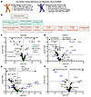
BACKGROUND. Infection is an important complication of implanted devices and prosthetics. Identifying infections sufficiently early to salvage implants and avoid reconstructive failure is a persistent medical challenge. METHODS. Two female cohorts >21 years undergoing breast implant reconstruction were recruited. Seroma fluid (82 breasts, 70 patients) was collected upon implant removal for infectious or non-infectious causes. Post-implantation drain fluid (100 samples, 44 breasts, 32 patients) was collected at routine visits prior to implant removal. A liquid-chromatography/mass spectrometry-based metabolomic approach was used to identify infection correlates. RESULTS. In seroma fluid specimens, infection was associated with a diverse set of small molecules including acetylated polyamines, defensins, glucosyl-sphingosine, and several peptide-like features (all P<0.001, diagnostic areas under the receiver operating curve 0.82-0.93). Notably, a subset of these markers were significantly elevated (p<0.05) in post-implantation drain fluid before recorded infection symptoms and diagnosis. Pseudomonas aeruginosa and its specialized exometabolites in drain specimens were also associated with subsequent P. aeruginosa infections. CONCLUSION. Tissue fluid from infected patients has a distinctive metabolome reflecting human and bacterial physiologic processes that often precede clinical diagnoses. A diagnostic based on these findings has potential to improve patient outcomes through early recognition of infection. TRIAL REGISTRATION. Not applicable. FUNDING. Work was supported by U54CK000609 from the CDC and an unencumbered research gift to TMM from Sientra. Metabolomic approaches were supported by RO1DK125860 and RO1DK111930 to JPH. The contents are solely the responsibility of the authors and do not necessarily represent the official views of CDC.
Authors
John A. Wildenthal, Margaret A. Olsen, Hung D. Tran, John I. Robinson, Terence M. Myckatyn, David K. Warren, Keith E. Brandt, Marissa M. Tenenbaum, Joani M Christensen, Thomas H. Tung, Justin M. Sacks, Rachel A. Anolik, Katelin B. Nickel, Hideji Fujiwara, Peter J. Mucha, Jeffrey P. Henderson
Abstract
While current antivirals primarily target viral proteins, host-directed strategies remain underexplored. Here, we performed a genome-wide CRISPRi screening to identify the host protein, Hepatocyte Growth Factor-Regulated Tyrosine Kinase Substrate (HGS), as essential for the pan-coronaviruses infection both in vitro and in vivo. Mechanistically, HGS directly interacts with the viral membrane (M) protein, facilitating its trafficking to the ER-Golgi intermediate compartment (ERGIC) for virion assembly. Conversely, HGS deficiency caused M retention in the ER, blocking assembly. Leveraging this interaction, we designed M-derived peptides and screened over 5,000 FDA-approved drugs, identifying riboflavin tetrabutyrate (RTB). Both the peptides and RTB bind HGS and disrupt its interaction with the M protein, leading to M retention in the ER and subsequent blockade of virion assembly. These agents demonstrated broad anti-pan-coronavirus activity in vitro and in vivo. Collectively, our findings establish HGS as a druggable host target and identify RTB as a promising broad-spectrum antiviral candidate.
Authors
Xubing Long, Rongrong Chen, Rong Bai, Buyun Tian, Yu Cao, Kangying Chen, Fuyu Li, Yiliang Wang, Yongjie Tang, Qi Yang, Liping Ma, Fan Wang, Maoge Zhou, Xianjie Qiu, Yongzhi Lu, Jie Zheng, Peng Zhou, Xinwen Chen, Qian Liu, Xuepeng Wei, Yongxia Shi, Yanhong Xue, Jincun Zhao, Wei Ji, Liqiao Hu, Jinsai Shang, Tao Xu, Zonghong Li
Abstract
Authors
Isin Y. Comba, Tijs Louwies, Ruben A. Mars, Yang Xiao, Prabhjot Kaur Sekhon, Brian S. Edwards, Adam Willits, Robin R. Shields-Cutler, Shreya Bellampalli, Arnaldo Mercado-Perez, Dennis R. Tienter, Lisa M. Till, David R. Linden, Gianrico Farrugia, Arthur Beyder, Kristen M. Smith-Edwards, Purna C. Kashyap
Abstract
Single-cell studies have revealed that intestinal macrophages maintain gut homeostasis through the balanced actions of reactive (inflammatory) and tolerant (non-inflammatory) subpopulations. How such balance is impaired in inflammatory bowel diseases (IBD), including Crohn’s disease (CD) and ulcerative colitis (UC), remains unresolved. Here, we define colon-specific macrophage states and reveal the critical role of non-inflammatory colon-associated macrophages (niColAMs) in IBD recovery. Through trans-scale analyses—integrating computational transcriptomics, proteomics, and in vivo interventional studies—we identified GIV (CCDC88A) as a key regulator of niColAMs. GIV emerged as the top-ranked gene in niColAMs that physically and functionally interacts with NOD2, an innate immune sensor implicated in CD and UC. Myeloid-specific GIV depletion exacerbates infectious colitis, prolongs disease, and abolishes the protective effects of the NOD2 ligand, muramyl dipeptide, in colitis and sepsis models. Mechanistically, GIV’s C-terminus binds the terminal leucine-rich repeat (LRR#10) of NOD2 and is required for NOD2 to dampen inflammation and clear microbes. The CD-associated 1007fs NOD2-variant, which lacks LRR#10, cannot bind GIV—providing critical insights into how this clinically relevant variant impairs microbial sensing and clearance. These findings illuminate a critical GIV-NOD2 axis essential for gut homeostasis and highlight its disruption as a driver of dysbiosis and inflammation in IBD.
Authors
Gajanan D. Katkar, Mahitha Shree Anandachar, Stella-Rita C. Ibeawuchi, Ella G. McLaren, Megan L. Estanol, Kennith Carpio-Perkins, Shu-Ting Hsu, Celia R. Espinoza, Jane E. Coates, Yashaswat S. Malhotra, Madhubanti Mullick, Vanessa Castillo, Daniella Vo, Saptarshi Sinha, Pradipta Ghosh
No posts were found with this tag.


Copyright © 2026 American Society for Clinical Investigation
ISSN: 0021-9738 (print), 1558-8238 (online)