Bone biology
Abstract
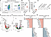
Intervertebral disc degeneration (IVDD) is a leading cause of low back pain, yet clinically, there remains no effective therapeutic approach to reverse its progression, imposing a substantial socioeconomic burden. While multiple factors contribute to IVDD pathogenesis, cellular senescence has emerged as a critical risk factor associated with both the incidence and progression of IVDD. Ageing and other damage factors drive nucleus pulposus cells (NPCs) towards a senescent phenotype characterized by increased secretion of proinflammatory factors, resulting in NPC dysfunction and tissue degeneration, which are hallmarks of IVDD. In this study, we demonstrated that PRMT2 deficiency disrupted arginine methylation‒ubiquitination crosstalk, driving NPC inflammatory senescence and accelerating IVDD progression. Mechanistically, PRMT2 loss reduced FBXO7 methylation at Arg 504, promoting the FBXO7-MED12 interaction to facilitate MED12 ubiquitination and subsequent proteasomal degradation. MED12 deficiency induced pathological R-loop accumulation, which activated the cytosolic DNA-sensing cGAS-STING axis, triggering inflammatory response cascades. Notably, engineered extracellular vesicles (EVs) delivering MED12-overexpressing plasmids effectively inhibited NP cell senescence and attenuated IVDD progression. Together, our findings establish that dysregulated methylation‒ubiquitination crosstalk critically drives IVDD progression and reveal MED12 as a promising therapeutic target for ameliorating the impact of IVDD.
Authors
Huaizhen Liang, Dingchao Zhu, Zhi Du, Xinyu Li, Rui Shi, Jie Lei, Bide Tong, Hanpeng Xu, Di Wu, Xingyu Zhou, Yifan Du, Zixuan Ou, Junyu Wei, Shuchang Peng, Wencan Ke, Zhiwei Liao, Bingjin Wang, Kun Wang, Xiaobo Feng, Yu Song, Cao Yang
Abstract
Obesity is associated with impaired wound healing, but the mechanisms linking excess adiposity to aberrant tissue repair remain unresolved. Heterotopic ossification (HO) is a severe example of pathologic tissue repair in which mesenchymal progenitor cells (MPCs) undergo aberrant osteochondral differentiation within soft tissue, leading to joint contractures and pain. Here, we show that accumulation of dietary omega-6 (ω-6) lipids in the injury site is a key mechanism linking obesity to HO. Specifically, in mice fed a high-fat diet (HFD), injured tissues were enriched in linoleic and arachidonic acids, providing substrate for myeloid cyclooxygenase-2 (COX-2)-dependent prostaglandin E2 (PGE2) production. PGE2 then drove a transcriptional program in mesenchymal progenitor cells that promoted osteochondral differentiation. An isocaloric, low linoleic acid HFD reduced HO despite comparable obesity, demonstrating that dietary lipid composition, rather than adiposity alone, drove pathological repair. Clinical data mirrored these findings, showing that obesity conferred increased HO risk, and COX-2 inhibition reduced HO exclusively in obese patients. Together, these findings identify injury site ω-6 lipid enrichment as the key signal linking the diet to MPC reprogramming, pointing to dietary lipid modulation as an actionable strategy to limit HO in obesity.
Authors
Stefanie L. Moye, Monisha Mittal, Tarun Srinivasan, Sneha Korlakunta, Chase A. Pagani, Ayelet Dar, Oromo Geshow, Dylan Feist, Lauren G. Zacharias, Zhao Li, Aaron W. James, Gerta Hoxhaj, Andrew M. Smith, Katherine A. Gallagher, Thomas P. Mathews, Robert J. Tower, Benjamin Levi
Abstract
The immune system is not only essential for host defense, but it is also involved in tissue maintenance and disease pathogenesis. Macrophages play a key role in tissue repair, fibrosis, and tumorigenesis, but the mechanisms underlying their multifunctionality have not been fully explored. Here, we identified Mrep (Ly6ChiCX3CR1loPDPN+CD9+) as a crucial subset of macrophages for muscle regeneration after muscle injury. Muscle regeneration required Mrep-derived activin A, which was produced via the TLR4/TIR domain–containing adapter-inducing interferon-β/TANK-binding kinase 1/interferon regulatory factor 3/7 signaling pathway in response to muscle injury. Mrep exerted pathological effects by secreting activin A in a model of genetically induced heterotopic ossification (HO), which was suppressed by TLR4 inhibition. Thus, this study elucidates the context-dependent functions of macrophages and the link between injury and HO, suggesting that Mrep is a potential therapeutic target for regenerating muscles and suppressing HO.
Authors
Wenqiang Yin, Kazuo Okamoto, Asuka Terashima, Warunee Pluemsakunthai, Takehito Ono, Taku Ito-Kureha, Shizuo Akira, Yoshinobu Hashizume, Roland Baron, Satoshi Ueha, Kouji Matsushima, Martin M. Matzuk, Yuji Mishina, Hiroshi Takayanagi
Abstract
Osteofibrous dysplasia (OFD) is a skeletal RASopathy presenting with periosteal bone lesions that may progress to fracture and delayed healing (pseudarthrosis). MET gene mutations reducing ubiquitin-mediated protein degradation via loss of the juxtamembrane domain (METΔJMD) were previously identified in OFD patients, resulting in ligand-dependent gain-of-function. The impact of METΔJMD expression on skeletal progenitor cell differentiation and the potential efficacy of targeted therapies remain unclear. We engineered MetΔJMD mice and showed that MetΔJMD expression inhibited osteogenic differentiation of skeletal progenitor cells in vitro and impaired cortical bone development and reduced bone stiffness in vivo. In contrast, conditional deletion of Met enhanced osteogenic differentiation of periosteal progenitor cells. Inhibition of MAPK signaling with MEK inhibitors restored osteogenic differentiation of mouse MetΔJMD skeletal progenitor cells and promoted activation of transcriptional signatures associated with skeletal development and osteoblast differentiation in OFD patient pseudarthrosis-derived primary cells. With this preclinical support, we treated with the MEK inhibitor mirdametinib a pediatric OFD patient suffering from a 3-year history of persistent pseudarthrosis, resulting in fracture union. Our findings demonstrate a bi-directional role for MET in regulating osteogenic differentiation of skeletal progenitor cells and a therapeutic avenue to improve clinical outcomes for this, and potential other, skeletal RASopathies.
Authors
Aysha Khalid, Kristin Denton, Nandina Paria, Ila Oxendine, Meghan Wassell, Reuel Cornelia, Sasidhar Uppuganti, Jeffry S. Nyman, G. Jayashree Jagadeesh, Carlos R. Ferreira, Simon J. Conway, Robert E. Hammer, John O. Ritter, Mylinh Nguyen, David A. Podeszwa, Laura J. Klesse, Carol A. Wise, Jonathan J. Rios
Abstract
Authors
Fadil M. Hannan, Mark Stevenson, Taha Elajnaf, Hussam Rostom, Kate E. Lines, Michelle Stewart, Sara Wells, Lee Moir, Thomas J. Gardella, Rajesh V. Thakker

Abstract
Although mammals generally demonstrate limited regenerative capacity compared with amphibians, the digit tip retains remarkable regenerative potential, providing a useful model to study successful mammalian regeneration. This process involves coordinated immune cell activity, vascular remodeling, and tissue reconstruction, yet the molecular checkpoints controlling regenerative versus fibrotic outcomes remain poorly understood. In mammals, regeneration of the digit tip (P3) proceeds through myeloid cell migration, early osteoclast-mediated osteolysis of the distal bone, and subsequent blastema-mediated regeneration. Here we test the hypothesis that lymphatic vessels regulate regenerative capacity by modulating local immune cell dynamics and osteoclast function. Using a lymphatic system–specific reporter line, we discovered that lymphatic vessels grow toward the nail region from the ventral side of the digit during quiescence and after amputation. These lymphatics closely surround, but do not invade, the native or regenerated bone. Unexpectedly, genetic, pharmacological, and surgical inhibition of lymphangiogenesis accelerated early osteolysis through enhanced transition of myeloid cells to osteoclasts, resulting in faster and more robust regeneration. These findings reveal a mechanism linking lymphatic vessel, immune regulation, and bone remodeling, suggesting that targeted manipulation of lymphatics dynamics may enhance regenerative outcomes after musculoskeletal injury.
Authors
Neda Vishlaghi, Trisha K. Ghotra, Monisha Mittal, Ji Hae L. Choi, Sneha Korlakunta, Mingquan Yan, Janna L. Crossley, Danielle Griswold-Wheeler, Elnaz Ghotbi, Conan Juan, Shiri Gur-Cohen, Babak Mehrara, David A. Brown, Michael T. Dellinger, Lindsay A. Dawson, Benjamin Levi
Abstract
Cellular senescence is a heterogeneous phenotype characterized primarily in mesenchymal cells, but the extent to which immune cells differ in their senescence phenotype, or “senotype”, is unclear. Here, we applied single-cell approaches alongside both global and cell-specific genetic senolytic mouse models to evaluate the senotype of immune cells in the bone marrow of aging mice. We found that myeloid-lineage cells exhibited the highest expression of p16 and senescence-associated secretory phenotype markers among all immune cell types. In contrast to clearance of p16+ senescent mesenchymal cells, targeted clearance of p16+ myeloid cells in aged mice only had minor effects on age-related bone loss in male mice, with no effects in females. In more detailed analyses, p16+ myeloid cells were only acutely cleared, being repopulated back to basal levels within a short time period. This led to a lack of long-lasting reduction in senescent cell burden, unlike when targeting bone mesenchymal cells. In vitro, myeloid-lineage cells differed markedly from mesenchymal cells in the development of a senescent phenotype. Collectively, our findings indicate that aged bone marrow myeloid cells do not achieve the fully developed senescent phenotype originally described in mesenchymal cells, justifying further characterization of senotypes of immune cells across tissues.
Authors
Madison L. Doolittle, Mitchell N. Froemming, Jennifer L. Rowsey, Ming Ruan, Leena Sapra, Joshua N. Farr, David G. Monroe, Sundeep Khosla

Abstract
Sympathetic tone is a central signaling axis inhibiting osteogenesis; however, the combination of durable local and systemic sympathetic effects on bone argues that multiple mechanisms, including yet-undiscovered pathways, are involved. Here, we found that sympathetic nerves constituted a component of the skeletal stem cell (SSC) niche: mice with conditional deletion of the classical axonal repellent Slit2 in sympathetic nerves (Slit2th mice), but not in bone stem/progenitor cells or sensory nerves, showed osteopenia due to an increase in sympathetic innervation and an associated decrease in SSCs. Mice with increased skeletal sympathetic innervation displayed impaired SSC niche function in an SSC orthotopic transplantation and engraftment system. Follistatin-like 1 (FSTL1) is a SLIT2-regulated soluble factor suppressing SSC self-renewal and osteogenic capacity. Accordingly, ablation of Fstl1 in sympathetic neurons enhanced SSC-driven osteogenesis and attenuated the bone loss seen in Slit2th mice. Together, the findings indicate that SLIT2 is a regulator of a sympathetic nerve–mediated SSC niche.
Authors
Zuoxing Wu, Na Li, Zhengqiong Luo, Zihan Chen, Xuemei He, Jie Han, Xixi Lin, Fan Shi, Haitao Huang, Baohong Shi, Yu Li, Xin Wang, Lin Meng, Dachuan Zhang, Lanfen Chen, Dawang Zhou, Weinan Cheng, Matthew B. Greenblatt, Ren Xu
Abstract
Osteoarthritis (OA) is the most common joint disease. Controlling the complex pathogenesis is challenging, thus disease-modifying OA drugs are not available. Forkhead box O (FOXO) transcription factors contribute to cartilage homeostasis through autophagy and oxidative stress resistance. Here, we sought to discover FOXO activators and found that cyproheptadine, a histamine H1 receptor (HRH1) inverse agonist, promoted FOXO3 nuclear translocation and increased FOXO target genes while suppressing inflammation. In a murine OA model, cyproheptadine reduced structural joint tissue damage and pain behaviors. Mechanistically, the inhibition of HRH1 constitutive activity mediated the effects of cyproheptadine on calcium balance between endoplasmic reticulum (ER) and cytoplasm, and FOXO activation was part of this mechanism. The anti-inflammatory effect of cyproheptadine involved the inhibition of protein kinase C/NF-κB pathway. HRH1 inhibition also suppressed osteogenesis in mesenchymal stem cells and nerve growth factor expression, which are mechanisms of osteophyte formation and pain behaviors. Moreover, cyproheptadine suppressed ER stress-induced lipogenesis by upregulating insulin-induced gene 1. Our findings suggest that HRH1 constitutive activity controls important OA-promoting mechanisms and indicate that HRH1 inverse agonists are promising drug repurposing candidates for structure and pain improvement in OA.
Authors
Ichiro Kurakazu, Merissa Olmer, Hannah Swahn, Kevin Myers, Chelsea Kenvisay, Yukio Akasaki, Yasuharu Nakashima, Martin K. Lotz
Abstract
Inflammatory diseases contribute to secondary osteoporosis. Hypertension is a highly prevalent inflammatory condition that is clinically associated with reduced bone mineral density and increased risk for fragility fracture. In this study, we showed that a significant loss in bone mass and strength occurs in two pre-clinical models of hypertension. This accompanied increases in immune cell populations, including monocytes, macrophages, and IL-17A-producing T cell subtypes in the bone marrow of hypertensive mice. Neutralizing IL-17A in angiotensin (ang) II-infused mice blunted hypertension-induced loss of bone mass and strength due to decreased osteoclastogenesis. Likewise, the inhibition of the CSF-1 receptor blunted loss of bone mass and prevented loss of bone strength in hypertensive mice. In an analysis of UK Biobank data, circulating bone remodeling markers exhibited striking associations with blood pressure and bone mineral density in > 27,000 humans. These findings illustrate a potential mechanism by which hypertension activates immune cells in the bone marrow, encouraging osteoclastogenesis and eventual loss in bone mass and strength.
Authors
Elizabeth M. Hennen, Sasidhar Uppuganti, Néstor de la Visitación, Wei Chen, Jaya Krishnan, Lawrence A. Vecchi III, David M. Patrick, Mateusz Siedlinski, Matteo Lemoli, Rachel Delgado, Mark P. de Caestecker, Wenhan Chang, Tomasz J. Guzik, Rachelle W. Johnson, David G. Harrison, Jeffry S. Nyman


Copyright © 2026 American Society for Clinical Investigation
ISSN: 0021-9738 (print), 1558-8238 (online)