
Advertisement
Research ArticleCell biologyVascular biology
Open Access |  10.1172/JCI193385
Mechanoreceptor plexin D1 regulates lymphatic valve morphogenesis and lymphedema pathogenesis
Kar-Lai Pang,1,2 Vedanta Mehta,1,2 Claire Aitken,1,2 Sara E. Dobbins,3 Jing Yu,2,4 Gabriele Bonetti,5 Adam N. Keen,1,2 Feiran Zhang,1,2 Amélie Sabine,6 Tatiana V. Petrova,6 Paul R. Riley,7 E. Yvonne Jones,2,4 Sandro Michelini,8 Matteo Bertelli,5 John S. Reader,1,2 Pia Ostergaard,3 and Ellie Tzima1,2
1Division of Cardiovascular Medicine, Radcliffe Department of Medicine, and
2Centre for Human Genetics, University of Oxford, Oxford, United Kingdom.
3School of Health and Medical Sciences, City St George’s University of London, London, United Kingdom.
4Division of Structural Biology, Centre for Human Genetics, University of Oxford, Oxford, United Kingdom.
5MAGI Euregio, Bolzano, Italy.
6Department of Oncology, University of Lausanne and Ludwig Institute for Cancer Research, Lausanne, Switzerland.
7Department of Physiology, Anatomy and Genetics, University of Oxford, Oxford, United Kingdom.
8Diagnostic and rehabilitative Service, San Giovanni Battista Hospital, Rome, Italy.
Address correspondence to: Ellie Tzima, Centre for Human Genetics, Roosevelt Drive, OX3 7BN, Oxford, United Kingdom. Phone: 44.0.1865.287588; Email: ellie.tzima@cardiov.ox.ac.uk.
Find articles by Pang, K. in: PubMed | Google Scholar
1Division of Cardiovascular Medicine, Radcliffe Department of Medicine, and
2Centre for Human Genetics, University of Oxford, Oxford, United Kingdom.
3School of Health and Medical Sciences, City St George’s University of London, London, United Kingdom.
4Division of Structural Biology, Centre for Human Genetics, University of Oxford, Oxford, United Kingdom.
5MAGI Euregio, Bolzano, Italy.
6Department of Oncology, University of Lausanne and Ludwig Institute for Cancer Research, Lausanne, Switzerland.
7Department of Physiology, Anatomy and Genetics, University of Oxford, Oxford, United Kingdom.
8Diagnostic and rehabilitative Service, San Giovanni Battista Hospital, Rome, Italy.
Address correspondence to: Ellie Tzima, Centre for Human Genetics, Roosevelt Drive, OX3 7BN, Oxford, United Kingdom. Phone: 44.0.1865.287588; Email: ellie.tzima@cardiov.ox.ac.uk.
Find articles by Mehta, V. in: PubMed | Google Scholar
1Division of Cardiovascular Medicine, Radcliffe Department of Medicine, and
2Centre for Human Genetics, University of Oxford, Oxford, United Kingdom.
3School of Health and Medical Sciences, City St George’s University of London, London, United Kingdom.
4Division of Structural Biology, Centre for Human Genetics, University of Oxford, Oxford, United Kingdom.
5MAGI Euregio, Bolzano, Italy.
6Department of Oncology, University of Lausanne and Ludwig Institute for Cancer Research, Lausanne, Switzerland.
7Department of Physiology, Anatomy and Genetics, University of Oxford, Oxford, United Kingdom.
8Diagnostic and rehabilitative Service, San Giovanni Battista Hospital, Rome, Italy.
Address correspondence to: Ellie Tzima, Centre for Human Genetics, Roosevelt Drive, OX3 7BN, Oxford, United Kingdom. Phone: 44.0.1865.287588; Email: ellie.tzima@cardiov.ox.ac.uk.
Find articles by Aitken, C. in: PubMed | Google Scholar
1Division of Cardiovascular Medicine, Radcliffe Department of Medicine, and
2Centre for Human Genetics, University of Oxford, Oxford, United Kingdom.
3School of Health and Medical Sciences, City St George’s University of London, London, United Kingdom.
4Division of Structural Biology, Centre for Human Genetics, University of Oxford, Oxford, United Kingdom.
5MAGI Euregio, Bolzano, Italy.
6Department of Oncology, University of Lausanne and Ludwig Institute for Cancer Research, Lausanne, Switzerland.
7Department of Physiology, Anatomy and Genetics, University of Oxford, Oxford, United Kingdom.
8Diagnostic and rehabilitative Service, San Giovanni Battista Hospital, Rome, Italy.
Address correspondence to: Ellie Tzima, Centre for Human Genetics, Roosevelt Drive, OX3 7BN, Oxford, United Kingdom. Phone: 44.0.1865.287588; Email: ellie.tzima@cardiov.ox.ac.uk.
Find articles by Dobbins, S. in: PubMed | Google Scholar
1Division of Cardiovascular Medicine, Radcliffe Department of Medicine, and
2Centre for Human Genetics, University of Oxford, Oxford, United Kingdom.
3School of Health and Medical Sciences, City St George’s University of London, London, United Kingdom.
4Division of Structural Biology, Centre for Human Genetics, University of Oxford, Oxford, United Kingdom.
5MAGI Euregio, Bolzano, Italy.
6Department of Oncology, University of Lausanne and Ludwig Institute for Cancer Research, Lausanne, Switzerland.
7Department of Physiology, Anatomy and Genetics, University of Oxford, Oxford, United Kingdom.
8Diagnostic and rehabilitative Service, San Giovanni Battista Hospital, Rome, Italy.
Address correspondence to: Ellie Tzima, Centre for Human Genetics, Roosevelt Drive, OX3 7BN, Oxford, United Kingdom. Phone: 44.0.1865.287588; Email: ellie.tzima@cardiov.ox.ac.uk.
Find articles by Yu, J. in: PubMed | Google Scholar
1Division of Cardiovascular Medicine, Radcliffe Department of Medicine, and
2Centre for Human Genetics, University of Oxford, Oxford, United Kingdom.
3School of Health and Medical Sciences, City St George’s University of London, London, United Kingdom.
4Division of Structural Biology, Centre for Human Genetics, University of Oxford, Oxford, United Kingdom.
5MAGI Euregio, Bolzano, Italy.
6Department of Oncology, University of Lausanne and Ludwig Institute for Cancer Research, Lausanne, Switzerland.
7Department of Physiology, Anatomy and Genetics, University of Oxford, Oxford, United Kingdom.
8Diagnostic and rehabilitative Service, San Giovanni Battista Hospital, Rome, Italy.
Address correspondence to: Ellie Tzima, Centre for Human Genetics, Roosevelt Drive, OX3 7BN, Oxford, United Kingdom. Phone: 44.0.1865.287588; Email: ellie.tzima@cardiov.ox.ac.uk.
Find articles by Bonetti, G. in: PubMed | Google Scholar
1Division of Cardiovascular Medicine, Radcliffe Department of Medicine, and
2Centre for Human Genetics, University of Oxford, Oxford, United Kingdom.
3School of Health and Medical Sciences, City St George’s University of London, London, United Kingdom.
4Division of Structural Biology, Centre for Human Genetics, University of Oxford, Oxford, United Kingdom.
5MAGI Euregio, Bolzano, Italy.
6Department of Oncology, University of Lausanne and Ludwig Institute for Cancer Research, Lausanne, Switzerland.
7Department of Physiology, Anatomy and Genetics, University of Oxford, Oxford, United Kingdom.
8Diagnostic and rehabilitative Service, San Giovanni Battista Hospital, Rome, Italy.
Address correspondence to: Ellie Tzima, Centre for Human Genetics, Roosevelt Drive, OX3 7BN, Oxford, United Kingdom. Phone: 44.0.1865.287588; Email: ellie.tzima@cardiov.ox.ac.uk.
Find articles by Keen, A. in: PubMed | Google Scholar
1Division of Cardiovascular Medicine, Radcliffe Department of Medicine, and
2Centre for Human Genetics, University of Oxford, Oxford, United Kingdom.
3School of Health and Medical Sciences, City St George’s University of London, London, United Kingdom.
4Division of Structural Biology, Centre for Human Genetics, University of Oxford, Oxford, United Kingdom.
5MAGI Euregio, Bolzano, Italy.
6Department of Oncology, University of Lausanne and Ludwig Institute for Cancer Research, Lausanne, Switzerland.
7Department of Physiology, Anatomy and Genetics, University of Oxford, Oxford, United Kingdom.
8Diagnostic and rehabilitative Service, San Giovanni Battista Hospital, Rome, Italy.
Address correspondence to: Ellie Tzima, Centre for Human Genetics, Roosevelt Drive, OX3 7BN, Oxford, United Kingdom. Phone: 44.0.1865.287588; Email: ellie.tzima@cardiov.ox.ac.uk.
Find articles by Zhang, F. in: PubMed | Google Scholar
1Division of Cardiovascular Medicine, Radcliffe Department of Medicine, and
2Centre for Human Genetics, University of Oxford, Oxford, United Kingdom.
3School of Health and Medical Sciences, City St George’s University of London, London, United Kingdom.
4Division of Structural Biology, Centre for Human Genetics, University of Oxford, Oxford, United Kingdom.
5MAGI Euregio, Bolzano, Italy.
6Department of Oncology, University of Lausanne and Ludwig Institute for Cancer Research, Lausanne, Switzerland.
7Department of Physiology, Anatomy and Genetics, University of Oxford, Oxford, United Kingdom.
8Diagnostic and rehabilitative Service, San Giovanni Battista Hospital, Rome, Italy.
Address correspondence to: Ellie Tzima, Centre for Human Genetics, Roosevelt Drive, OX3 7BN, Oxford, United Kingdom. Phone: 44.0.1865.287588; Email: ellie.tzima@cardiov.ox.ac.uk.
Find articles by Sabine, A. in: PubMed | Google Scholar
1Division of Cardiovascular Medicine, Radcliffe Department of Medicine, and
2Centre for Human Genetics, University of Oxford, Oxford, United Kingdom.
3School of Health and Medical Sciences, City St George’s University of London, London, United Kingdom.
4Division of Structural Biology, Centre for Human Genetics, University of Oxford, Oxford, United Kingdom.
5MAGI Euregio, Bolzano, Italy.
6Department of Oncology, University of Lausanne and Ludwig Institute for Cancer Research, Lausanne, Switzerland.
7Department of Physiology, Anatomy and Genetics, University of Oxford, Oxford, United Kingdom.
8Diagnostic and rehabilitative Service, San Giovanni Battista Hospital, Rome, Italy.
Address correspondence to: Ellie Tzima, Centre for Human Genetics, Roosevelt Drive, OX3 7BN, Oxford, United Kingdom. Phone: 44.0.1865.287588; Email: ellie.tzima@cardiov.ox.ac.uk.
Find articles by Petrova, T. in: PubMed | Google Scholar
1Division of Cardiovascular Medicine, Radcliffe Department of Medicine, and
2Centre for Human Genetics, University of Oxford, Oxford, United Kingdom.
3School of Health and Medical Sciences, City St George’s University of London, London, United Kingdom.
4Division of Structural Biology, Centre for Human Genetics, University of Oxford, Oxford, United Kingdom.
5MAGI Euregio, Bolzano, Italy.
6Department of Oncology, University of Lausanne and Ludwig Institute for Cancer Research, Lausanne, Switzerland.
7Department of Physiology, Anatomy and Genetics, University of Oxford, Oxford, United Kingdom.
8Diagnostic and rehabilitative Service, San Giovanni Battista Hospital, Rome, Italy.
Address correspondence to: Ellie Tzima, Centre for Human Genetics, Roosevelt Drive, OX3 7BN, Oxford, United Kingdom. Phone: 44.0.1865.287588; Email: ellie.tzima@cardiov.ox.ac.uk.
Find articles by Riley, P. in: PubMed | Google Scholar
1Division of Cardiovascular Medicine, Radcliffe Department of Medicine, and
2Centre for Human Genetics, University of Oxford, Oxford, United Kingdom.
3School of Health and Medical Sciences, City St George’s University of London, London, United Kingdom.
4Division of Structural Biology, Centre for Human Genetics, University of Oxford, Oxford, United Kingdom.
5MAGI Euregio, Bolzano, Italy.
6Department of Oncology, University of Lausanne and Ludwig Institute for Cancer Research, Lausanne, Switzerland.
7Department of Physiology, Anatomy and Genetics, University of Oxford, Oxford, United Kingdom.
8Diagnostic and rehabilitative Service, San Giovanni Battista Hospital, Rome, Italy.
Address correspondence to: Ellie Tzima, Centre for Human Genetics, Roosevelt Drive, OX3 7BN, Oxford, United Kingdom. Phone: 44.0.1865.287588; Email: ellie.tzima@cardiov.ox.ac.uk.
Find articles by Jones, E. in: PubMed | Google Scholar
1Division of Cardiovascular Medicine, Radcliffe Department of Medicine, and
2Centre for Human Genetics, University of Oxford, Oxford, United Kingdom.
3School of Health and Medical Sciences, City St George’s University of London, London, United Kingdom.
4Division of Structural Biology, Centre for Human Genetics, University of Oxford, Oxford, United Kingdom.
5MAGI Euregio, Bolzano, Italy.
6Department of Oncology, University of Lausanne and Ludwig Institute for Cancer Research, Lausanne, Switzerland.
7Department of Physiology, Anatomy and Genetics, University of Oxford, Oxford, United Kingdom.
8Diagnostic and rehabilitative Service, San Giovanni Battista Hospital, Rome, Italy.
Address correspondence to: Ellie Tzima, Centre for Human Genetics, Roosevelt Drive, OX3 7BN, Oxford, United Kingdom. Phone: 44.0.1865.287588; Email: ellie.tzima@cardiov.ox.ac.uk.
Find articles by Michelini, S. in: PubMed | Google Scholar
1Division of Cardiovascular Medicine, Radcliffe Department of Medicine, and
2Centre for Human Genetics, University of Oxford, Oxford, United Kingdom.
3School of Health and Medical Sciences, City St George’s University of London, London, United Kingdom.
4Division of Structural Biology, Centre for Human Genetics, University of Oxford, Oxford, United Kingdom.
5MAGI Euregio, Bolzano, Italy.
6Department of Oncology, University of Lausanne and Ludwig Institute for Cancer Research, Lausanne, Switzerland.
7Department of Physiology, Anatomy and Genetics, University of Oxford, Oxford, United Kingdom.
8Diagnostic and rehabilitative Service, San Giovanni Battista Hospital, Rome, Italy.
Address correspondence to: Ellie Tzima, Centre for Human Genetics, Roosevelt Drive, OX3 7BN, Oxford, United Kingdom. Phone: 44.0.1865.287588; Email: ellie.tzima@cardiov.ox.ac.uk.
Find articles by Bertelli, M. in: PubMed | Google Scholar
1Division of Cardiovascular Medicine, Radcliffe Department of Medicine, and
2Centre for Human Genetics, University of Oxford, Oxford, United Kingdom.
3School of Health and Medical Sciences, City St George’s University of London, London, United Kingdom.
4Division of Structural Biology, Centre for Human Genetics, University of Oxford, Oxford, United Kingdom.
5MAGI Euregio, Bolzano, Italy.
6Department of Oncology, University of Lausanne and Ludwig Institute for Cancer Research, Lausanne, Switzerland.
7Department of Physiology, Anatomy and Genetics, University of Oxford, Oxford, United Kingdom.
8Diagnostic and rehabilitative Service, San Giovanni Battista Hospital, Rome, Italy.
Address correspondence to: Ellie Tzima, Centre for Human Genetics, Roosevelt Drive, OX3 7BN, Oxford, United Kingdom. Phone: 44.0.1865.287588; Email: ellie.tzima@cardiov.ox.ac.uk.
Find articles by Reader, J. in: PubMed | Google Scholar
1Division of Cardiovascular Medicine, Radcliffe Department of Medicine, and
2Centre for Human Genetics, University of Oxford, Oxford, United Kingdom.
3School of Health and Medical Sciences, City St George’s University of London, London, United Kingdom.
4Division of Structural Biology, Centre for Human Genetics, University of Oxford, Oxford, United Kingdom.
5MAGI Euregio, Bolzano, Italy.
6Department of Oncology, University of Lausanne and Ludwig Institute for Cancer Research, Lausanne, Switzerland.
7Department of Physiology, Anatomy and Genetics, University of Oxford, Oxford, United Kingdom.
8Diagnostic and rehabilitative Service, San Giovanni Battista Hospital, Rome, Italy.
Address correspondence to: Ellie Tzima, Centre for Human Genetics, Roosevelt Drive, OX3 7BN, Oxford, United Kingdom. Phone: 44.0.1865.287588; Email: ellie.tzima@cardiov.ox.ac.uk.
Find articles by
Ostergaard, P.
in:
PubMed
|
Google Scholar
|

1Division of Cardiovascular Medicine, Radcliffe Department of Medicine, and
2Centre for Human Genetics, University of Oxford, Oxford, United Kingdom.
3School of Health and Medical Sciences, City St George’s University of London, London, United Kingdom.
4Division of Structural Biology, Centre for Human Genetics, University of Oxford, Oxford, United Kingdom.
5MAGI Euregio, Bolzano, Italy.
6Department of Oncology, University of Lausanne and Ludwig Institute for Cancer Research, Lausanne, Switzerland.
7Department of Physiology, Anatomy and Genetics, University of Oxford, Oxford, United Kingdom.
8Diagnostic and rehabilitative Service, San Giovanni Battista Hospital, Rome, Italy.
Address correspondence to: Ellie Tzima, Centre for Human Genetics, Roosevelt Drive, OX3 7BN, Oxford, United Kingdom. Phone: 44.0.1865.287588; Email: ellie.tzima@cardiov.ox.ac.uk.
Find articles by Tzima, E. in: PubMed | Google Scholar
Published July 1, 2026 - More info
J Clin Invest. 2026;136(13):e193385. https://doi.org/10.1172/JCI193385.
© 2026 Pang et al. This work is licensed under the Creative Commons Attribution 4.0 International License. To view a copy of this license, visit http://creativecommons.org/licenses/by/4.0/.
Received: March 13, 2025; Accepted: May 8, 2026


-
Results
Cell-autonomous Plxnd1 signaling controls lymphatic valve morphogenesis. Single-cell transcriptomics data (18) show expression of Plxnd1 in LECs, particularly valve LECs (Supplemental Figure 1, A and B; supplemental material available online with this article; https://doi.org/10.1172/JCI193385DS1). We confirmed these data by whole-mount immunostaining, superresolution microscopy, and 3D image reconstruction of mesenteric lymphatic valves at E18.5, which showed that PLXND1 protein was prominently expressed in valve LECs and colocalized with the lymphatic transcription factor PROX1 and the junctional adhesion molecule VE-cadherin (Figure 1A). To study the role of Plxnd1 in the lymphatic vasculature, we crossed Plxnd1fl/fl mice with the tamoxifen-inducible lymphatic endothelial- cell specific CreERT2 (Prox1) mice to generate Plxnd1fl/fl controls and Plxnd1fl/fl Prox1CreERT2 mice (referred to hereafter as Plxnd1iLECKO) (Figure 1B). To ensure that there were no effects on embryonic development due to maternal Plxnd1 deletion, we administered tamoxifen to control Plxnd1fl/fl pregnant dams (that had mated with Plxnd1fl/fl Prox1CreERT2 males and therefore had a mix of Plxnd1fl/fl control embryos and Plxnd1iLECKO embryos). We chose to induce recombination (and therefore deletion of Plxnd1 in LECs) at E14.5, when lymph flow starts and after lymphatic vessels have formed, as we wanted to decouple possible roles of Plxnd1 in lymph flow–specific events from roles in early LEC specification (10). We observed specific deletion of Plxnd1 in E18.5 LECs within lymphatic, but not blood vessels, thereby validating Cre efficiency and specificity (Supplemental Figure 1, C and D). Whole-mount staining of the mesentery showed regularly spaced lymphatic valves in control embryos, with characteristic V-shape morphology at high magnification, as shown by PROX1 and VEGFR3 staining (Figure 1, C and D) and integrin α 9 (ITGA9) (Supplemental Figure 2). In contrast, Plxnd1iLECKO embryos had markedly fewer valves made up of PROX1+ cells that failed to remodel into defined lymphatic valves (Figure 1, C and D). We also studied lymphatic valve formation in the embryonic dorsal skin, which also revealed defective and reduced numbers of lymphatic valves with loss of Plxnd1 (Supplemental Figure 3, A and B). Next, we determined the developmental stage at which the lymphatic valve defect appears in Plxnd1iLECKO embryos, using PROX1hi-expressing cells as a marker for prospective valve leaflet cells (13). In stage 1, a few PROX1hi LECs appeared in the vessel; by stage 2, their number increased and they formed a circumferential ring. As development proceeds, these cells reoriented perpendicularly to the vessel axis (stage 2.5) and migrated toward the lumen, forming disk-like protrusions and a local constriction (stage 3). The valves then elongated to form V-shaped leaflets with a dense extracellular matrix core (stage 4, Supplemental Figure 2). Examination of valve-forming regions (VFRs) in E18.5 mesenteries revealed a shift toward stage 2 and reduced stages 3–4 in Plxnd1iLECKO embryos compared with Plxnd1fl/fl embryos (Figure 1, E–G). In control embryos, PROXhi nuclei and VE-cadherin were detected at both the vessel wall and centrally, consistent with collective cell migration and valve protrusion (13, 19, 20) (Supplemental Figure 4). In contrast, Plxnd1iLECKO embryos showed PROX1hi nuclei and VE-cadherin restricted to the vessel wall, indicating defective cell migration (Supplemental Figure 4). Analysis of lymphatic vascular patterning in the skin showed that the lymphatic vessels of Plxnd1iLECKO embryos were similar to those of Plxnd1fl/fl controls with regard to vessel width, branch points, and surface area in both the mesentery and embryonic dorsal skin (Supplemental Figure 5, A and B, and Supplemental Figure 6, A–D). These data therefore show that loss of Plxnd1 in LECs, at a time when lymph flow starts, led to specific defects in lymphatic valve development. To understand whether PLXND1 is also required for valve maintenance, we deleted Plxnd1 postnatally using tamoxifen-inducible, endothelial-cell specific CreERT2 mice (Plxnd1iECKO). Similar to the findings in Cdk5-KO pups using endothelial-cell specific CreERT2 mice (21), lymphatic vessels and valves appeared to be normal, and chylothorax was not observed in the Plxnd1iECKO pups (Supplemental Figure 7, A and B). Furthermore, we observed normal smooth coverage in the Plxnd1iECKO pups compared with control littermates (Supplemental Figure 8, A and B).
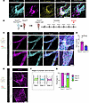 Figure 1
LEC Plxnd1 is indispensable for lymphatic valve development. (A) Whole-mount immunostaining and superresolution imaging of PLXND1 (cyan), PROX1 (magenta), and VE-cadherin (yellow) of E18.5 mesenteric lymphatic valves. 3D image reconstruction is also shown. n = 3. Scale bar: 40 μm. (B) Experimental strategy for Plxnd1 deletion in embryonic LECs. (C) Whole-mount immunostaining of E18.5 mesentery from Plxnd1fl/fl and Plxnd1iLECKO embryos stained for PROX1 and VEGFR3 as markers of lymphatic valves and lymphatic vessels, respectively. Scale bars: 200 μm and 40 μm (magnified images). (D) Quantification of lymphatic valves per millimeter of lymphatic collecting vessel in Plxnd1fl/fl and Plxnd1iLECKO E18.5 mesenteries (n = 4, data are the mean ± SEM; 2-tailed Student’s t test, *P < 0.05). (E) Representative images of E18.5 mesenteric lymphatic valves stained for PROX1 in Plxnd1fl/fl and Plxnd1iLECKO embryos. Scale bar: 50 μm. (F) Schematic diagram showing different stages of lymphatic valve formation. (G) Stage distribution within VFRs in E18.5 mesenteries. (n = 3; 10 VFRs from each embryo were analyzed; 2-tailed Student’s t test, **P < 0.01).
PLXND1 regulates the lymphatic valve program via an unconventional pathway. Mechanotransduction due to lymph flow is a critical regulator of the lymphatic valve transcriptional program (3, 4). To determine if the lymphatic valve defects observed with loss of lymphatic Plxnd1 are associated with altered mechanotransduction, we assayed the expression of key lymphatic regulators in human LECs (transfected with either scrambled or PLXND1 siRNAs) (Supplemental Figure 9A) in response to lymphatic valve shear stress (oscillatory shear stress [OSS]; ± 4 dynes/cm2, ¼ Hz) (10, 22). Our results showed that loss of PLXND1 did not affect the mRNA levels (Supplemental Figure 9, B–D), protein levels, or nuclear localization (Supplemental Figure 10, A–C) of the key lymphatic transcriptional regulators GATA2, PROX1, or FOXC2; however, expression of the downstream target gene GJA4 (which codes for CX37) was dramatically reduced in PLXND1-depleted LECs in response to lymphatic valve shear stress in vitro (Figure 2A). Notably, we confirmed reduced CX37 expression at the protein level in vitro (Figure 2B) and in vivo in lymphatic valves, but not arteries, of Plxnd1iLECKO mice (Figure 2C).
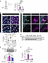 Figure 2
PLXND1 regulates the lymphatic valve program via an unconventional pathway. (A) Schematic of the lymphatic shear stress profile. Human LECs (HLECs) transfected with either scrambled or PLXND1 siRNAs were left static or exposed to shear stress for 48 hours, RNA was isolated, and quantitative PCR (qPCR) was performed to quantify the expression of GJA4 (n = 7, data are the mean ± SEM; 2-way ANOVA with Tukey’s multiple-comparison test, ****P < 0.0001). (B) HLECs transfected with scrambled or PLXND1 siRNAs were left static or exposed to shear stress for 48 hours. Cells were fixed and immunostained to quantify the expression of CX37 (300–350 cells were analyzed per condition; n = 5 biological replicates; 2-way ANOVA with Tukey’s multiple-comparison test, *P < 0.05). Scale bar: 50 μm. (C) Immunofluorescence staining of Plxnd1fl/fl and Plxnd1iLECKO E18.5 mesentery for CX37 (cyan) and PROX1 (magenta). A, artery, L, lymphatic vessel. Arrowheads indicate lymphatic valves (n = 4, scale bar: 40 μm). (D) HLECs transfected with either scrambled, PLXND1, or CDK5 siRNAs were left static or exposed to shear stress before immunoprecipitating FOXC2 and analyzing serine phosphorylation (n = 3). Immunoprecipitation with nonspecific IgG was used as a control. WCL, whole-cell lysate. (E) Phosphorylation of CDK5 (pY15) in HLECs transfected with scrambled or PLXND1 siRNAs and exposed to shear stress (n = 4, data are the mean ± SEM; 2-way ANOVA with Tukey’s multiple-comparison test, *P < 0.05, **P < 0.01). Dashed line indicates where the blot was spliced. (F) HLECs transfected with either scrambled or CDK5 siRNAs were left static or exposed to shear stress for 48 hours, and qPCR was performed to quantify the expression of GJA4 (n = 3, data are the mean ± SEM; 2-way ANOVA with Tukey’s multiple-comparison test, *P < 0.05, **P < 0.01).
To explore the molecular mechanisms by which PLXND1 regulates mechanosensitive GJA4 expression, we assayed serine phosphorylation of FOXC2, as it is critical for the transcriptional activation of this protein (10, 23). The results showed that shear stress increased serine phosphorylation of FOXC2 in control LECs, but this was abrogated with loss of PLXND1 (Figure 2D). FOXC2 protein levels remained unchanged upon PLXND1 deletion in vitro (Supplemental Figure 10C) and in vivo (Supplemental Figure 11, A and B). To explore this further, we investigated the kinase responsible for FOXC2 serine phosphorylation in response to lymphatic valve shear stress. We examined the role of CDK5, whose loss in LECs (21) phenocopies the Plxnd1iLECKO mice, by assaying its phosphorylation on Tyr15; our results showed that PLXND1 was required for activation of CDK5 in response to lymphatic valve shear stress (Figure 2E). In agreement with a role for CDK5 in this pathway, we found that CDK5 itself was essential for flow-induced serine phosphorylation of FOXC2 (Figure 2D) and upregulation of GJA4 at both the mRNA level and CX37 at the protein level (Figure 2F and Supplemental Figure 12, A and B) (21). These results, therefore, demonstrate that PLXND1 regulates the lymphatic valve gene program via an unconventional mechanism that involves posttranslational modification of FOXC2 via CDK5-dependent phosphorylation and downstream CX37 upregulation.
PLXND1 is a mechanosensor in LECs. The requirement for PLXND1 in LEC mechanotransduction raised the question of whether PLXND1 functions as a mechanosensor in these cells. To address this question, we applied tensional force on endogenous PLXND1 using magnetic beads coated with an antibody that recognizes the PLXND1 ectodomain (17). Tensional force on PLXND1 in LECs induced phosphorylation of CDK5, the kinase responsible for phosphorylation (and activation) of FOXC2 (Figure 3, A and D). This mechanosensing ability was specific to PLXND1, as force application on its coreceptor, NRP1, did not elicit a response (Figure 3, B and E). Crucially, the force-induced activation of the CDK5 pathway was unique to PLXND1, as force on another mechanosensor also expressed in LECs, PECAM-1, did not induce CDK5 phosphorylation (Figure 3, C and F). Instead, force on PECAM-1 induced activation of ERK1/2 (Figure 3I), whereas force on PLXND1 or NRP1 did not (Figure 3, G and H). These data therefore identify PLXND1 as a mechanosensor in LECs that is uniquely capable of activating the CDK5/FOXC2/CX37 pathway in response to mechanical force.
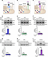 Figure 3
PLXND1 is a lymphatic force sensor. Schematic showing the tensional force experimental strategy using magnetic beads coated with antibodies against the extracellular domain of PLXND1 (A), NRP1 (B), or PECAM1 (C) in HLECs. HLECs were incubated with anti-PLXND1 (D and G), anti-NRP1 (E and H), or anti–PECAM-1 (F and I) antibody magnetic beads and subjected to 10 pN force for the indicated durations. Phosphorylation of CDK5 (pY15) and ERK1/2 (pT202/Y204) were determined by Western blotting, and quantification of the Western blots is shown (n = 3, data are the mean ± SEM; 1-way ANOVA with Kruskal-Wallis test, **P < 0.01).
A lymphatic mechanocomplex induced by lymph flow. An emerging paradigm in mechanosensation is that mechanosensors form complexes with auxiliary receptors (24); however, to our knowledge, no mechanically induced complex has ever been reported in lymphatics. In considering possible interactors of PLXND1, we first assessed lymphatic valves in vivo and human LECs in vitro for the expression of known PLXND1 coreceptors. Consistent with previous reports, we found that coreceptors of PLXND1, including VEGFR2 (25) and NRP1 (26) (Figure 4A), NRP2 (27), and VEGFR3 (28) (Supplemental Figure 13) were also expressed in lymphatic valves, although expression of NRP2 was much lower in the valve compared with the lymphangion (Supplemental Figure 13) (10). We found that ITGA9, a signature integrin of LECs required for lymphatic valve formation (29), was also highly expressed (Figure 4A). Human LECs express VEGFR2, NRP1, NRP2, and VEGFR3 (at varying levels compared with human aortic ECs); expression of ITGA9 was exclusive to LECs (Supplemental Figure 14). To determine whether PLXND1 forms mechanical force–induced interactions in LECs, PLXND1 immunoprecipitates from static and sheared LECs were immunoprobed for candidate coreceptors. Surprisingly, in response to lymphatic valve shear stress, PLXND1 formed a mechanocomplex with NRP1 and VEGFR2 (Figure 4B), but not with NRP2 and VEGFR3 (Supplemental Figure 15), despite the well-recognized roles of NRP2 and VEGFR3 in LECs (27, 30, 31). The kinase CDK5 and ITGA9 were also part of the mechanocomplex (Figure 4B). These interactions were not observed in static cells or when a control IgG antibody was used for immunoprecipitations. To solidify the existence of this lymphatic mechanocomplex, we also performed reverse immunoprecipitations of both VEGFR2 and CDK5 and saw the same flow-dependent complex formation (Figure 4, C and D). Knockdown of the PLXND1 coreceptors VEGFR2 or NRP1 abrogated the formation of the flow-dependent mechanocomplex (Supplemental Figure 16, A and B). Notably, SEMA3E treatment did not induce formation of the mechanocomplex (Supplemental Figure 17). These results therefore demonstrate the discovery of a PLXND1-based lymphatic mechanocomplex that formed in response to lymphatic valve shear stress.
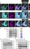 Figure 4
Force induces the formation of a lymphatic mechanocomplex. (A) Whole-mount immunostaining and superresolution imaging of E18.5 mesentery lymphatic valves for VEGFR2, NRP1, and ITGA9 and staining for the lymphatic valve marker PROX1 (n = 3. Scale bar: 40 μm). Orthogonal views in 2 planes are also shown. (B) HLECs were left static or exposed to shear stress before immunoprecipitating PLXND1 and analyzing for the presence of VEGFR2, NRP1, CDK5, and ITGA9 (n = 2). Immunoprecipitation with nonspecific IgG was used as a control. HLECs were left as static or exposed to shear stress before immunoprecipitating VEGFR2 (C) or CDK5 (D) and analyzed for its association with PLXND1, NRP1, CDK5 and ITGA9, or PLXND1, VEGFR2, ITGA9 and NRP1, respectively. Immunoprecipitation with nonspecific IgG was used as a control. n = 2 (C and D).
Rare variants in the PLXND1 gene in patients with lymphedema. Patients with primary lymphedema often present with variants in genes important for lymphatic valve formation (2, 6). We hypothesized that since Plxnd1 regulates lymphatic valve formation in mice, we might identify variants in this gene in humans with lymphatic disease. We examined exome sequencing data from 235 individuals from hospitals in Italy; crucially, these patients tested negative for variants in known lymphedema and lymphatic malformation–causing genes. We identified potential candidate variants in the PLXND1 gene in 2 patients with primary lymphedema (Supplemental Table 4, variants 1 and 5, which was confirmed by Sanger sequencing (Supplemental Figure 18, A and B). Patient RX626.2021 was diagnosed with bilateral (right>left) lower limb lymphedema at 59 years of age, while patient RX528.2021 was diagnosed with bilateral (right>left) toe and dorsal foot edema at birth (Supplemental Figure 18). Both patients tested positive on the Stemmer sign (toe skin pinch and lift) test and showed more edema in the orthostatic (standing) position than in the clinostatic (lying down) position; clinical data and lymphoscintigraphy results are shown in Supplemental Figure 18, C–E. In silico analysis revealed that the c.533G>A; pSer178Asn variant is located within the semaphorin (SEMA) binding domain of PLXND1, and the c.2026C>G; p.Gln676Glu variant is distal from its SEMA domain (Figure 5A). The 2 variants were chosen for further investigation and functional validation.
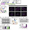 Figure 5
PLXND1 variants associated with lymphedema selectively disrupt ligand versus mechanical activation of the receptor. (A) Lollipop plot of PLXND1 variants showing a 2D representation of their location in the PLXND1 protein. The red variant (S178N) resides within the SEMA binding domain, the green variant (Q676E) is located distal from this domain, and the blue variants correspond to mutations that have not yet been functionally characterized. PSI, plexin-semaphorin-integrin homology domains; IPT/TIG, immunoglobulin plexin transcription/transcription factor immunoglobulin domains; TM, transmembrane domain. (B) Schematic showing ligand activation of PLXND1 via SEMA3E and the downstream signaling investigated. (C) SEMA3E-induced cell collapse assays. HLECs in which endogenous PLXND1 was knocked down were transduced with adenoviruses expressing WT or mutant Q676E or S178N PLXND1 and were subsequently treated with SEMA3E and immunostained with anti-vinculin antibody (cyan) and phalloidin (magenta). Representative images from 3 biological replicates are shown. Scale bar: 20 μm. (D) HLECs in which endogenous PLXND1 was knocked down were transduced with adenoviruses expressing WT or mutant PLXND1 and treated with SEMA3E before assaying phosphorylation of FAK (pY397) and cofilin (pS3) (n = 3, data are the ± SEM; 2-way ANOVA with Tukey’s multiple-comparison test, ****P < 0.0001). (E) Schematic showing mechanical activation of PLXND1 via tensional force application using magnetic beads and the downstream signaling investigated. (F) HLECs in which endogenous PLXND1 was knocked down were transduced with adenoviruses expressing WT or mutant PLXND1 and incubated with anti-PLXND1 magnetic beads followed by force application. Phosphorylation of CDK5 (pY15) was determined (n = 3, data are the mean ± SEM; 2-way ANOVA with Tukey’s multiple-comparison test, *P < 0.05).
To further determine the relevance of this finding, we examined exome sequencing data from 272 patients with primary lymphedema from St George’s University Hospitals NHS Foundation Trust, London, in conjunction with whole-genome data from 163 additional patients recruited for the 100,000 Genomes Project. An additional 7 potential candidate variants in the PLXND1 gene were identified (Figure 5A and Supplemental Table 4), which supports a potential link between PLXND1 variants and susceptibility to lymphedema.
Function of lymphedema variants in ligand and mechanical activation of PLXND1. To investigate the functional significance of the putative c.533G>A; pSer178Asn and c.2026C>G; p.Gln676Glu lymphedema variants, we transduced human LECs (depleted of endogenous PLXND1 via siRNAs) with adenoviruses expressing either WT PLXND1 or the PLXND1 Ser178Asn (S178N) or Gln676Glu (Q676E) mutants. Immunoblotting revealed similar expression levels of WT and mutant PLXND1 in LECs (Supplemental Figure 19A). To examine possible effects of the mutations on the subcellular localization of PLXND1, we performed confocal imaging of transduced Cos7 cells. Both WT and mutant PLXND1 proteins showed similar subcellular localization at the plasma membrane in Cos7 cells (Supplemental Figure 19B), suggesting that the mutations do not affect the expression or subcellular localization of PLXND1. We therefore carried out assays that focused on assessing the effect of the mutations on both the canonical, semaphorin-binding role of PLXND1 and its newly identified mechanical role in lymphatics.
To assay possible effects of the mutations on the canonical ligand activation of PLXND1, we examined responses to semaphorin 3E (SEMA3E) (Figure 5B). LECs (depleted of endogenous PLXND1 via siRNAs) transduced with adenovirus expressing WT PLXND1 displayed the typical cell collapse/retraction in response to ligand activation of PLXND1, also observed in other cell types (32–34) (Figure 5C); LECs expressing mutant Q676E PLXND1 also collapsed in response to SEMA3E, suggesting that the mutation does not interfere with the ligand activation of the receptor. In contrast, LECs expressing the S178N PLXND1 mutant failed to collapse, thus showing that this mutation interferes with the normal ligand activation response. To further explore the role of the mutations in PLXND1 ligand activation, we established 2 additional assays to measure ligand activation of PLXND1: we reasoned that SEMA3E-induced cell collapse is preceded by changes in the activation of cytoskeletal proteins. The actin-severing protein cofilin, which is activated through dephosphorylation at Ser3, regulates RhoA-dependent actin stress fiber formation (35, 36), while the focal adhesion protein FAK (autophosphorylated at Tyr397) regulates focal adhesion growth (37, 38). In response to SEMA3E, both WT and Q676E PLXND1 mutant–expressing LECs showed similar changes to the phosphorylation of cofilin and FAK (Figure 5D), consistent with similar cytoskeleton remodeling, ultimately leading to cell collapse (Figure 5C). Strikingly, these responses were absent in the S178N PLXND1–expressing LECs. Collectively, these experiments showed that ligand activation of PLXND1 was specifically affected by the S178N mutation (located within the SEMA domain of PLXND1) but not the Q676E mutation, away from the SEMA domain.
To analyze the effects of the lymphedema variants on the mechanical activation of PLXND1, we used the tensional force system to apply mechanical force on PLXND1 (Figure 5E). LECs depleted of endogenous PLXND1 and transduced with adenoviruses expressing WT, S178N, or Q676E PLXND1 were subjected to force, and the phosphorylation of CDK5 was assayed by Western blotting using phosphorylated Y15 (pY15) CDK5–specific antibodies. Like endogenous PLXND1, the WT PLXND1 transduced in LECs was able to detect mechanical force and activate the CDK5 pathway, as evidenced by increased phosphorylation of CDK5 on Y15 (Figure 5F). The S178N mutant showed a similar response. In contrast, Q676E PLXND1–expressing LECs failed to activate CDK5 in response to force, indicating that this lymphedema variant suppressed the mechanical activation of PLXND1. Taken together, these results show that 1 lymphedema variant selectively affected the ligand activation of PLXND1, while the other variant selectively affected the mechanical activation of the receptor.
Lymphedema variants interfere with lymphatic mechanocomplex formation. Given that lymphatic valve shear stress induces formation of a PLXND1-NRP1-VEGFR2-ITGA9 lymphatic mechanocomplex and the Q676E PLXND1 variant (Figure 6A) disrupts the mechanical activation of PLXND1, we tested the hypothesis that this variant affects formation of the lymphatic mechanocomplex. We transduced LECs (depleted of endogenous PLXND1) with adenoviruses expressing either WT, Q676E, or S178N PLXND1 and subjected them to lymphatic shear stress before immunoprecipitating PLXND1. Western blotting of immunoprecipitates revealed that, while WT PLXND1 associated with NRP1, VEGFR2, and ITGA9 in response to shear stress, the Q676E PLXND1 mutant did not support formation of the mechanocomplex (Figure 6B). To further explore the role of PLXND1 variants in the mechanosensitive CDK5/FOXC2/CX37 pathway, we examined 2 key downstream events: FOXC2 phosphorylation and GJA4 expression. WT PLXND1 induced serine phosphorylation of FOXC2 (Figure 6C) and GJA4 expression (Supplemental Figure 20, A and B), however, Q676E PLXND1 failed to support FOXC2 phosphorylation (Figure 6C) and upregulate GJA4 expression (Supplemental Figure 20B). Neither FOXC2 phosphorylation nor GJA4 expression was affected by S178N PLXND1 (Figure 6C and Supplemental Figure 20A). Thus, this mutation not only abolished the mechanical activation of the receptor (Figure 5F), but also disrupted formation of the lymphatic mechanocomplex. Crucially, the S178N mutant, required for the ligand response, did not interfere with the formation of the mechanocomplex.
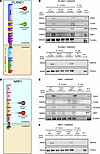 Figure 6
Lymphedema-associated variants disrupt lymphatic mechanocomplex formation and impair FOXC2 phosphorylation. (A) Schematic of the PLXND1 domain structure showing the positions of the lymphedema-associated S178N and Q676E mutations. (B) HLECs in which endogenous PLXND1 was knocked down were transduced with adenoviruses expressing WT or mutant PLXND1 and subjected to shear stress before immunoprecipitating PLXND1 and analyzing for its association with CDK5, ITGA9, VEGFR2, and NRP1 (n = 3). (C) HLECs in which endogenous PLXND1 was knocked down were transduced with adenoviruses expressing WT or mutant PLXND1 and exposed to shear stress before immunoprecipitating FOXC2 and analyzing for serine phosphorylation. (D) Schematic of the NRP1 domain structure showing position of the lymphedema-associated R552Q mutation and the VEGF-A D320K mutation. (E) HLECs in which endogenous NRP1 was knocked down were transduced with adenoviruses expressing WT or mutant NRP1 were subjected to shear stress before immunoprecipitating PLXND1 and analyzing for its association with CDK5, ITGA9, VEGFR2, and NRP1 (n = 3). (F) HLECS in which endogenous NRP1 was knocked down were transduced with adenoviruses expressing WT or mutant NRP1 and exposed to shear stress before immunoprecipitating FOXC2 and analyzing for serine phosphorylation.
In addition to PLXND1, variants in another member of the lymphatic mechanocomplex have also been associated with lymphatic diseases; a missense variant c.1655G>A, corresponding to R552Q in NRP1 segregates with lymphedema (39), although its functional relevance and mechanism of action have not been investigated. To test if the NRP1 lymphedema-associated variant affects the formation of the lymphatic mechanocomplex, we generated this variant in a human NRP1 adenovirus expression vector. We also generated an additional NRP1 mutation (D320K) (Figure 6D), which inhibits its ability to bind to VEGF-A but does not affect its expression (40), as we hypothesized that disruption of NRP1-VEGFA binding would not interfere with mechanical responses. We transduced LECs depleted of endogenous NRP1 with adenoviruses encoding WT, R552Q, or D320K NRP1 and analyzed expression levels of the various constructs by Western blotting. Their subcellular localization was assessed in Cos7 cells by confocal imaging. We found that both the lymphedema R552Q variant and the VEGF-A D320K NRP1 mutant were expressed at levels similar to those of WT NRP1 (Supplemental Figure 21A). Similarly, the surface expression was not affected by the mutations (Supplemental Figure 21B). We therefore tested the effects of the mutants on formation of the lymphatic mechanocomplex. LECs depleted of endogenous NRP1 and transduced with adenoviruses expressing WT, R552Q, or D320K NRP1 were subjected to lymphatic shear stress, and formation of the mechanocomplex was assessed following immunoprecipitation of PLXND1. Our data showed that LECs expressing WT NRP1 formed the mechanocomplex, however, the lymphedema missense variant R552Q led to a complete loss of the mechanocomplex. Interestingly, LECs expressing the D320K NRP1 mutant showed normal mechanocomplex formation in response to lymphatic shear stress, suggesting that the NRP1 mutation that interferes with VEGF-A binding does not affect the mechanical response (Figure 6E). We next examined how NRP1 mutants affect the mechanosensitive CDK5/FOXC2/CX37 cascade. WT NRP1 induced robust FOXC2 serine phosphorylation and GJA4 expression, whereas the R552Q variant failed to induce FOXC2 phosphorylation (Figure 6F) or GJA4 expression (Supplemental Figure 20C). PLXND1 expression level remained unchanged by the NRP1 mutation (Supplemental Figure 20D). Collectively, these results identify disruption of the lymphatic mechanocomplex as the shared biological mechanism underlying rare variants in PLXND1 and NRP1.


Copyright © 2026 American Society for Clinical Investigation
ISSN: 0021-9738 (print), 1558-8238 (online)