
Go to JCI Insight
  • About
  • Editors
  • Consulting Editors
  • For authors
  • Journal stats
  • Publication ethics
  • Publication alerts by email
  • Advertising
  • Job board
  • Contact
  • Clinical Research and Public Health
  • Current issue
  • Past issues
  • By specialty
    • COVID-19
    • Cardiology
    • Gastroenterology
    • Immunology
    • Metabolism
    • Nephrology
    • Neuroscience
    • Oncology
    • Pulmonology
    • Vascular biology
    • All ...
  • Videos
    • ASCI Milestone Awards
    • Video Abstracts
    • Conversations with Giants in Medicine
  • Reviews
    • View all reviews ...
    • Emerging therapeutic strategies in breast cancer (Oct 2026)
    • The cGAS-STING pathway: DNA sensing in health and disease (Jun 2026)
    • Neurodegeneration (Mar 2026)
    • Clinical innovation and scientific progress in GLP-1 medicine (Nov 2025)
    • Pancreatic Cancer (Jul 2025)
    • Complement Biology and Therapeutics (May 2025)
    • Evolving insights into MASLD and MASH pathogenesis and treatment (Apr 2025)
    • View all review series ...
  • Viewpoint
  • Collections
    • In-Press Preview
    • Clinical Research and Public Health
    • Research Letters
    • Letters to the Editor
    • Editorials
    • Commentaries
    • Editor's notes
    • Reviews
    • Viewpoints
    • 100th anniversary
    • Top read articles

  • Current issue
  • Past issues
  • Specialties
  • Reviews
  • Review series
  • ASCI Milestone Awards
  • Video Abstracts
  • Conversations with Giants in Medicine
  • In-Press Preview
  • Clinical Research and Public Health
  • Research Letters
  • Letters to the Editor
  • Editorials
  • Commentaries
  • Editor's notes
  • Reviews
  • Viewpoints
  • 100th anniversary
  • Top read articles
  • About
  • Editors
  • Consulting Editors
  • For authors
  • Journal stats
  • Publication ethics
  • Publication alerts by email
  • Advertising
  • Job board
  • Contact
Top
  • View PDF
  • Download citation information
  • Send a comment
  • Terms of use
  • Standard abbreviations
  • Need help? Email the journal
  • Top
  • Abstract
  • Introduction
  • Currently approved and emerging ADCs in breast cancer
  • Structure, design principles, and mechanisms of action
  • Current challenges and pressing translational questions
  • Future generations of ADCs in breast cancer
  • Concluding perspective
  • Conflict of interest
  • Funding support
  • Footnotes
  • References
  • Version history
  • Article usage
  • Citations to this article

Advertisement

Review Series Open Access | 10.1172/JCI207630

Antibody-drug conjugates in breast cancer: redefining targeted therapy

Chenxu Guo1,2 and Leif W. Ellisen1,2,3

1Mass General Brigham Cancer Institute, Boston, Massachusetts, USA.

2Harvard Medical School, Boston, Massachusetts, USA.

3Ludwig Center at Harvard, Boston, Massachusetts, USA.

Address correspondence to: Leif W. Ellisen, CPZN 4204, 185 Cambridge Street, Boston, Massachusetts, 02114, USA. Email: LELLISEN@mgh.harvard.edu.

Find articles by Guo, C. in: PubMed | Google Scholar |

1Mass General Brigham Cancer Institute, Boston, Massachusetts, USA.

2Harvard Medical School, Boston, Massachusetts, USA.

3Ludwig Center at Harvard, Boston, Massachusetts, USA.

Address correspondence to: Leif W. Ellisen, CPZN 4204, 185 Cambridge Street, Boston, Massachusetts, 02114, USA. Email: LELLISEN@mgh.harvard.edu.

Find articles by Ellisen, L. in: PubMed | Google Scholar |

Published October 1, 2026 - More info

Published in Volume 136, Issue 19 on October 1, 2026
J Clin Invest. 2026;136(19):e207630. https://doi.org/10.1172/JCI207630.
© 2026 Guo, et al. This work is licensed under the Creative Commons Attribution 4.0 International License. To view a copy of this license, visit http://creativecommons.org/licenses/by/4.0/.
Published October 1, 2026 - Version history
View PDF
Abstract

Antibody-drug conjugates (ADCs) have transformed the treatment landscape of breast cancer and redefined the conceptual distinction between targeted therapy and conventional chemotherapy. Originally conceived as “magic bullets” that selectively deliver cytotoxic warheads to antigen-expressing tumor cells, clinical and mechanistic evidence indicates that ADC activity depends on a broader interplay of target-dependent and target-independent mechanisms, including extracellular payload release, bystander killing, off-tumor uptake, and immune modulation. Here, we examine ADCs in breast cancer as a distinct therapeutic paradigm. We discuss how antigen biology, linker chemistry, payload features, and drug-to-antibody ratio collectively determine efficacy, toxicity, and therapeutic index. We then compare currently approved and emerging HER2- and TROP2-directed ADCs, highlighting how differences in linker stability, payload pharmacology, and bystander capacity can affect clinical outcomes in ADCs sharing the same target. We further discuss the biological basis and translational challenges of de novo and acquired resistance related to targets, payloads, and tumor microenvironmental constraints, as well as the implications of these mechanisms for biomarker development, sequencing rationales, and combination strategies with immune checkpoint inhibitors and DNA repair–targeting therapies. Finally, we outline future directions of ADC development, including expansion of the target space, novel payload modalities, and next-generation antibody and conjugation engineering.

Introduction

Paul Ehrlich’s century-old “magic bullet” concept envisioned drugs that selectively eradicate diseased cells (1). Antibody-drug conjugates (ADCs) have the potential to realize this vision by coupling highly specific monoclonal antibodies to potent cytotoxic payloads, preferentially directing killing effects to cancer cells. By concentrating potent drugs in tumors while sparing normal tissues, ADCs are designed to expand the narrow therapeutic index of conventional chemotherapies and other bioactive compounds. This effect is achieved through target-selective delivery of a higher maximum tolerated dose (MTD) and use of potent toxins with a lower minimum effective dose that are otherwise too toxic for systemic administration.

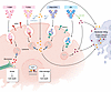
Structurally, a typical ADC comprises three core components: a target-specific antibody, a chemical linker, and a cytotoxic small molecule payload (Figure 1). Upon target engagement, ADCs are proposed to operate through a multistep process involving antigen binding, internalization, endolysosomal trafficking, and lysosomal degradation, leading to payload release and ultimately tumor cell death (Figure 1). Despite extensive investigation across preclinical and clinical settings, no consensus exists on optimal design and characteristics of these individual components, and approved breast cancer ADCs span a wide range of successful engineering strategies (2, 3).
Distinct structural features and mechanisms of action of FDA-approved ADCsFigure 1

Distinct structural features and mechanisms of action of FDA-approved ADCs in breast cancer. Trastuzumab emtansine (T-DM1), trastuzumab deruxtecan (T-DXd), datopotamab deruxtecan (Dato-DXd), and sacituzumab govitecan (SG) differ in target, linker chemistry, payload class, and drug-to-antibody ratio, which together shape intracellular processing, bystander activity, and downstream cytotoxic effects. T-DM1 carries the noncleavable maytansinoid payload DM1 and primarily requires antigen binding, internalization, lysosomal degradation, and intracellular release of a membrane-impermeable catabolite that disrupts tubulin dynamics and induces mitotic arrest, with minimal bystander killing. In contrast, T-DXd and Dato-DXd contain cleavable tetrapeptide linkers and membrane-permeable topoisomerase 1 inhibitor (TOP1i) payloads that can be released after cathepsin-mediated cleavage, diffuse into neighboring antigen-low or antigen-negative cells, and promote bystander killing. SG similarly delivers a TOP1i payload through a pH-sensitive linker that can undergo hydrolysis in acidic conditions, further supporting extracellular payload liberation and bystander effects.

The emerging consensus from both preclinical and clinical studies is that ADCs constitute a hybrid modality whose payload delivery is variably target dependent and independent (4). Of note, ADCs are targeted primarily in their biodistribution rather than necessarily in their cytotoxic effects. Following payload release, particularly for membrane-permeable payloads, cytotoxic activity may extend beyond antigen-expressing cells through bystander killing of adjacent cells and extracellular payload diffusion. ADC efficacy and toxicity are governed by unique biological principles including antigen biology, intracellular trafficking, payload pharmacology, and tumor microenvironment (TME) context, motivating a mechanistic and translationally focused assessment to optimize their application for breast cancer therapy.
Currently approved and emerging ADCs in breast cancer

Overview of FDA-approved ADCs in breast cancer. Currently, four ADCs are FDA approved for breast cancer treatment, utilizing two molecular targets, two payload classes, and distinct linkers (Table 1). T-DM1 couples the human epidermal growth factor receptor 2 (HER2) antibody trastuzumab to the tubulin inhibitor emtansine (DM1) via a noncleavable MCC linker, restricting cytotoxicity to HER2-overexpressing cells without bystander activity (5). T-DXd uses a cathepsin-cleavable linker to deliver the topoisomerase 1 inhibitor (TOP1i) deruxtecan (DXd), enabling lysosomal release and membrane-permeable diffusion to neighboring cells (6). SG targets tumor-associated calcium signal transducer 2–expressing (TROP2-expressing) cells with a pH-sensitive hydrolyzable linker to deliver the TOP1i SN-38, generating sustained intracellular and extracellular payload release within the TME (7, 8). Dato-DXd also targets TROP2 but employs a cleavable linker with DXd, conferring bystander capacity analogous to T-DXd (9, 10).

Table 1

Comparison of FDA-approved ADCs in breast cancer

Notably, T-DXd, Dato-DXd, and SG share TOP1i payloads with cleavable or hydrolyzable linkers, a commonality in design that confers bystander killing through extracellular release of membrane-permeable payloads. These ADCs span a broad range of drug-to-antibody ratios (DARs) (~4 to ~8), suggesting that optimized linker-payload chemistry can preserve tolerability and therapeutic index despite varying DAR.

HER2-directed ADCs: a shared target and divergent designs. T-DM1 and T-DXd exemplify how ADCs sharing the same antibody diverge in efficacy and toxicity due to differences in linker cleavability, payload type, and bystander capacity. While T-DM1 became a standard treatment for HER2-positive disease through the EMILIA study in the metastatic setting (11) and the KATHERINE trial in the post-neoadjuvant setting (12), T-DXd was subsequently proved to be generally more effective. In head-to-head trials, T-DXd dramatically outperformed T-DM1 for both HER2-positive metastatic disease (DESTINY-Breast03; median progression-free survival [PFS] 25.1 vs. 7.2 months) (13) and post-neoadjuvant residual disease (DESTINY-Breast05; 3-year invasive disease–free survival 92.4% vs. 83.7%) (14). The mechanistic basis for T-DXd’s superiority to T-DM1 is likely multifactorial: its higher DAR delivers more payload per antibody molecule; its DXd payload is more potent than DM1’s; and its cleavable linker with membrane-permeable payload confers bystander killing that T-DM1 lacks (6, 15).

The success of T-DXd has also expanded the scope of HER2-directed therapy beyond clinically HER2-positive disease to tumors with lower levels of HER2 expression that lack HER2 oncogene addiction. Indeed, T-DXd demonstrated significant PFS and overall survival (OS) benefits compared with chemotherapy in HER2-low (DESTINY-Breast04) (16) and HER2-ultralow (DESTINY-Breast06) (17) breast cancers. Meanwhile, newer HER2-ADCs currently in trials illustrate persistent trade-offs between efficacy and distinct toxicities of these agents. Trastuzumab duocarmazine improved PFS over physician’s choice therapy in pretreated HER2-positive disease in the TULIP trial but faces ocular toxicity challenges (18). Another HER2-ADC, ARX788, showed clinical viability with improved PFS over lapatinib-capecitabine in the ACE-Breast-02 trial (19). Finally, the third-generation TOP1i-based trastuzumab pamirtecan is reported to have met its phase III primary PFS endpoint against T-DM1, though its peer-reviewed efficacy and safety details remain to be reported (20).

TROP2-directed ADCs: extending targeted therapy. Treatment of triple-negative breast cancer (TNBC), a breast cancer subtype historically lacking in target-selective therapies, has been revolutionized by the development of ADCs (21). SG was the first ADC displaying a significant OS benefit in pretreated metastatic TNBC (mTNBC). In the pivotal ASCENT trial, SG improved median PFS (5.6 vs. 1.7 months) and OS (12.1 vs. 6.7 months) compared with chemotherapy (22). The SG indication has since expanded to earlier treatment lines based on the subsequent ASCENT-03 study (23), while ASCENT-04 demonstrated PFS benefits when SG plus anti–programmed cell death protein 1 (anti–PD-1) pembrolizumab was compared with chemo-pembrolizumab in first-line anti–programmed cell death ligand 1–positive (anti–PD-L1–positive) TNBC (24). In addition, SG showed significant PFS (25) and OS (26) benefits over chemotherapy in advanced hormone receptor–positive (HR-positive)/HER2-negative breast cancer.

Similar clinical progress has been achieved with the other approved TROP2-directed ADC, Dato-DXd. In TROPION-Breast01, Dato-DXd improved PFS over chemotherapy in pretreated HR-positive/HER2-negative disease (27). Final OS analysis did not reach statistical significance, potentially confounded by postprogression crossover to other ADCs (28). Dato-DXd is also approved for first line treatment of metastatic TNBC based on results of the phase 3 TROPION-Breast02 trial (29). As with HER2-directed ADCs, emerging TROP2-ADCs show potential promise. Sacituzumab tirumotecan employs an innovative dual-cleavage linker and belotecan-derived TOP1i payload and achieved notable PFS (6.7 vs. 2.5 months) and objective response rate (ORR; 45.4% vs. 12.0%) improvements over chemotherapy in pretreated mTNBC in the phase III OptiTROP-Breast01 trial (30).

Toxicity patterns and implications. Individual ADCs typically demonstrate distinct toxicity signatures reflecting a composite of their structural and pharmacological properties rather than that of the payload alone. A recent meta-analysis correlates linker cleavability, DAR, and free payload levels with hematologic toxicity severity (31). Such hematologic toxicity reflects systemic payload exposure from premature linker cleavage in circulation, nonspecific uptake by hematopoietic progenitors, and target-independent FcγR-mediated internalization by myeloid cells (32, 33). SG predominantly causes neutropenia and diarrhea, consistent with its distinct linker chemistry and payload mechanism (22, 34). By contrast, T-DXd, with its more stable peptide cleavable linker, exhibits roughly half the rate of neutropenia seen with SG; instead, its most insidious toxicity is interstitial lung disease (ILD), a potentially life-threatening complication occurring in up to 15% of patients (35). Although generally poorly understood, ILD is a target-independent effect rather than an on-target consequence, driven by ADC uptake in alveolar macrophages rather than HER2-expressing lung epithelium (36). T-DM1 exhibits its unique profile of thrombocytopenia and hepatotoxicity, driven by megakaryocyte uptake via FcγRIIa binding or micropinocytosis (37, 38) and hepatic ADC processing (39, 40), respectively. Since the precise mechanisms driving these varied target-independent toxicities are not fully understood, rational ADC design to “engineer out” dose-limiting toxicities remains an ongoing challenge.

Beyond mechanistic understanding, proactive toxicity monitoring and management are increasingly important for maximizing the therapeutic index of ADCs in clinical practice. For TOP1i-based ADCs such as T-DXd and SG, nausea and vomiting often require guideline-directed antiemetic prophylaxis, while early recognition of ILD/pneumonitis and prompt corticosteroid treatment are critical for preventing severe outcomes (41). Ocular toxicities associated with certain ADCs may benefit from ophthalmologic monitoring and timely supportive interventions (18). Together, these observations underscore that many ADC-associated toxicities are manageable when recognized early and addressed through multidisciplinary supportive care (41).

Structure, design principles, and mechanisms of action

Target antigen biology. Effective ADC target selection extends beyond surface expression level. Traditional, ideal antigens combine high tumor specificity with efficient internalization kinetics, including rapid endocytic uptake, lysosomal trafficking, and limited shedding of extracellular domains that could sequester circulating ADCs (42, 43). Specifically, antigen recycling further increases payload delivery, as repeated cycles of internalization and resurfacing enable a single antigen molecule to shuttle multiple ADC molecules into the cell (44). HER2 and TROP2 exemplify such targets, combining high expression with rapid internalization and recycling to support intracellular payload accumulation (42, 43, 45).

However, not all ADC design strategies depend on target antigen internalization. A distinct class of noninternalizing ADCs in clinical development targets extracellular antigens or stromal components, such as tumor-associated extracellular matrix proteins and secreted factors such as Gal-3BP (46). These targets are stably displayed on the tumor cell surface or within the peritumoral space but do not undergo efficient endocytic uptake (47). Rather than relying on lysosomal processing, noninternalizing ADCs exploit extracellular linker cleavage or local pH changes within the TME to release membrane-permeable payloads that diffuse into surrounding tumor cells (48–50). This strategy expands ADC targets beyond antigens with favorable internalization kinetics and may be particularly relevant in breast cancer, where computational analyses have identified such candidate targets that exhibit suitable tumor-selective expression (51).

Intra- and intertumoral antigen heterogeneity restrains conventional ADC efficacy. However, this constraint can be partially mitigated via bystander effects of ADCs with cleavable linkers and membrane-permeable payloads. For instance, due to its limited bystander effect, T-DM1 is highly effective in HER2-positive breast cancer but lacks meaningful clinical activity in HER2-low or HER2-ultralow populations where heterogeneity may play a larger role (52, 53). By contrast, T-DXd can successfully eliminate adjacent HER2-negative cells in HER2-heterogeneous tumors (15, 49). Clinically, the DESTINY-Breast04 trial established substantial T-DXd survival benefits in HER2-low patients previously excluded from HER2-targeted therapies (16). However, bystander effects may also decrease tumor cell spatial specificity and enhance off-target toxicity, highlighting ADCs’ dual identity as targeted yet systemic therapies.

Payloads as mechanistic drivers. As noted, approved ADCs for breast cancer employ two dominant payload classes: tubulin-binding agents (e.g., DM1) and DNA-targeting TOP1i agents (e.g., DXd and SN-38) (4). One element of the success of TOP1i-associated ADCs in breast cancer is the fact that these drugs are not typically used as standard systemic chemotherapy. Since tumors are treatment naive to this payload class, cross-resistance related to prior therapy is limited (54).

While ADC payloads share cytotoxic mechanisms with conventional chemotherapy, their intracellular exposure kinetics differ due to antibody-mediated delivery and lysosomal processing compared with systemically administered free drug (2). ADC antitumor efficacy heavily depends on payload potency and mechanisms of action: the more favorable profile of DXd over DM1 likely contributes to the clinical superiority of T-DXd over T-DM1 despite incorporation of the same antibody component (6). Beyond potency, payload class shapes the broader tumor response, as exemplified by TOP1i agents that engage the DNA damage response and induce immunogenic cell death (ICD), creating potential synergies with immune checkpoint inhibitors (ICIs) and DNA repair–targeting therapies (4). Critically, payload membrane permeability dictates bystander killing capacity. Permeable payloads like DXd and SN-38 can diffuse across cell membranes to eliminate antigen-negative neighbors, whereas charged, impermeable DM1 is confined to the originally targeted cell. Increased permeability may overcome heterogenous target expression in breast cancer, further explaining why T-DXd outperforms T-DM1 despite targeting the same antigen (15, 55).

Linkers and DAR. Linker chemistry governs the site and timing of payload release, fundamentally shaping both efficacy and off-tumor toxicity. Noncleavable linkers as in T-DM1 require complete antibody degradation in the lysosome, restricting payload cytotoxicity to antigen-positive cells. Cleavable linkers, by contrast, liberate payloads via protease cleavage (T-DXd and Dato-DXd), acidic pH (SG), or reducing conditions, allowing subsequent diffusion and bystander killing (56). Additionally, pH-sensitive hydrolyzable linkers enable gradual extracellular payload release within the acidic TME even in the absence of internalization, providing sustained local drug exposure (8). Recent evidence further indicates that TME extracellular proteases can cleave cathepsin-sensitive peptide linkers prior to internalization, potentially contributing to ADC activity even without direct antigen engagement (57).

The DAR critically influences ADC structural homogeneity, aggregation propensity, and efficacy. While high DAR increases payload delivery, it elevates overall ADC hydrophobicity, promoting aggregation, accelerating clearance, and reducing tolerability (58). The “saturation front” hypothesis further highlights DAR trade-offs: high-DAR ADCs rapidly saturate perivascular tumor regions, creating steep concentration gradients that impede deep tumor penetration and thus reduce efficacy despite higher payload delivery per antibody (59). In preclinical studies, coadministering unconjugated antibodies can improve penetration by competing for surface binding, effectively reducing the local DAR and overcoming the saturation front problem (60). Clinical analyses align with these findings, as lower DAR ADCs permit dose escalation and correlate with improved response rates, supporting a therapeutic index advantage (61).

Current challenges and pressing translational questions

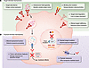
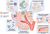
The exact mechanisms and optimal design characteristics of ADCs remain actively debated (2). On one hand, preclinical and clinical evidence suggests that target-dependent delivery of ADC is at least partially required for efficacy. On the other hand, on-target, on-tumor delivery is not the sole driver of ADC activity. Pharmacodynamic analyses suggest ADCs do not increase the MTD of conventional chemotherapy, while pharmacokinetic analyses suggest on-tumor ADC distribution is typically less than 1% of the administered dose (62). Beyond bystander effects, uptake of ADCs by either receptor-dependent (FcγRs, FcRn, and C-type lectin receptors) or receptor-independent (nonspecific endocytosis) mechanisms also contributes to off-target toxicity in normal cells (32, 37). Meanwhile, ADCs also actively modulate the immune landscape through engagement of FcγRs to drive antibody-dependent cellular phagocytosis and payload-induced ICD and myeloid cell activation through TLR4 and STING pathways (57). The sections below discuss ADC features driving response and resistance, mechanisms of acquired resistance, and rational therapeutic combination strategies to overcome them (Figures 2 and 3).
Target- and payload-mediated determinants of ADC response and resistance inFigure 2

Target- and payload-mediated determinants of ADC response and resistance in breast cancer. ADC activity is governed by coordinated target-directed and payload-related mechanisms that influence response, de novo resistance, and acquired resistance. (A) Target-directed mechanisms include antigen surface abundance, intratumoral heterogeneity with spatially variable antigen expression, binding site mutations that alter epitopes and reduce antibody binding, target downregulation resulting in loss of antigen expression, and altered target trafficking that reduces surface localization and limits ADC accessibility. (B) Payload-related mechanisms include altered lysosomal function leading to defective ADC processing and payload release, tumor cell–intrinsic features such as growth capacity and apoptosis escape (e.g., via signal transducer and activator of transcription 3 [STAT3] and polo-like kinase 1 [PLK1]), mutations in payload targets (e.g., TOP1) that impair drug engagement, and active payload clearance mediated by efflux transporters. These processes collectively determine intracellular payload delivery and cytotoxic effects, informing biomarker development, treatment sequencing, and rational combination strategies.
Tumor microenvironmental regulation of ADC activity and resistance.Figure 3

Tumor microenvironmental regulation of ADC activity and resistance. The tumor microenvironment (TME) critically influences ADC distribution, payload activity, and therapeutic outcome. Physical barriers, including stromal architecture and extracellular matrix composition, restrict antibody penetration, while tight junction–mediated immune exclusion (e.g., TROP2 and claudin-7 expression) may further impede lymphocyte infiltration. Disordered tumor vasculature impairs ADC perfusion and distribution compared with normal vasculature. Extracellular payload release, driven by linker instability or extracellular cleavage, can enhance bystander killing of neighboring antigen-negative tumor cells, particularly for membrane-permeable payloads. The immune context contributes through Fc-dependent effector functions, including antibody-dependent cellular cytotoxicity (ADCC) mediated by NK cells and antibody-dependent cellular phagocytosis (ADCP) mediated by macrophages. In addition, ADC-induced immunogenic cell death promotes dendritic cell activation and CD8+ T cell–mediated antitumor immunity. These interconnected TME features collectively shape ADC sensitivity and resistance and provide a rationale for combination strategies targeting stromal and immune components.

Determinants of ADC response and de novo resistance. Target expression variably predicts response to HER2-directed ADCs (53) (Figure 2). For T-DM1, higher HER2 abundance correlates with significantly prolonged PFS and improved ORR (52). In the DAISY trial, T-DXd response duration correlated with HER2 expression (63), and resistance emerged through HER2 loss and ERBB2 binding site mutations that impair ADC internalization and increase IC50 (64). Analogous findings have been reported for SG: TROP2 expression correlates to some degree with benefit in mTNBC (65), while an acquired TROP2 mutation that disrupts antibody binding has been associated with resistance (66). For Dato-DXd, clinical predictive utility remains to be established, though preclinical models suggest that higher TROP2 expression correlates with response and restores sensitivity in resistant tumors (67).

An additional challenge is that target antigen expression is not static but evolves during therapy. Exposure to HER2-directed therapies, endocrine therapy, and chemotherapy may alter antigen density through selective pressure, transcriptional reprogramming, and clonal evolution, potentially shifting tumors between HER2-zero, HER2-low, and HER2-positive states (64, 68, 69). Such temporal variability complicates patient selection for ADC treatment when therapeutic decisions rely on archival tissue samples, as antigen assessment needs to be viewed as a dynamic rather than fixed tumor characteristic.

Beyond antigen expression, ADC efficacy hinges on intracellular payload trafficking, processing, and cellular vulnerability. De novo resistance can emerge from defective lysosomal processing, even when target expression is preserved. For instance, preclinical models show T-DM1–resistant HER2-positive cells have normal internalization but elevated lysosomal pH, which impairs proteolytic payload release (70). Additionally, loss of the SLC46A3 transporter prevents DM1 transfer to cytoplasm (71, 72), while upregulated caveolin-1 reroutes ADCs away from productive lysosomal pathways via caveolae-mediated endocytosis (73, 74). Moreover, the disproportionate success of ADCs in breast cancer compared with several other solid tumor types is not strictly related to differential target expression but more likely to the disease’s high intrinsic chemosensitivity (75).

Furthermore, heterogenous tumor-intrinsic mechanisms governing cell cycle progression, DNA repair, and apoptosis fundamentally dictate payload sensitivity within breast cancer (76). For example, aberrant STAT3 activation confers T-DM1 resistance in HER2-positive breast cancer (77). Multiomic profiling identified the mitotic kinase PLK1 as a primary driver of both de novo and acquired resistance to T-DM1, correlating with accelerated cell cycle progression and enhanced DNA repair (78). Inhibiting PLK1 overcomes T-DM1 resistance in preclinical models by triggering mitotic arrest, caspase activation, and DNA damage through CDK1-dependent phosphorylation and inactivation of BCL-2/BCL-xL (78). Finally, drug efflux transporters are known to mediate payload resistance. In a large cohort of T-DXd–treated patients, lower expression of ABCC1, which effluxes DXd, correlated with longer OS independently of HER2 status (79), while increased ABCC1 has been identified as a primary mediator for T-DM1 resistance in preclinical models (80).

Though less clinically validated than tumor-intrinsic determinants, the TME is increasingly recognized as a modulator of ADC response and resistance through pharmacokinetic and immunological mechanisms (Figure 3). The breast cancer TME is generally permissive to macromolecular delivery, lacking the extreme desmoplasia, poor or disordered vascularization, and profound immunosuppression that restrict ADC penetration in stroma-dense tumors such as pancreatic ductal adenocarcinoma (81, 82). Furthermore, extracellular proteases such as cathepsin L secreted by both tumor and stromal cells in the breast cancer TME can actively drive ADC efficacy, as discussed above (57). In contrast, physical barriers within the TME can impose restrictions on ADC distribution. As ~150 kDa macromolecules, ADCs are hindered by dense extracellular matrices, high interstitial fluid pressure, and the binding site barrier. In xenografts, transient competitive inhibition improved T-DM1 penetration into deeper tumor tissues and enhanced efficacy, as high-affinity antigen binding near perivascular regions creates the “saturation front” problem that limits ADC infiltration (83).

Finally, the TME immune context adds further complexity. While direct clinical evidence in breast cancer remains limited, syngeneic TNBC models reveal that TROP2 actively shapes physical barriers to T cell infiltration, driving immune exclusion via claudin-7–associated tight junction regulation (84). Preclinically, T-DXd induces ICD, promoting DC activation and durable immune memory responses (85). However, biomarker analyses from the DAISY trial found no significant increase in granzyme B–positive CD8+ T lymphocytes in patient biopsies during T-DXd treatment, indicating that ICD-mediated immune activation may be less robust in patients than preclinical models predict (86). These findings highlight an important translational gap between preclinical demonstrations of ADC-induced immunogenicity and clinically measurable immune activation, warranting caution when extrapolating preclinical ICD signals to the clinical efficacy of ADC–ICI combinations.

These various ADC response determinants have not yet been successfully developed as clinical biomarkers, and current target-based patient selection in breast cancer has notable limitations. For T-DXd, HER2 immunohistochemistry (IHC) suffers from interobserver variability, spatiotemporal heterogeneity, and a categorical scoring system originally designed for trastuzumab eligibility rather than for ADCs that shows activity across a broader expression continuum (87). Similarly, TROP2 IHC fails to reliably predict clinical benefit. In the TROPiCS-02 trial, SG improved OS regardless of TROP2 expression, suggesting static target expression–based selection alone is insufficient (26). Emerging approaches to target assessment include quantitative immunofluorescence, spatial antigen mapping, and AI-assisted digital pathology for standardized, observer-independent scoring (88).

Acquired resistance mechanisms and implications for ADC sequencing. Antigen downregulation is a recurrent mechanism associated with acquired ADC resistance in the clinic. Among patients treated with HER2-directed ADCs, 49% showed HER2 downregulation in paired patient samples following T-DXd progression, with over half of these experiencing complete HER2 loss, which correlates with reduced ADC internalization and increased IC50 in isogenic models (64). Alternatively, resistance can emerge via acquired HER2 binding site mutations (V597M and P593R) despite preserved surface expression. These mutations reduce trastuzumab binding, impairing ADC internalization through pharmacological rather than transcriptional mechanisms (64). Importantly, resistance driven by either of these mechanisms can be overcome using a TROP2-directed ADC carrying the same DXd payload, providing a strong rationale for antigen-switching sequencing strategies in these cases (64). Target-mediated acquired resistance also affects TROP2-directed ADCs. In a TNBC patient with a prolonged response and subsequent acquired resistance to SG, postprogression analysis identified a novel TROP2 T256R missense mutation in the dominant resistant clone (66). This single point mutation conferred SG resistance by disrupting TROP2’s plasma membrane localization and reducing cell surface binding to the hRS7 antibody (66).

Acquired payload target mutations represent another clinical mechanism driving ADC resistance. In breast cancer patients progressing on TOP1i-based ADCs, circulating tumor DNA analysis in a small series detected distinct TOP1 missense mutations in 12.9% of cases versus 0.7% in ADC-naive patients treated for breast cancer (89). These mutant proteins exhibited reduced enzymatic activity, attenuated covalent DNA binding, and functional resistance to both DXd and SN-38 payloads, and patients harboring TOP1 mutations experienced dramatically shorter responses to subsequent TOP1i-based ADCs (median 52 vs. 455 days) (89). Another acquired resistance pathway involves upregulation of the ABCC1 drug efflux pump via the NFE2L2/KEAP1 axis, whereby KEAP1 negatively regulates the NFE2L2 transcription factor that drives ABCC1 expression. Post–T-DXd tumor specimens showed enriched mutations in NFE2L2 and KEAP1 relative to T-DXd–naive samples, suggesting transcriptional upregulation of DXd efflux is actively selected for under ADC pressure, driving resistance independently of target antigen loss (79).

For patients who typically receive multiple lines of therapy in the metastatic setting, the question of optimal ADC timing relative to chemotherapy has been addressed by comparisons across trials. Results from DESTINY-Breast04 (second-line T-DXd in HER2-low) and DESTINY-Breast06 (first/second-line T-DXd in HER2-low/ultralow) collectively suggest that earlier ADC deployment produces longer PFS, potentially suggesting that preserved target expression and reduced preexisting payload resistance at earlier treatment lines favor ADC efficacy (16, 17). By contrast, defining the optimal sequencing strategy for use of multiple ADCs remains a central, unresolved clinical challenge. While the goal is to rationally switch ADC targets or payloads to preserve efficacy, current evidence is largely retrospective and highly heterogeneous, revealing substantial but variable cross-resistance (90). Despite these limitations, several provisional sequencing principles have emerged, including earlier deployment of effective ADCs and switching payload classes or antigen targets when feasible (Table 2). A multicenter study found that switching to a second ADC (ADC2) with a different payload class yields higher response rates (~22.6% ORR) than same-payload sequences (~5.3% ORR), but PFS for ADC2 remained similar regardless of payload class (91). Similarly, real-world analyses indicate that results with sequential use of currently approved ADCs are generally unsatisfactory, as time to treatment failure and PFS are consistently shorter for ADC2 regardless of sequence order, HR status, or intervening chemotherapy (92–94).
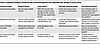
Table 2

Proposed biological framework and unresolved questions for sequential ADC therapy in breast cancer

Nonetheless, cross-resistance appears most pronounced when sequential ADCs share payload mechanisms. A database analysis of post–T-DXd patients showed that subsequent SG yielded inferior real-world PFS across subtypes, supporting the rationale to switch to non-TOP1i therapies after T-DXd progression rather than substituting another TOP1i-based ADC (95). Conversely, switching payloads while retaining the same target has proven effective for use of T-DXd after T-DM1 exposure. In the DESTINY-Breast02 trial, T-DXd produced a median PFS of 17.8 versus 6.9 months and an ORR of 70% versus 29% over chemotherapy after prior T-DM1 treatment (96), consistent with the absence of cross-resistance between maytansinoid and TOP1i payloads. Furthermore, despite frequent ADC2 resistance, a clinically meaningful subset still achieves durable responses, including patients who switch antibody targets (97), experience prolonged initial ADC responses, or receive ADC2 in an earlier treatment line (98). Ongoing and impending clinical trials have been designed to systematically test sequencing and biomarkers for currently approved ADCs. Nonetheless, innovation and implementation of new ADC classes with distinct targets and payloads are needed to increase options and improve outcomes associated with sequential ADC use.

Combination strategies to enhance response and overcome resistance. The rationale for combining ADCs with ICIs centers in part on the ability of TOP1i payloads to induce ICD, thereby priming an immune response that can be further augmented via ICI (99). Payloads including DXd and SN-38 trigger ICD hallmarks such as calreticulin surface exposure, high mobility group protein B1 release, and ATP secretion that promote antigen cross-presentation and T cell priming, while ICIs reinvigorate exhausted T cells to amplify the adaptive immune response (100–102). Potential synergy is supported by compelling preclinical data, as the combination of T-DM1 with dual immune checkpoint blockade nearly eradicates tumors in HER2-positive models (103). Clinically, TROP2-directed SG plus the anti–PD-1 antibody pembrolizumab demonstrated superior PFS compared with chemotherapy plus pembrolizumab for first-line treatment of PD-L1–positive advanced TNBC in the randomized phase III ASCENT-04 trial (24). Similarly, in the phase Ib/II BEGONIA trial, first-line Dato-DXd plus durvalumab achieved approximately 80% ORR regardless of PD-L1 expression (104). Clinical data supporting the addition of an ICI to HER2-directed ADCs have to date been less convincing. In the phase Ib DS8201-A-U105 study, T-DXd plus nivolumab achieved a 66% ORR in pretreated HER2-positive metastatic breast cancer (105). This is comparable to historical T-DXd monotherapy data, which showed ORRs of 61% in DESTINY-Breast01 (106), 70% in DESTINY-Breast02 (96), and 80% in DESTINY-Breast03 (13). Similarly, T-DXd plus nivolumab achieved 50% ORR in HER2-low breast cancer, which is again comparable to T-DXd monotherapy in DESTINY-Breast04 (52%) (16). As with other immunotherapy applications in oncology, improved predictive markers and alternative immune activation strategies remain unmet needs.

The hypothesized synergy between TOP1i-based ADCs and PARP inhibitors (PARPis) operates through three convergent mechanisms: (a) PARP1 normally PARylates TOP1 cleavage complex (TOP1cc) to release it from DNA, while PARP inhibition prevents this release, stabilizing TOP1cc and prolonging DNA damage; (b) PARP inhibition blocks tyrosyl-DNA-phosphodiesterase 1, the phosphodiesterase required to excise covalently trapped TOP1 from DNA, further preventing TOP1cc repair; and (c) PARP inhibition impairs repair of replication-associated double-strand breaks that arise when replication forks collide with TOP1i-stabilized TOP1cc (107).

This combination was initially tested in 6 patients (SEASTAR trial), where SG plus the PARPi rucaparib demonstrated antitumor activity regardless of homologous recombination mutation status or PARPi preexposure (108). A subsequent clinical trial evaluated the hypothesis that ADC-mediated delivery of the TOP1i creates a high tumor/normal cell dose gradient that enables a temporal window for sequential rather than concurrent ADC/PARPi dosing. In the phase Ib trial of SG plus PARPi talazoparib in mTNBC, this strategy was shown to mitigate the dose-limiting myelosuppression that has plagued systemic TOP1i/PARPi combinations (109). Further testing of this strategy is likely to involve newer PARP1-selective inhibitors, as currently approved PARPis inhibit both PARP1 and PARP2, while it is PARP2 that disproportionately drives hematological toxicity (110).
Future generations of ADCs in breast cancer

Expanding the target space. ADC target development in breast cancer is expanding beyond HER2 and TROP2 toward both tumor-associated antigens and nontumor components of the TME (Figure 4). To overcome the limitations of heterogeneous antigen expression and acquired resistance, a diverse array of novel tumor-associated membrane proteins is under active preclinical and clinical investigation (Table 3). While these novel targets offer distinct tumor selectivity and internalization kinetics, they also warrant rigorous evaluation of their respective safety and efficacy profiles. An alternative, emerging paradigm shifts ADC targeting from tumor cells to the TME (111). Utilizing noninternalizing ADCs with cleavable linkers directed against nontumor cells circumvents the need for uniform tumor antigen expression and relies on extracellular payload release within the TME (47). Key targets include cancer-associated fibroblasts and endothelia, allowing for localized payload release within the TME to effectively ablate tumor-promoting stromal and vascular compartments as well as adjacent breast cancer cells (Table 3).
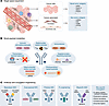
Future generations of ADCs in breast cancer.Figure 4

Future generations of ADCs in breast cancer. Next-generation ADC development in breast cancer is advancing along three major axes. (A) Target space expansion extends beyond established tumor-associated antigens to include novel tumor cell targets, such as HER3, Nectin-4, mesothelin (MSLN), and folate receptor alpha (FOLR1), as well as nontumor antigens expressed by cancer-associated stromal compartments, including fibroblasts and endothelial cells, exemplified by LRRC5, FAP, TEM8, and CD105. (B) Novel payload modalities broaden the functional repertoire of ADCs beyond conventional cytotoxic warheads to include immune-stimulating payloads (for example, STING and TLR7/8 agonists), transcription-directed payloads (for example, α-amanitin), oligonucleotide-based payloads (for example, siRNA), targeted protein-degrading payloads (including proteolysis-targeting chimeras [PROTACs] and molecular glues), immunotoxins (for example, PE24 and PE25), radionuclide-based payloads (for example, 177Lu and 67Cu), and photosensitizer-based payloads (for example, IR700). (C) Antibody and conjugation engineering aims to improve selectivity, stability, and therapeutic index through biparatopic ADCs, dual-payload ADCs, probody ADCs, Fc engineering, bispecific ADCs, and site-specific conjugation strategies, including engineered cysteines, glycan remodeling, Fc-affinity peptide-mediated conjugation, unnatural amino acids, and enzymatic ligation.

Table 3

Next-generation ADCs relevant to breast cancer with novel targets

New payload modalities. Payload modality innovation represents a major frontier in ADC design (Figure 4 and Table 4). Degrader-antibody conjugates merge targeted delivery with proteolysis-targeting chimeras or molecular glues to eliminate oncoproteins via the ubiquitin-proteasome system and have shown sensitive and specific efficacy in breast cancer models (112, 113). Another approach utilizes immune-stimulating antibody conjugates (ISACs) to deliver immune agonists and convert immunologically cold breast tumors into inflamed niches, promoting localized myeloid activation and T cell priming (114). However, clinical translation remains challenging; despite strong mechanistic rationales, ISACs inherently suffer from a narrow therapeutic index, as maximizing antitumor potency frequently exacerbates immune-related systemic toxicity (114, 115).

Table 4

Next-generation ADCs relevant to breast cancer with nonchemotherapy payload modalities

In addition, several other novel payload modalities remain primarily in proof-of-concept stages (115). These include transcription-directed payloads that exploit cytotoxicity outside the canonical tubulin or TOP1 axes to kill nondividing tumor cells through transcriptional arrest. Antibody-oligonucleotide conjugates extend the ADC concept to enable sequence-specific knockdown of oncogenic transcripts or resistance-mediating genes (116). Immunotoxins, an exploratory class fusing targeting antibodies to bacterial toxins, eradicate both dividing and nondividing tumor cells by inhibiting protein synthesis across various breast cancer subtypes (117). Antibody-radionuclide conjugates leverage antibody specificity to deliver localized radiation with theranostic potential, though clinical progress is hindered by dose-limiting myelosuppression, heterogeneous radionuclide distribution, and the absence of prospective randomized trials (118). Finally, antibody-photosensitizer conjugates are activated by focal light to induce targeted ICD and durable antitumor immune memory, providing a strong rationale for combination with immune checkpoint blockade (119).

Antibody and conjugation engineering. Antibody and conjugation engineering represents another driver for future ADC design in breast cancer (Figure 4 and Table 5). Biparatopic ADCs, which bind nonoverlapping epitopes of the same antigen, enforce receptor clustering and accelerate internalization, while bispecific ADCs simultaneously engage two independent antigens to mitigate antigen escape and enhance tumor selectivity (120). Additionally, dual-payload ADCs address tumor heterogeneity and acquired resistance by codelivering two distinct cytotoxic warheads with nonoverlapping mechanisms, raising the barrier to resistance and achieving synergistic cytotoxicity (121).
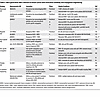
Table 5

Next-generation ADCs relevant to breast cancer with innovative antibody and conjugation engineering

Conjugation technologies are also rapidly evolving. Conventional ADCs rely on stochastic coupling, creating heterogeneous mixtures with variable DARs that complicate pharmacokinetics and clinical consistency (122). By contrast, site-specific conjugation technologies enable precise positional control of payload attachment (123). This approach generates homogeneous ADCs with defined DARs, improving pharmacokinetic consistency, reducing toxicity, enhancing reproducibility, and widening the therapeutic index, as seen in approved ADCs like T-DXd (124).

Moreover, orthogonal engineering strategies enable further spatial and pharmacokinetic control. Masked or “probody” ADCs utilize auto-inhibitory peptides that block antigen binding on normal cells in the TME but are cleaved exclusively by tumor-enriched proteases, thereby sparing normal tissues and widening the therapeutic window (3). Furthermore, Fc backbone engineering provides another dimension for optimization (125), allowing for enhancement of immune-mediated cytotoxicity or extension of plasma half-life for prolonged tumor exposure.
Concluding perspective

The emerging paradigm of ADCs in breast cancer represents not only an incremental advance in targeted drug delivery but also a conceptual evolution of chemotherapy. Clinical and mechanistic data suggest ADC activity arises from a complex interplay of antibody-mediated distribution, linker stability, payload pharmacology, and microenvironmental release, involving both target-dependent and target-independent mechanisms such as bystander killing and off-tumor uptake. Breast cancer has become the archetype for ADC development, with HER2- and TROP2-directed ADCs transforming treatment paradigms, expanding therapeutic eligibility, and displacing conventional chemotherapy. Realizing next-generation ADC potential will require mechanism-informed biomarkers beyond target-based IHC, biology-driven sequential treatment strategies, rational combinations, and continued innovation in targets, payloads, antibodies, and conjugation. Translational evaluation should focus on improved therapeutic index through better patient selection, adaptive treatment strategies, and reduced systemic and off-tumor toxicity. Together, these priorities define a translational roadmap aligned with the biological complexity of ADC mechanisms.

Conflict of interest

LWE has received research support from Sanofi.

Funding support

This work was supported by NIH/NCI grant 1R01CA260890 and NIH/NCI SPORE grant 2P50CA168504 (LWE).

Footnotes

Copyright: © 2026, Guo et al. This is an open access article published under the terms of the Creative Commons Attribution 4.0 International License.

Reference information: J Clin Invest. 2026;136(19):e207630. https://doi.org/10.1172/JCI207630.

References
  1. Strebhardt K, Ullrich A. Paul Ehrlich’s magic bullet concept: 100 years of progress. Nat Rev Cancer. 2008;8(6):473–480.
    View this article via: CrossRef PubMed Google Scholar
  2. Colombo R, Rich JR. The therapeutic window of antibody drug conjugates: a dogma in need of revision. Cancer Cell. 2022;40(11):1255–1263.
    View this article via: CrossRef PubMed Google Scholar
  3. Tsuchikama K, et al. Exploring the next generation of antibody-drug conjugates. Nat Rev Clin Oncol. 2024;21(3):203–223.
    View this article via: CrossRef PubMed Google Scholar
  4. Dumontet C, et al. Antibody-drug conjugates come of age in oncology. Nat Rev Drug Discov. 2023;22(8):641–661.
    View this article via: CrossRef PubMed Google Scholar
  5. Lewis Phillips GD, et al. Targeting HER2-positive breast cancer with trastuzumab-DM1, an antibody-cytotoxic drug conjugate. Cancer Res. 2008;68(22):9280–9290.
    View this article via: CrossRef PubMed Google Scholar
  6. Ogitani Y, et al. DS-8201a, A novel HER2-targeting ADC with a novel DNA topoisomerase i inhibitor, demonstrates a promising antitumor efficacy with differentiation from T-DM1. Clin Cancer Res. 2016;22(20):5097–5108.
    View this article via: CrossRef PubMed Google Scholar
  7. Cardillo TM, et al. Humanized anti-Trop-2 IgG-SN-38 conjugate for effective treatment of diverse epithelial cancers: preclinical studies in human cancer xenograft models and monkeys. Clin Cancer Res. 2011;17(10):3157–3169.
    View this article via: CrossRef PubMed Google Scholar
  8. Santi DV, et al. Does sacituzumab-govitecan act as a conventional antibody drug conjugate (ADC), a prodrug of SN-38 or both? Ann Transl Med. 2021;9(14):1113.
    View this article via: CrossRef PubMed Google Scholar
  9. Forveille S, et al. Datopotamab deruxtecan induces hallmarks of immunogenic cell death. Cell Stress. 2025;9:194–200.
    View this article via: CrossRef PubMed Google Scholar
  10. Okajima D, et al. Datopotamab deruxtecan, a novel TROP2-directed antibody-drug conjugate, demonstrates potent antitumor activity by efficient drug delivery to tumor cells. Mol Cancer Ther. 2021;20(12):2329–2340.
    View this article via: CrossRef PubMed Google Scholar
  11. Verma S, et al. Trastuzumab emtansine for HER2-positive advanced breast cancer. N Engl J Med. 2012;367(19):1783–1791.
    View this article via: CrossRef PubMed Google Scholar
  12. Geyer CE Jr. Survival with trastuzumab emtansine in residual HER2-positive breast cancer. N Engl J Med. 2025;392(3):249–257.
    View this article via: CrossRef PubMed Google Scholar
  13. Cortes J, et al. Trastuzumab deruxtecan versus trastuzumab emtansine for breast cancer. N Engl J Med. 2022;386(12):1143–1154.
    View this article via: CrossRef PubMed Google Scholar
  14. Loibl S, et al. Trastuzumab deruxtecan in residual HER2-positive early breast cancer. N Engl J Med. 2026;394(9):845–857.
    View this article via: CrossRef PubMed Google Scholar
  15. Ogitani Y, et al. Bystander killing effect of DS-8201a, a novel anti-human epidermal growth factor receptor 2 antibody-drug conjugate, in tumors with human epidermal growth factor receptor 2 heterogeneity. Cancer Sci. 2016;107(7):1039–1046.
    View this article via: CrossRef PubMed Google Scholar
  16. Modi S, et al. Trastuzumab deruxtecan in previously treated HER2-low advanced breast cancer. N Engl J Med. 2022;387(1):9–20.
    View this article via: CrossRef PubMed Google Scholar
  17. Bardia A, et al. Trastuzumab deruxtecan after endocrine therapy in metastatic breast cancer. N Engl J Med. 2024;391(22):2110–2122.
    View this article via: CrossRef PubMed Google Scholar
  18. Turner N, et al. Trastuzumab duocarmazine in pretreated human epidermal growth factor Receptor 2-positive advanced or metastatic breast cancer: an open-label, randomized, phase III trial (TULIP). J Clin Oncol. 2025;43(5):513–523.
    View this article via: CrossRef PubMed Google Scholar
  19. Hu X, et al. ACE-Breast-02: a randomized phase III trial of ARX788 versus lapatinib plus capecitabine for HER2-positive advanced breast cancer. Signal Transduct Target Ther. 2025;10(1):56.
    View this article via: CrossRef PubMed Google Scholar
  20. BioNTech. BioNTech and DualityBio announce phase 3 trial of ADC candidate BNT323/DB-1303 met primary endpoint of progression free survival in HER2-positive metastatic or unresectable breast cancer. https://investors.biontech.de/news-releases/news-release-details/biontech-and-dualitybio-announce-phase-3-trial-adc-candidate Accessed July 2, 2026.
  21. Dri A, et al. Breaking barriers in triple negative breast cancer (TNBC) - unleashing the power of antibody-drug conjugates (ADCs). Cancer Treat Rev. 2024;123:102672.
    View this article via: CrossRef PubMed Google Scholar
  22. Bardia A, et al. Sacituzumab govitecan in metastatic triple-negative breast cancer. N Engl J Med. 2021;384(16):1529–1541.
    View this article via: CrossRef PubMed Google Scholar
  23. Cortes J, et al. Sacituzumab govitecan in untreated, advanced triple-negative breast cancer. N Engl J Med. 2025;393(19):1912–1925.
    View this article via: CrossRef PubMed Google Scholar
  24. Tolaney SM, et al. Sacituzumab govitecan plus pembrolizumab for advanced triple-negative breast cancer. N Engl J Med. 2026;394(4):354–366.
    View this article via: CrossRef PubMed Google Scholar
  25. Rugo HS, et al. Sacituzumab govitecan in hormone receptor-positive/human epidermal growth factor receptor 2-negative metastatic breast cancer. J Clin Oncol. 2022;40(29):3365–3376.
    View this article via: CrossRef PubMed Google Scholar
  26. Rugo HS, et al. Overall survival with sacituzumab govitecan in hormone receptor-positive and human epidermal growth factor receptor 2-negative metastatic breast cancer (TROPiCS-02): a randomised, open-label, multicentre, phase 3 trial. Lancet. 2023;402(10411):1423–1433.
    View this article via: CrossRef PubMed Google Scholar
  27. Bardia A, et al. Datopotamab deruxtecan versus chemotherapy in previously treated inoperable/metastatic hormone receptor-positive human epidermal growth factor receptor 2-negative breast cancer: primary results from TROPION-Breast01. J Clin Oncol. 2025;43(3):285–296.
    View this article via: CrossRef PubMed Google Scholar
  28. Pistilli B, et al. Datopotamab deruxtecan versus chemotherapy in previously treated inoperable/metastatic hormone receptor-positive, HER2-negative breast cancer: final overall survival analysis of the phase III TROPION-Breast01 study. Ann Oncol. 2025;37(5):663–674.
    View this article via: CrossRef PubMed Google Scholar
  29. Dent R, et al. Datopotamab deruxtecan in patients with untreated, advanced triple-negative breast cancer (TROPION-Breast02): a randomised, open-label, international, phase III trial. Ann Oncol. 2026;37:1066–1080.
    View this article via: CrossRef PubMed Google Scholar
  30. Yin Y, et al. Sacituzumab tirumotecan in previously treated metastatic triple-negative breast cancer: a randomized phase 3 trial. Nat Med. 2025;31(6):1969–1975.
    View this article via: CrossRef PubMed Google Scholar
  31. Tang SC, et al. Influence of antibody-drug conjugate cleavability, drug-to-antibody ratio, and free payload concentration on systemic toxicities: a systematic review and meta-analysis. Cancer Metastasis Rev. 2024;44(1):18.
    View this article via: CrossRef PubMed Google Scholar
  32. Mahalingaiah PK, et al. Potential mechanisms of target-independent uptake and toxicity of antibody-drug conjugates. Pharmacol Ther. 2019;200:110–125.
    View this article via: CrossRef PubMed Google Scholar
  33. Tarantino P, et al. Optimizing the safety of antibody-drug conjugates for patients with solid tumours. Nat Rev Clin Oncol. 2023;20(8):558–576.
    View this article via: CrossRef PubMed Google Scholar
  34. Premji S, et al. Abstract PO4-12-06: Interstitial lung disease (ILD) in sequential antibody drug conjugate (ADC) use in patients with metastatic breast cancer (MBC): a multi-institutional experience. Cancer Res. 2024;84(9_suppl):PO4-12-06.
  35. Powell CA, et al. Pooled analysis of drug-related interstitial lung disease and/or pneumonitis in nine trastuzumab deruxtecan monotherapy studies. ESMO Open. 2022;7(4):100554.
    View this article via: CrossRef PubMed Google Scholar
  36. Kumagai K, et al. Interstitial pneumonitis related to trastuzumab deruxtecan, a human epidermal growth factor receptor 2-targeting Ab-drug conjugate, in monkeys. Cancer Sci. 2020;111(12):4636–4645.
    View this article via: CrossRef PubMed Google Scholar
  37. Uppal H, et al. Potential mechanisms for thrombocytopenia development with trastuzumab emtansine (T-DM1). Clin Cancer Res. 2015;21(1):123–133.
    View this article via: CrossRef PubMed Google Scholar
  38. Zhao H, et al. Inhibition of megakaryocyte differentiation by antibody-drug conjugates (ADCs) is mediated by macropinocytosis: implications for ADC-induced thrombocytopenia. Mol Cancer Ther. 2017;16(9):1877–1886.
    View this article via: CrossRef PubMed Google Scholar
  39. Endo Y, et al. Payload of T-DM1 binds to cell surface cytoskeleton-associated protein 5 to mediate cytotoxicity of hepatocytes. Oncotarget. 2018;9(98):37200–37215.
    View this article via: CrossRef PubMed Google Scholar
  40. Yan H, et al. Ado-trastuzumab emtansine targets hepatocytes via human epidermal growth factor receptor 2 to induce hepatotoxicity. Mol Cancer Ther. 2016;15(3):480–490.
    View this article via: CrossRef PubMed Google Scholar
  41. Bianchini G, et al. Emetogenicity of antibody-drug conjugates (ADCs) in solid tumors with a focus on trastuzumab deruxtecan: insights from an Italian expert panel. Cancers (Basel). 2022;14(4):1022.
    View this article via: CrossRef PubMed Google Scholar
  42. Hammood M, et al. Impact of endocytosis mechanisms for the receptors targeted by the currently approved antibody-drug conjugates (ADCs)-a necessity for future adc research and development. Pharmaceuticals (Basel). 2021;14(7):674.
    View this article via: CrossRef PubMed Google Scholar
  43. Esapa B, et al. Target antigen attributes and their contributions to clinically approved antibody-drug conjugates (ADCs) in haematopoietic and solid cancers. Cancers (Basel). 2023;15(6):1845.
    View this article via: CrossRef PubMed Google Scholar
  44. Durbin KR, et al. Mechanistic modeling of antibody-drug conjugate internalization at the cellular level reveals inefficient processing steps. Mol Cancer Ther. 2018;17(6):1341–1351.
    View this article via: CrossRef PubMed Google Scholar
  45. Tong Y, et al. Advances in Trop-2 targeted antibody-drug conjugates for breast cancer: mechanisms, clinical applications, and future directions. Front Immunol. 2024;15:1495675.
    View this article via: CrossRef PubMed Google Scholar
  46. Giansanti F, et al. Secreted Gal-3BP is a novel promising target for non-internalizing antibody-drug conjugates. J Control Release. 2019;294:176–184.
    View this article via: CrossRef PubMed Google Scholar
  47. Ashman N, et al. Non-internalising antibody-drug conjugates. Chem Soc Rev. 2022;51(22):9182–9202.
    View this article via: CrossRef PubMed Google Scholar
  48. Dal Corso A, et al. A non-internalizing antibody-drug conjugate based on an anthracycline payload displays potent therapeutic activity in vivo. J Control Release. 2017;264:211–218.
    View this article via: CrossRef PubMed Google Scholar
  49. Staudacher AH, Brown MP. Antibody drug conjugates and bystander killing: is antigen-dependent internalisation required? Br J Cancer. 2017;117(12):1736–1742.
    View this article via: CrossRef PubMed Google Scholar
  50. Feng C, et al. Extracellular payload release from non-internalizing antibody-drug conjugates: mechanisms and linker technologies. Drug Deliv. 2026;33(1):2645769.
    View this article via: CrossRef PubMed Google Scholar
  51. Fauteux F, et al. Computational selection of antibody-drug conjugate targets for breast cancer. Oncotarget. 2016;7(3):2555–2571.
    View this article via: CrossRef PubMed Google Scholar
  52. Burris HA, et al. Phase II study of the antibody drug conjugate trastuzumab-DM1 for the treatment of human epidermal growth factor receptor 2 (HER2)-positive breast cancer after prior HER2-directed therapy. J Clin Oncol. 2011;29(4):398–405.
    View this article via: CrossRef PubMed Google Scholar
  53. Krop IE, et al. A phase II study of trastuzumab emtansine in patients with human epidermal growth factor receptor 2-positive metastatic breast cancer who were previously treated with trastuzumab, lapatinib, an anthracycline, a taxane, and capecitabine. J Clin Oncol. 2012;30(26):3234–3241.
    View this article via: CrossRef PubMed Google Scholar
  54. Kumler I, et al. A systematic review on topoisomerase 1 inhibition in the treatment of metastatic breast cancer. Breast Cancer Res Treat. 2013;138(2):347–358.
    View this article via: CrossRef PubMed Google Scholar
  55. Giugliano F, et al. Bystander effect of antibody-drug conjugates: fact or fiction? Curr Oncol Rep. 2022;24(7):809–817.
    View this article via: CrossRef PubMed Google Scholar
  56. Su D, Zhang D. Linker design impacts antibody-drug conjugate pharmacokinetics and efficacy via modulating the stability and payload release efficiency. Front Pharmacol. 2021;12:687926.
    View this article via: CrossRef PubMed Google Scholar
  57. Tsao LC, et al. Effective extracellular payload release and immunomodulatory interactions govern the therapeutic effect of trastuzumab deruxtecan (T-DXd). Nat Commun. 2025;16(1):3167.
    View this article via: CrossRef PubMed Google Scholar
  58. Hamblett KJ, et al. Effects of drug loading on the antitumor activity of a monoclonal antibody drug conjugate. Clin Cancer Res. 2004;10(20):7063–7070.
    View this article via: CrossRef PubMed Google Scholar
  59. Cilliers C, et al. Multiscale modeling of antibody-drug conjugates: connecting tissue and cellular distribution to whole animal pharmacokinetics and potential implications for efficacy. AAPS J. 2016;18(5):1117–1130.
    View this article via: CrossRef PubMed Google Scholar
  60. Cilliers C, et al. Improved tumor penetration and single-cell targeting of antibody-drug conjugates increases anticancer efficacy and host survival. Cancer Res. 2018;78(3):758–768.
    View this article via: CrossRef PubMed Google Scholar
  61. Rubahamya B, et al. Clinical translation of antibody drug conjugate dosing in solid tumors from preclinical mouse data. Sci Adv. 2024;10(22):eadk1894.
    View this article via: CrossRef PubMed Google Scholar
  62. Colombo R, et al. The journey of antibody-drug conjugates: lessons learned from 40 years of development. Cancer Discov. 2024;14(11):2089–2108.
    View this article via: CrossRef PubMed Google Scholar
  63. Mosele F, et al. Trastuzumab deruxtecan in metastatic breast cancer with variable HER2 expression: the phase 2 DAISY trial. Nat Med. 2023;29(8):2110–2120.
    View this article via: CrossRef PubMed Google Scholar
  64. Chen W, et al. Trastuzumab deruxtecan resistance via loss of HER2 expression and binding. Cancer Discov. 2026;16(2):235–249.
    View this article via: CrossRef PubMed Google Scholar
  65. Bardia A, et al. Biomarker analyses in the phase III ASCENT study of sacituzumab govitecan versus chemotherapy in patients with metastatic triple-negative breast cancer. Ann Oncol. 2021;32(9):1148–1156.
    View this article via: CrossRef PubMed Google Scholar
  66. Coates JT, et al. Parallel genomic alterations of antigen and payload targets mediate polyclonal acquired clinical resistance to sacituzumab govitecan in triple-negative breast cancer. Cancer Discov. 2021;11(10):2436–2445.
    View this article via: CrossRef PubMed Google Scholar
  67. Meric-Bernstam F, et al. Antitumor activity and biomarker analysis for TROP2 antibody-drug conjugate datopotamab deruxtecan in patient-derived breast cancer xenograft models. Clin Cancer Res. 2025;31(3):573–587.
    View this article via: CrossRef PubMed Google Scholar
  68. Gurau AM, et al. HER2-low breast cancer: biological framework and determinants of HER2 instability. Medicina (Kaunas). 2026;62(2):304.
    View this article via: CrossRef PubMed Google Scholar
  69. Pegram M, et al. Estrogen/HER2 receptor crosstalk in breast cancer: combination therapies to improve outcomes for patients with hormone receptor-positive/HER2-positive breast cancer. NPJ Breast Cancer. 2023;9(1):45.
    View this article via: CrossRef PubMed Google Scholar
  70. Rios-Luci C, et al. Resistance to the antibody-drug conjugate T-DM1 is based in a reduction in lysosomal proteolytic activity. Cancer Res. 2017;77(17):4639–4651.
    View this article via: CrossRef PubMed Google Scholar
  71. Hamblett KJ, et al. SLC46A3 is required to transport catabolites of noncleavable antibody maytansine conjugates from the lysosome to the cytoplasm. Cancer Res. 2015;75(24):5329–5340.
    View this article via: CrossRef PubMed Google Scholar
  72. Li G, et al. Mechanisms of acquired resistance to trastuzumab emtansine in breast cancer cells. Mol Cancer Ther. 2018;17(7):1441–1453.
    View this article via: CrossRef PubMed Google Scholar
  73. Pereira PMR, et al. Caveolin-1 temporal modulation enhances antibody drug efficacy in heterogeneous gastric cancer. Nat Commun. 2022;13(1):2526.
    View this article via: CrossRef PubMed Google Scholar
  74. Sung M, et al. Caveolae-mediated endocytosis as a novel mechanism of resistance to trastuzumab emtansine (T-DM1). Mol Cancer Ther. 2018;17(1):243–253.
    View this article via: CrossRef PubMed Google Scholar
  75. Harbeck N, et al. Breast cancer. Nat Rev Dis Primers. 2019;5(1):66.
    View this article via: CrossRef PubMed Google Scholar
  76. Chang HL, et al. Antibody-drug conjugates in breast cancer: overcoming resistance and boosting immune response. J Clin Invest. 2023;133(18):e172156.
    View this article via: JCI CrossRef PubMed Google Scholar
  77. Wang L, et al. STAT3 activation confers trastuzumab-emtansine (T-DM1) resistance in HER2-positive breast cancer. Cancer Sci. 2018;109(10):3305–3315.
    View this article via: CrossRef PubMed Google Scholar
  78. Saatci O, et al. Targeting PLK1 overcomes T-DM1 resistance via CDK1-dependent phosphorylation and inactivation of Bcl-2/xL in HER2-positive breast cancer. Oncogene. 2018;37(17):2251–2269.
    View this article via: CrossRef PubMed Google Scholar
  79. Sledge GW, et al. Mechanisms of resistance to trastuzumab deruxtecan in breast cancer elucidated by multi-omic molecular profiling. NPJ Breast Cancer. 2025;12(1):1.
    View this article via: CrossRef PubMed Google Scholar
  80. Loganzo F, et al. Tumor cells chronically treated with a trastuzumab-maytansinoid antibody-drug conjugate develop varied resistance mechanisms but respond to alternate treatments. Mol Cancer Ther. 2015;14(4):952–963.
    View this article via: CrossRef PubMed Google Scholar
  81. Hartupee C, et al. Pancreatic cancer tumor microenvironment is a major therapeutic barrier and target. Front Immunol. 2024;15:1287459.
    View this article via: CrossRef PubMed Google Scholar
  82. Yajaman DR, et al. Advancing antibody-drug conjugates: precision oncology approaches for breast and pancreatic cancers. Cancers (Basel). 2025;17(11):1792.
    View this article via: CrossRef PubMed Google Scholar
  83. Bordeau BM, et al. Transient competitive inhibition bypasses the binding site barrier to improve tumor penetration of trastuzumab and enhance T-DM1 efficacy. Cancer Res. 2021;81(15):4145–4154.
    View this article via: CrossRef PubMed Google Scholar
  84. Wu B, et al. TROP2/claudin program mediates immune exclusion to impede checkpoint blockade in breast cancer. J Immunother Cancer. 2026;14(4):e012265.
    View this article via: CrossRef PubMed Google Scholar
  85. Iwata TN, et al. A HER2-targeting antibody-drug conjugate, trastuzumab deruxtecan (DS-8201a), enhances antitumor immunity in a mouse model. Mol Cancer Ther. 2018;17(7):1494–1503.
    View this article via: CrossRef PubMed Google Scholar
  86. Mosele MF, et al. 343MO Unraveling the mechanisms of action and resistance to trastuzumab deruxtecan (T-DXd): Supplementary biomarker analyses from DAISY trial. Ann Oncol. 2024;35:S359.
    View this article via: CrossRef Google Scholar
  87. Tarantino P, et al. ESMO expert consensus statements (ECS) on the definition, diagnosis, and management of HER2-low breast cancer. Ann Oncol. 2023;34(8):645–659.
    View this article via: CrossRef PubMed Google Scholar
  88. Davis AA, et al. Novel treatment approaches utilizing antibody-drug conjugates in breast cancer. NPJ Breast Cancer. 2025;11(1):42.
    View this article via: CrossRef PubMed Google Scholar
  89. Abelman RO, et al. TOP1 mutations and cross-resistance to antibody-drug conjugates in patients with metastatic breast cancer. Clin Cancer Res. 2025;31(10):1966–1974.
    View this article via: CrossRef PubMed Google Scholar
  90. de Almeida LFC, et al. Sequencing antibody-drug conjugates in metastatic breast cancer: a systematic review. Cancer Treat Rev. 2026;142:103067.
    View this article via: CrossRef PubMed Google Scholar
  91. Chen M, et al. Optimal sequential strategies for antibody-drug conjugate in metastatic breast cancer: evaluating efficacy and cross-resistance. Oncologist. 2024;29(8):e957–e966.
    View this article via: CrossRef PubMed Google Scholar
  92. Huppert LA, et al. Multicenter retrospective cohort study of the sequential use of the antibody-drug conjugates (ADCs) trastuzumab deruxtecan (T-DXd) and sacituzumab govitecan (SG) in patients with HER2-low metastatic breast cancer (MBC). NPJ Breast Cancer. 2025;11(1):34.
    View this article via: CrossRef PubMed Google Scholar
  93. Poumeaud F, et al. Efficacy of administration sequence: sacituzumab govitecan and trastuzumab deruxtecan in HER2-low metastatic breast cancer. Br J Cancer. 2024;131(4):702–708.
    View this article via: CrossRef PubMed Google Scholar
  94. Tan J, et al. Real-world antibody-drug conjugate (ADC) sequential use in metastatic breast cancer. J Clin Oncol. 2025;43(16_suppl):e13122.
  95. Tarantino P, et al. Outcomes of subsequent treatment regimens after trastuzumab deruxtecan in patients with metastatic breast cancer. J Natl Cancer Inst. 2025;117(11):2327–2335.
    View this article via: CrossRef PubMed Google Scholar
  96. Andre F, et al. Trastuzumab deruxtecan versus treatment of physician’s choice in patients with HER2-positive metastatic breast cancer (DESTINY-Breast02): a randomised, open-label, multicentre, phase 3 trial. Lancet. 2023;401(10390):1773–1785.
    View this article via: CrossRef PubMed Google Scholar
  97. Abelman RO, et al. Sequential use of antibody-drug conjugate after antibody-drug conjugate for patients with metastatic breast cancer: ADC after ADC (A3) study. J Clin Oncol. 2023;41(16_suppl):1022.
    View this article via: CrossRef Google Scholar
  98. Mai N, et al. Sequential antibody-drug conjugate therapy in patients with metastatic breast cancer treated with sacituzumab govitecan and trastuzumab deruxtecan. JCO Precis Oncol. 2025;9:e2400898.
    View this article via: CrossRef PubMed Google Scholar
  99. Zippelius A, et al. Unveiling the molecular and immunological drivers of antibody-drug conjugates in cancer treatment. Nat Rev Cancer. 2025;25(12):925–944.
    View this article via: CrossRef PubMed Google Scholar
  100. Adekiya TA, Adesina SK. Leveraging synergy: a review of the therapeutic potential of SN-38 and immune checkpoint blockade in breast and prostate cancer treatment. J Pers Med. 2025;15(11):512.
    View this article via: CrossRef PubMed Google Scholar
  101. Gong Y, et al. Injectable reactive oxygen species-responsive SN38 prodrug scaffold with checkpoint inhibitors for combined chemoimmunotherapy. ACS Appl Mater Interfaces. 2020;12(45):50248–50259.
    View this article via: CrossRef PubMed Google Scholar
  102. Jiang X, et al. Two-stage SN38 release from a core-shell nanoparticle enhances tumor deposition and antitumor efficacy for synergistic combination with immune checkpoint blockade. ACS Nano. 2022;16(12):21417–21430.
    View this article via: CrossRef PubMed Google Scholar
  103. Muller P, et al. Trastuzumab emtansine (T-DM1) renders HER2+ breast cancer highly susceptible to CTLA-4/PD-1 blockade. Sci Transl Med. 2015;7(315):315ra188.
    View this article via: CrossRef PubMed Google Scholar
  104. Schmid P, et al. 555MO Datopotamab deruxtecan (Dato-DXd) + durvalumab (D) as first-line (1L) treatment (tx) for unresectable locally advanced/metastatic triple-negative breast cancer (a/mTNBC): Final results from the phase Ib/II BEGONIA study. Ann Oncol. 2025;36:S413–S414.
    View this article via: CrossRef Google Scholar
  105. Hamilton E, et al. Trastuzumab deruxtecan with nivolumab in HER2-expressing metastatic breast or urothelial cancer: analysis of the phase Ib DS8201-A-U105 study. Clin Cancer Res. 2024;30(24):5548–5558.
    View this article via: CrossRef PubMed Google Scholar
  106. Modi S, et al. Trastuzumab deruxtecan in previously treated HER2-positive breast cancer. N Engl J Med. 2020;382(7):610–621.
    View this article via: CrossRef PubMed Google Scholar
  107. Chowdhuri SP, Das BB. Top1-PARP1 association and beyond: from DNA topology to break repair. NAR Cancer. 2021;3(1):zcab003.
    View this article via: CrossRef PubMed Google Scholar
  108. Yap TA, et al. Phase Ib SEASTAR Study: combining rucaparib and sacituzumab govitecan in patients with cancer with or without mutations in homologous recombination repair genes. JCO Precis Oncol. 2022;6:e2100456.
    View this article via: CrossRef PubMed Google Scholar
  109. Bardia A, et al. Antibody-drug conjugate sacituzumab govitecan enables a sequential TOP1/PARP inhibitor therapy strategy in patients with breast cancer. Clin Cancer Res. 2024;30(14):2917–2924.
    View this article via: CrossRef PubMed Google Scholar
  110. Ray A, Opyrchal M. Targeting PARP1: a promising approach for next-generation poly (ADP-ribose) polymerase inhibitors. Curr Breast Cancer Rep. 2025;17(1):22.
    View this article via: CrossRef PubMed Google Scholar
  111. Xu T, et al. Reprogramming the tumor immune microenvironment via antibody-drug conjugates. Expert Opin Pharmacother. 2026;27(3):237–252.
    View this article via: CrossRef PubMed Google Scholar
  112. Hong KB, An H. Degrader-antibody conjugates: emerging new modality. J Med Chem. 2023;66(1):140–148.
    View this article via: CrossRef PubMed Google Scholar
  113. Dragovich PS. Degrader-antibody conjugates. Chem Soc Rev. 2022;51(10):3886–3897.
    View this article via: CrossRef PubMed Google Scholar
  114. Zhao M, et al. Next-generation drug conjugates for breast cancer: Clinical prospects and mechanism-based strategies to overcome resistance. Drug Discov Today. 2026;31(2):104599.
    View this article via: CrossRef PubMed Google Scholar
  115. Izzo D, et al. Innovative payloads for ADCs in cancer treatment: moving beyond the selective delivery of chemotherapy. Ther Adv Med Oncol. 2025;17:17588359241309461.
    View this article via: CrossRef PubMed Google Scholar
  116. Dovgan I, et al. Antibody-oligonucleotide conjugates as therapeutic, imaging, and detection agents. Bioconjug Chem. 2019;30(10):2483–2501.
    View this article via: CrossRef PubMed Google Scholar
  117. Alewine C, et al. Advances in anticancer immunotoxin therapy. Oncologist. 2015;20(2):176–185.
    View this article via: CrossRef PubMed Google Scholar
  118. Hasan M, et al. Drug conjugates-an emerging approach to treat breast cancer. Pharmacol Res Perspect. 2018;6(4):e00417.
    View this article via: CrossRef PubMed Google Scholar
  119. Cui Y, et al. Antibody drug conjugates of near-infrared photoimmunotherapy (NIR-PIT) in breast cancers. Technol Cancer Res Treat. 2023;22:15330338221145992.
    View this article via: CrossRef PubMed Google Scholar
  120. Pegram MD, et al. HER2-overexpressing/amplified breast cancer as a testing ground for antibody-drug conjugate drug development in solid tumors. Clin Cancer Res. 2020;26(4):775–786.
    View this article via: CrossRef PubMed Google Scholar
  121. Wang Z, et al. Antibody-drug conjugates: recent advances in payloads. Acta Pharm Sin B. 2023;13(10):4025–4059.
    View this article via: CrossRef PubMed Google Scholar
  122. Tsuchikama K, An Z. Antibody-drug conjugates: recent advances in conjugation and linker chemistries. Protein Cell. 2018;9(1):33–46.
    View this article via: CrossRef PubMed Google Scholar
  123. Fan Q, et al. A review of conjugation technologies for antibody drug conjugates. Antib Ther. 2025;8(2):157–170.
    View this article via: PubMed Google Scholar
  124. Matsuda Y, Mendelsohn BA. An overview of process development for antibody-drug conjugates produced by chemical conjugation technology. Expert Opin Biol Ther. 2021;21(7):963–975.
    View this article via: CrossRef PubMed Google Scholar
  125. Cheung A, et al. Antibody-drug conjugate design and mechanisms of action for cancer treatment: state of the art and beyond. Physiol Rev. 2026;106(3):1681–1756.
    View this article via: CrossRef PubMed Google Scholar
  126. FDA. FDALabel Database: KADCYLA. https://nctr-crs.fda.gov/fdalabel/services/spl/set-ids/23f3c1f4-0fc8-4804-a9e3-04cf25dd302e/spl-doc?hl=kadcyla Updated March 2026. Accessed July 2, 2026.
  127. FDA. FDALabel Database: ENHERTU. https://nctr-crs.fda.gov/fdalabel/services/spl/set-ids/7e67e73e-ddf4-4e4d-8b50-09d7514910b6/spl-doc?hl=enhertu Updated May 2026. Accessed July 2, 2026.
  128. FDA. FDALabel Database: DATROWAY. https://nctr-crs.fda.gov/fdalabel/services/spl/set-ids/2950227c-6230-4ca4-a135-46e44d9424a0/spl-doc?hl=DATROWAY Updated May 2026. Accessed July 2, 2026.
  129. FDA. FDALabel Database: TRODELVY. https://nctr-crs.fda.gov/fdalabel/services/spl/set-ids/57a597d2-03f0-472e-b148-016d7169169d/spl-doc?hl=TRODELVY Updated March 2025. Accessed July 2, 2026.
  130. Cerci B, et al. Mechanisms of resistance to antibody-drug conjugates in breast cancer. Drug Resist Updat. 2026;85:101353.
    View this article via: CrossRef PubMed Google Scholar
  131. Sankyo D. HERTHENA-Breast04 Phase 3 Trial of Patritumab Deruxtecan Initiated in Patients with Metastatic Hormone Receptor Positive, HER2 Negative Breast Cancer Previously Treated with Endocrine Therapy. Accessed April 10, 2026.
  132. Bili Newman A, et al. Antibody-drug conjugates in breast cancer: progress and future directions. Cell Rep Med. 2026;7(5):102742.
    View this article via: CrossRef PubMed Google Scholar
  133. Zhang J, et al. Bulumtatug Fuvedotin (BFv, 9MW2821), a next-generation Nectin-4 targeting antibody-drug conjugate, in patients with advanced solid tumors: a first-in-human, open-label, multicenter, phase I/II study. Ann Oncol. 2025;36(8):934–943.
    View this article via: CrossRef PubMed Google Scholar
  134. Wang Y, et al. Targeted therapeutic strategies for Nectin-4 in breast cancer: recent advances and future prospects. Breast. 2025;79:103838.
    View this article via: CrossRef PubMed Google Scholar
  135. Hassan R, et al. First-in-human, multicenter, phase I dose-escalation and expansion study of anti-mesothelin antibody-drug conjugate anetumab ravtansine in advanced or metastatic solid tumors. J Clin Oncol. 2020;38(16):1824–1835.
    View this article via: CrossRef PubMed Google Scholar
  136. Schlam I, et al. Next-generation antibody-drug conjugates for breast cancer: moving beyond HER2 and TROP2. Crit Rev Oncol Hematol. 2023;190:104090.
    View this article via: CrossRef PubMed Google Scholar
  137. Liu Y, et al. Folate receptor alpha for cancer therapy: an antibody and antibody-drug conjugate target coming of age. MAbs. 2025;17(1):2470309.
    View this article via: CrossRef PubMed Google Scholar
  138. Ma Y, et al. A B7H3-targeting antibody-drug conjugate in advanced solid tumors: a phase 1/1b trial. Nat Med. 2025;31(6):1949–1957.
    View this article via: CrossRef PubMed Google Scholar
  139. Corti C, et al. Future potential targets of antibody-drug conjugates in breast cancer. Breast. 2023;69:312–322.
    View this article via: CrossRef PubMed Google Scholar
  140. Katoh M, et al. Antibody-drug conjugates targeting the cadherin, claudin and nectin families of adhesion molecules. Front Mol Med. 2025;5:1661016.
    View this article via: CrossRef PubMed Google Scholar
  141. Demetri GD, et al. First-in-human phase I study of ABBV-085, an antibody-drug conjugate targeting lrrc15, in sarcomas and other advanced solid tumors. Clin Cancer Res. 2021;27(13):3556–3566.
    View this article via: CrossRef PubMed Google Scholar
  142. Torres-Jiménez J, et al. First-in-human phase 1 dose escalation trial of OMTX705, a novel anti-fibroblast activation protein (FAP) antibody drug conjugate (ADC), in monotherapy and in combination with pembrolizumab in patients with solid tumors. J Clin Oncol. 2025;43(16_suppl):3028.
    View this article via: CrossRef Google Scholar
  143. Maneiro MA, et al. Antibody-PROTAC conjugates enable HER2-dependent targeted protein degradation of BRD4. ACS Chem Biol. 2020;15(6):1306–1312.
    View this article via: CrossRef PubMed Google Scholar
  144. Dragovich PS, et al. Antibody-mediated delivery of chimeric protein degraders which target estrogen receptor alpha (ERα). Bioorg Med Chem Lett. 2020;30(4):126907.
    View this article via: CrossRef PubMed Google Scholar
  145. Palacino J, et al. Abstract 3933: ORM-5029: A first-in-class targeted protein degradation therapy using antibody neodegrader conjugate (AnDC) for HER2-expressing breast cancer. Cancer Res. 2022;82(12_suppl):3933.
  146. Hurvitz SA, et al. A phase 1, first-in-human, open label, escalation and expansion study of ORM-5029, a highly potent GSPT1 degrader targeting HER2, in patients with HER2-expressing advanced solid tumors. J Clin Oncol. 2023;41(suppl 16):TPS1114-TPS.
    View this article via: CrossRef Google Scholar
  147. Janku F, et al. Preclinical characterization and phase I study of an Anti-HER2-TLR7 immune-stimulator antibody conjugate in patients with HER2+ malignancies. Cancer Immunol Res. 2022;10(12):1441–1461.
    View this article via: CrossRef PubMed Google Scholar
  148. Sharma MR, et al. 164P Preliminary results from a phase I/II study of BDC-1001, a novel HER2 targeting TLR7/8 immune-stimulating antibody conjugate (ISAC), in patients (pts) with advanced HER2-expressing solid tumors. Ann Oncol. 2021;32:S1453–S1454.
    View this article via: CrossRef Google Scholar
  149. Ackerman SE, et al. Immune-stimulating antibody conjugates elicit robust myeloid activation and durable antitumor immunity. Nat Cancer. 2021;2(1):18–33.
    View this article via: CrossRef PubMed Google Scholar
  150. Metz H, et al. SBT6050, a HER2-directed TLR8 therapeutic, as a systemically administered, tumor-targeted human myeloid cell agonist. J Clin Oncol. 2020;38(15_supplal):3110.
    View this article via: CrossRef Google Scholar
  151. Bukhalid RA, et al. XMT-2056, a HER2-directed STING agonist antibody-drug conjugate, induces innate antitumor immune responses by acting on cancer cells and tumor-resident immune cells. Clin Cancer Res. 2025;31(9):1766–1782.
    View this article via: CrossRef PubMed Google Scholar
  152. Li Y, et al. Targeted immunotherapy for HER2-low breast cancer with 17p loss. Sci Transl Med. 2021;13(580):eabc6894.
    View this article via: CrossRef PubMed Google Scholar
  153. Moldenhauer G, et al. Therapeutic potential of amanitin-conjugated anti-epithelial cell adhesion molecule monoclonal antibody against pancreatic carcinoma. J Natl Cancer Inst. 2012;104(8):622–634.
    View this article via: CrossRef PubMed Google Scholar
  154. Yao YD, et al. Targeted delivery of PLK1-siRNA by ScFv suppresses Her2+ breast cancer growth and metastasis. Sci Transl Med. 2012;4(130):130ra48.
    View this article via: CrossRef PubMed Google Scholar
  155. Dou S, et al. Anti-Her2 single-chain antibody mediated DNMTs-siRNA delivery for targeted breast cancer therapy. J Control Release. 2012;161(3):875–883.
    View this article via: CrossRef PubMed Google Scholar
  156. Peng Y, et al. Manufacture and evaluation of a HER2-positive breast cancer immunotoxin 4D5Fv-PE25. Microb Cell Fact. 2023;22(1):100.
    View this article via: CrossRef PubMed Google Scholar
  157. Alewine C, et al. Efficacy of RG7787, a next-generation mesothelin-targeted immunotoxin, against triple-negative breast and gastric cancers. Mol Cancer Ther. 2014;13(11):2653–2661.
    View this article via: CrossRef PubMed Google Scholar
  158. Zhang J, et al. The immunotoxin targeting PRLR increases tamoxifen sensitivity and enhances the efficacy of chemotherapy in breast cancer. J Exp Clin Cancer Res. 2024;43(1):173.
    View this article via: CrossRef PubMed Google Scholar
  159. Denardo SJ, et al. Radioimmunotherapy for advanced breast cancer using I-131-ChL6 antibody. Anticancer Res. 1997;17(3b):1745–1751.
    View this article via: PubMed Google Scholar
  160. DeNardo SJ, et al. Radioimmunotherapy for breast cancer: treatment of a patient with I-131 L6 chimeric monoclonal antibody. Int J Biol Markers. 1991;6(4):221–230.
    View this article via: CrossRef PubMed Google Scholar
  161. Richman CM, et al. Radioimmunotherapy for breast cancer using escalating fractionated doses of 131I-labeled chimeric L6 antibody with peripheral blood progenitor cell transfusions. Cancer Res. 1995;55(23 suppl):5916s–5920s.
    View this article via: PubMed Google Scholar
  162. Bhusari P, et al. Development of Lu-177-trastuzumab for radioimmunotherapy of HER2 expressing breast cancer and its feasibility assessment in breast cancer patients. Int J Cancer. 2017;140(4):938–947.
    View this article via: CrossRef PubMed Google Scholar
  163. Pougoue Ketchemen J, et al. Effectiveness of [67Cu]Cu-trastuzumab as a theranostic against HER2-positive breast cancer. Eur J Nucl Med Mol Imaging. 2024;51(7):2070–2084.
    View this article via: CrossRef PubMed Google Scholar
  164. Nagaya T, et al. Near infrared photoimmunotherapy targeting EGFR positive triple negative breast cancer: optimizing the conjugate-light regimen. PLoS One. 2015;10(8):e0136829.
    View this article via: CrossRef PubMed Google Scholar
  165. Yamashita S, et al. Trastuzumab-based near-infrared photoimmunotherapy in xenograft mouse of breast cancer. Cancer Med. 2023;12(4):4579–4589.
    View this article via: CrossRef PubMed Google Scholar
  166. Maczynska J, et al. Immunomodulatory activity of IR700-labelled affibody targeting HER2. Cell Death Dis. 2020;11(10):886.
    View this article via: CrossRef PubMed Google Scholar
  167. Furusawa A, et al. EpCAM-targeted near-infrared photoimmunotherapy (NIR-PIT) for the treatment of breast cancer. Ann Med. 2025;57(1):2540599.
    View this article via: CrossRef PubMed Google Scholar
  168. Li JY, et al. A biparatopic HER2-targeting antibody-drug conjugate induces tumor regression in primary models refractory to or ineligible for HER2-targeted therapy. Cancer Cell. 2016;29(1):117–129.
    View this article via: CrossRef PubMed Google Scholar
  169. Pegram MD, et al. First-in-human, phase 1 dose-escalation study of biparatopic Anti-HER2 antibody-drug conjugate MEDI4276 in patients with HER2-positive advanced breast or gastric cancer. Mol Cancer Ther. 2021;20(8):1442–1453.
    View this article via: CrossRef PubMed Google Scholar
  170. Hamblett KJ, et al. Abstract P6-17-13: ZW49, a HER2 targeted biparatopic antibody drug conjugate for the treatment of HER2 expressing cancers. Cancer Res. 2019;79(suppl 4):P6-17-3.
  171. Jhaveri K, et al. 460MO Preliminary results from a phase I study using the bispecific, human epidermal growth factor 2 (HER2)-targeting antibody-drug conjugate (ADC) zanidatamab zovodotin (ZW49) in solid cancers. Ann Oncol. 2022;33:S749–S750.
    View this article via: CrossRef Google Scholar
  172. Xue Q, et al. A biparatopic HER2-targeting ADC constructed via site-specific glycan conjugation exhibits superior stability, safety, and efficacy. RSC Chem Biol. 2025;6(8):1284–1296.
    View this article via: CrossRef PubMed Google Scholar
  173. Park JJ, et al. Safety and efficacy of JSKN003 in patients with advanced/metastatic solid tumors: a first-in-human, dose-escalation, multicenter, open-label, phase I study. J Clin Oncol. 2024;42(suppl 16):3038.
    View this article via: CrossRef Google Scholar
  174. Zong HF, et al. A novel bispecific antibody drug conjugate targeting HER2 and HER3 with potent therapeutic efficacy against breast cancer. Acta Pharmacol Sin. 2024;45(8):1727–1739.
    View this article via: CrossRef PubMed Google Scholar
  175. Zhang J, et al. 302MO Phase I study of iza-bren (BL-B01D1), an EGFR x HER3 bispecific antibody-drug conjugate (ADC), in patients with locally advanced or metastatic breast cancer (BC). ESMO Open. 2025;10(suppl 4):104874.
    View this article via: CrossRef Google Scholar
  176. Ji X, et al. Recent advances in bispecific antibody-drug conjugates for breast cancer therapy. Cancer Chemother Pharmacol. 2026;96(1):15.
    View this article via: CrossRef PubMed Google Scholar
  177. Yao H, et al. First-in-human study of JSKN016, a bispecific anti-TROP2/HER3 antibody drug conjugate (ADC): Antitumor activity in patients (pts) with metastatic triple-negative breast cancer (mTNBC) and safety results. J Clin Oncol. 2025;43(suppl 16):e13138.
  178. Yamazaki CM, et al. Antibody-drug conjugates with dual payloads for combating breast tumor heterogeneity and drug resistance. Nat Commun. 2021;12(1):3528.
    View this article via: CrossRef PubMed Google Scholar
  179. Zhou ZZ, et al. A dual-payload antibody-drug conjugate targeting CD276/B7-H3 elicits cytotoxicity and immune activation in triple-negative breast cancer. Cancer Res. 2024;84(22):3848–3863.
    View this article via: CrossRef PubMed Google Scholar
  180. Barok M, et al. ARX788, a novel anti-HER2 antibody-drug conjugate, shows anti-tumor effects in preclinical models of trastuzumab emtansine-resistant HER2-positive breast cancer and gastric cancer. Cancer Lett. 2020;473:156–163.
    View this article via: CrossRef PubMed Google Scholar
  181. Skidmore L, et al. ARX788, a site-specific anti-HER2 antibody-drug conjugate, demonstrates potent and selective activity in HER2-low and T-DM1-resistant breast and gastric cancers. Mol Cancer Ther. 2020;19(9):1833–1843.
    View this article via: CrossRef PubMed Google Scholar
  182. Singh S, et al. Nonclinical efficacy and safety of CX-2029, an Anti-CD71 probody-drug conjugate. Mol Cancer Ther. 2022;21(8):1326–1336.
    View this article via: CrossRef PubMed Google Scholar
  183. Johnson M, et al. Phase I, first-in-human study of the probody therapeutic CX-2029 in adults with advanced solid tumor malignancies. Clin Cancer Res. 2021;27(16):4521–4530.
    View this article via: CrossRef PubMed Google Scholar
  184. Scribner JA, et al. Preclinical evaluation of IMGC936, a next-generation maytansinoid-based antibody-drug conjugate targeting ADAM9-expressing tumors. Mol Cancer Ther. 2022;21(7):1047–1059.
    View this article via: CrossRef PubMed Google Scholar
  185. Montes de Oca R, et al. Belantamab mafodotin (GSK2857916) drives immunogenic cell death and immune-mediated antitumor responses in vivo. Mol Cancer Ther. 2021;20(10):1941–1955.
    View this article via: CrossRef PubMed Google Scholar
Version history
  • Version 1 (October 1, 2026): Electronic publication

Article tools

  • View PDF
  • Download citation information
  • Send a comment
  • Terms of use
  • Standard abbreviations
  • Need help? Email the journal

Review Series

Emerging therapeutic strategies in breast cancer

  • Toward targeted therapeutics for lobular breast cancer
    Kristina A. Fanucci et al.
  • Antibody-drug conjugates in breast cancer: redefining targeted therapy
    Chenxu Guo et al.
  • Expanding the therapeutic scope of PARP inhibitors in breast cancer: current indications and future directions
    Charlotte S. Walmsley et al.
  • Breast cancer immunogenicity as a spectrum: evolving concepts and therapeutic implications
    Jasmine Kay et al.

Metrics

  • Article usage
  • Citations to this article

Go to

  • Top
  • Abstract
  • Introduction
  • Currently approved and emerging ADCs in breast cancer
  • Structure, design principles, and mechanisms of action
  • Current challenges and pressing translational questions
  • Future generations of ADCs in breast cancer
  • Concluding perspective
  • Conflict of interest
  • Funding support
  • Footnotes
  • References
  • Version history
Advertisement
Advertisement

Copyright © 2026 American Society for Clinical Investigation
ISSN: 0021-9738 (print), 1558-8238 (online)

Sign up for email alerts