
Advertisement
Review Series
Open Access |  10.1172/JCI207634
Breast cancer immunogenicity as a spectrum: evolving concepts and therapeutic implications
Jasmine Kay,1,2 Julia R. Dixon-Douglas,1,2 Michael A. Harris,1,2 Courtney T. van Geelen,1,2 and Sherene Loi1,2
1The Peter MacCallum Cancer Institute, Melbourne, Victoria, Australia.
2The Peter MacCallum Department of Oncology, University of Melbourne, Melbourne, Victoria, Australia.
Address correspondence to: Sherene Loi, Peter MacCallum Cancer Centre, 305 Grattan Street, Victoria Melbourne, Australia. Email: sherene.loi@petermac.org.
Find articles by Kay, J. in: PubMed | Google Scholar
1The Peter MacCallum Cancer Institute, Melbourne, Victoria, Australia.
2The Peter MacCallum Department of Oncology, University of Melbourne, Melbourne, Victoria, Australia.
Address correspondence to: Sherene Loi, Peter MacCallum Cancer Centre, 305 Grattan Street, Victoria Melbourne, Australia. Email: sherene.loi@petermac.org.
Find articles by Dixon-Douglas, J. in: PubMed | Google Scholar
1The Peter MacCallum Cancer Institute, Melbourne, Victoria, Australia.
2The Peter MacCallum Department of Oncology, University of Melbourne, Melbourne, Victoria, Australia.
Address correspondence to: Sherene Loi, Peter MacCallum Cancer Centre, 305 Grattan Street, Victoria Melbourne, Australia. Email: sherene.loi@petermac.org.
Find articles by Harris, M. in: PubMed | Google Scholar
1The Peter MacCallum Cancer Institute, Melbourne, Victoria, Australia.
2The Peter MacCallum Department of Oncology, University of Melbourne, Melbourne, Victoria, Australia.
Address correspondence to: Sherene Loi, Peter MacCallum Cancer Centre, 305 Grattan Street, Victoria Melbourne, Australia. Email: sherene.loi@petermac.org.
Find articles by van Geelen, C. in: PubMed | Google Scholar
1The Peter MacCallum Cancer Institute, Melbourne, Victoria, Australia.
2The Peter MacCallum Department of Oncology, University of Melbourne, Melbourne, Victoria, Australia.
Address correspondence to: Sherene Loi, Peter MacCallum Cancer Centre, 305 Grattan Street, Victoria Melbourne, Australia. Email: sherene.loi@petermac.org.
Find articles by
Loi, S.
in:
PubMed
|
Google Scholar
|

Published October 1, 2026 - More info
J Clin Invest. 2026;136(19):e207634. https://doi.org/10.1172/JCI207634.
© 2026 Kay et al. This work is licensed under the Creative Commons Attribution 4.0 International License. To view a copy of this license, visit http://creativecommons.org/licenses/by/4.0/.

-
TNBC as the model for breast cancer immunotherapy
Given the prognostic significance of immune infiltration in TNBC, coupled with the success of anti–PD-(L)1 immune checkpoint blockade (ICB) in other immunogenic malignancies, TNBC was an attractive option for ICB trials in breast cancer.
Initial studies in previously treated advanced and metastatic TNBC showed only modest response rates to anti–PD-1 monotherapy with pembrolizumab (objective response rate [ORR] 5%–6% in KEYNOTE-086) (30, 31) and no significant improvement compared to chemotherapy in KEYNOTE-119 (32). Despite these modest results, the long duration of response to treatment and longer overall survival in patients with tumors that were PD-L1 positive by IHC indicated that a subset of patients with TNBC may derive benefit from immunotherapy. The KEYNOTE-355 randomized, phase III study reported a statistically significant improvement in overall survival with the addition of pembrolizumab to first-line chemotherapy in metastatic, PD-L1–positive TNBC, leading to regulatory approvals for pembrolizumab for this indication (12). The anti–PD-L1 antibody atezolizumab is also approved in some regions for TNBC, based on similar results from the IMpassion130 trial (33). Other clinical trials in the metastatic setting have likewise observed some efficacy signals in PD-L1–positive cohorts. However, the overall response rates in the advanced setting remain minimal, and side effects remain a major challenge (Table 1).
Following these landmark trials, the combination of pembrolizumab and chemotherapy has been implemented in early-stage TNBC as well, with improved pCR rates, recurrence-free survival, and overall survival in the neoadjuvant setting. The KEYNOTE-522 trial was the first randomized, phase III trial to show a significant improvements in pCR event-free survival, and overall survival at the cost of increased treatment related toxicity in early-stage TNBC with a 5% increase in grade 3 treatment-related events (73% vs. 78%) (34). Importantly, these results were observed independent of the PD-L1 status of the patients. These results have since been reproduced in other similarly designed neoadjuvant clinical trials, establishing ICB as the standard of care for TNBC (Table 2). Improving the response rates further will require better understanding of biomarker inconsistencies across different trials and efforts to standardize assays in future applications (35).


-
Immune landscape and heterogeneity of HR+/HER2– BC
HR+ BC represents the vast majority of diagnoses (6, 45), many of them in women under the age of 50 (2, 4). These tumors generally depend on ER signaling for growth and survival (46), though HR+ BC is also incredibly common in postmenopausal women (47).
In clinical practice, all HR+ tumors are classified under the same umbrella based on IHC testing and are often treated with the same regimens. However, HR+ BC subtypes have vastly different biologies and patient outcomes. PAM50 is a 50-gene signature that has been used to categorize breast cancers into five intrinsic subtypes that have distinct clinical outcomes (48). By PAM50 analysis, more than 60% of HR+ BCs are classified luminal A, with luminal B cancers making up the second most common subtype (~25%) (45). The minor remaining cases are made up of basal-like, HER2-enriched, and normal-like phenotypes (45, 48). Basal-like and luminal B subtypes are known to be more aggressive than luminal A tumors because of a gene signature that more closely resembles a TNBC-like phenotype, presenting with higher grade or stage (49, 50). Histologically, luminal A tumors can be categorized by high PR expression, low nuclear grade, and low Ki67 (proliferation marker), while luminal B tumors typically have lower PR expression, higher grade, and high Ki67, making them more proliferative and more likely to benefit from chemotherapy (49, 51).
In addition to heterogeneity based on molecular classification, other clinical factors appear to affect disease outcomes and may reflect different underlying disease biology. For example, younger women with HR+ BC have worse outcomes than their older counterparts, independent of genomic risk, with some data showing worse outcomes even when matched according to genomic risk scores (52, 53). The TAILORx and RxPONDER studies have also indicated that younger women may derive more benefit from chemotherapy rather than endocrine therapy alone compared with postmenopausal patients (54, 55). This suggests that the benefit from chemotherapy may be driven by early induction of menopause, rendering younger premenopausal women at higher risk of recurrences and mortality.
As noted above, HR+ BC has generally been considered nonimmunogenic, mostly based on direct comparisons with the other subtypes (56). However, given the vast heterogeneity of HR+ BCs, it is perhaps unsurprising that immune infiltration would also be highly heterogenous. Indeed, a subset of early-stage HR+ tumors exhibit a more TNBC-like phenotype, with lower ER expression, correlating with increased TILs (26, 57).
Although the median TILs in HR+/HER2– BC is low compared with TNBC, a minority of HR+/HER2– tumors have above 30% TILs (26, 58). Higher TIL levels are also associated with lower PR expression in HR+ BCs, increased lymph node involvement, and higher tumor grade, correlating to a more luminal B and biologically aggressive phenotype (Figure 1A) (58, 59). This is also true at the transcriptomic level, with expression of ESR1 negatively correlated with TILs and PD-L1 expression (60). Interestingly, spatial distribution of CD8+ TILs has a positive impact on ER+ BC patient outcomes (61). Other studies have also highlighted high spatial heterogeneity in the TIL landscape between HR+ samples (62), again challenging the notion that HR+ BC is a “cold” subtype and encouraging more in-depth characterization of the TME among this subtype.
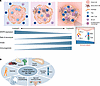 Figure 1
Immunogenicity spectrum within early-stage HR+ BC and host factors that may influence the TME. (A) The vast heterogeneity of HR+ BC leads to a broad spectrum for immunogenicity, which correlates with receptor expression, prognosis, and tumor grade. High-immunogenicity tumors can be more susceptible to ICB. (B) Numerous host factors affect tumor immunogenicity.
Homologous recombination deficiency (HRD) has also been linked to the immunogenicity of HR+ BCs and their biology (63). HRD refers to the loss of DNA damage proteins, such as BRCA1/2, RAD50/51, and PALB2, or associated genes, such as ATM and CHEK1/2, through either germline variants or acquired mutations within tumor cells (64–66). Patients harboring mutations in core homologous recombination pathways are highly responsive to DNA repair–targeted treatments, such as poly (ADP-ribose) polymerase (PARP) inhibition (67). HRD tumors have been associated with more inflamed and immunogenic TMEs in breast cancer (68, 69). This may be because HRD is correlated with increased tumor mutation burden (TMB) and thus higher neoantigen load (70). High TMB has been correlated with increased immunogenicity and with APOBEC-driven mutagenic signatures in breast cancers (71). Furthermore, deficiency in BRCA1/2 has also been correlated with high levels of large genomic rearrangements (72) that may increase the repertoire of potentially immunogenic alterations, including structural variants that are incompletely captured by conventional TMB measures. Neoantigen load and presentation via HLA molecules are essential for T cell engagement, potentially leading to a “hot” immune environment (73). Interestingly, HR+/HER2– BCs have very high levels of HLA downregulation (74, 75), an established mechanism of immune escape (76), often as a result of strong immune activation (77). This could indicate that HR+ BCs induce a very strong immune reaction in the early stages of development, which then pressures immune escape and ultimately leads to a less inflamed microenvironment.
-
Clinical immune modulation in HR+/HER2– BC
The prognostic role of TILs in HR+/HER2– BCs has been somewhat controversial. While some studies have reported a lack of a correlation between TILs and disease-free survival outcomes in HR+ disease (19, 78), the association between higher TILs and more aggressive biology (luminal B, higher grade, and nodal involvement) needs to be considered.
Several early clinical trials evaluating ICB in pretreated HR+/HER2– metastatic breast cancer failed to show clinically meaningful response rates, even in PD-L1–positive selected populations (Tables 3 and 4). Nonetheless, some signals for activity were observed, with an ORR of 19% in the ICON study in a cohort who received the anti-CTLA4 antibody ipilimumab in combination with the anti–PD-1 antibody nivolumab sequentially after chemotherapy (79). More recently, efforts have also focused on identifying subgroups more likely to benefit from ICB. The phase II Nimbus study evaluated a combination of ipilimumab and nivolumab in patients with high TMB (TMB > 9 mutations per megabase) in HER2– metastatic breast cancer (both TNBC and HR+ BC), with an ORR of 20% (80).
Metastatic disease is associated with a more suppressive immune TME, as well as larger tumor burden. Multiomic analysis of matched primary and metastatic samples in the AURORA studies of relapse in metastatic breast cancer have highlighted the vast heterogeneity of metastatic TMEs, which are heavily context dependent on subtype and site (81, 82). For instance, lymph node or skin metastases were shown to have higher immune scores relative to other metastatic sites, such as the liver (81). Lymph node–only metastases have also been correlated with more durable responses to anti–PD-1 blockade (83), indicating the cellular composition of the metastatic site (particularly the immune cell composition) determines the level of efficacy of ICB. While this constrains our ability to identify broad biomarkers of immunogenicity in metastatic patients, there is also an opportunity to improve our understanding of metastatic TMEs in breast cancer and how best to harness immunity in late-stage disease.
Higher response rates to immunotherapy are seen in early-stage HR+ disease, similar to what has been observed in TNBC, particularly when it is administered in the neoadjuvant setting. The GIADA trial was one of the first trials that tested ICB as a neoadjuvant therapy in early-stage HR+/HER2– disease (84). The trial showed moderate efficacy in a cohort of 43 luminal B–like early-stage breast cancers, with basal-like phenotypes showing the highest response rates to neoadjuvant nivolumab (84). Subsequently, two large, randomized, phase III studies have evaluated the efficacy of anti–PD-1 antibodies combined with neoadjuvant chemotherapy for HR+/HER2– BC: CHECKMATE-7FL (28), evaluating nivolumab, and KEYNOTE-756 (29), evaluating pembrolizumab. Both studies recruited high-grade or lymph node–positive patients with high Ki67, and included patients with low ER expression, to capture this biologically aggressive phenotype. Both studies showed a statistically significant improvement in pCR rate compared with chemotherapy. Most astonishingly, subgroup analysis from CHECKMATE-7FL showed that the addition of nivolumab nearly doubled the pCR rate (24.5% vs. 13.8%) in a BC subtype historically viewed as immune “cold” (28). This is further supported by the PD-L1 subgroup analysis from the KEYNOTE-756 trial showing similarly impressive response rates in this group (29). These two independent studies indicate preexisting immunity that is highly sensitive to the addition of ICB to chemotherapy. The findings are notable because pCR rates in HR+ BC have historically remained low with chemotherapy, ADCs, and endocrine therapies. They point to an underappreciated role for T cell biology in shaping prognosis and treatment response and suggest a therapeutic opportunity in a subgroup with historically poor outcomes.
Moreover, the broad variation in TIL within this cohort suggests an opportunity to further sensitize patients to ICB through synergistic combinations. Several studies have explored combinations of ICB with other targeted therapies that appear to (through various mechanisms) contribute to immune priming, including PARP inhibitors, CDK4/6 inhibitors, and ADCs. PARP inhibitors are thought to promote a proimmune microenvironment by activating the cGAS-STING pathway (85), and research has reported synergy with ICB (86). For instance, in the metastatic setting, the MEDIOLA study on HER2– disease showed promising efficacy in combining the PARP inhibitor olaparib with the anti–PD-L1 antibody durvalumab (87). In the early-stage setting, this combination was tested in one arm of the adaptive platform I-SPY2.2 study (88), and further evaluation of this strategy is ongoing in the OLIO trial (ACTRN12623000657628).
Immunomodulatory effects of CDK4/6 inhibitors have also been reported. Although mechanisms remain unclear, upregulation of antigen presentation machinery, downregulation of regulatory T cells, influx of CD8+ T cells, and induction of immunological memory have all been observed following treatment with CDK4/6 inhibitors (89–91). Preclinical studies and some limited clinical data have also shown synergy between CDK4/6 inhibitors and ICB (92). However, implementation of this approach has been limited by toxicity and questions of how best to harness the immunomodulatory effects of CDK4/6 inhibitors in the clinical setting.
ADCs are a novel drug class showing efficacy in HR+ BC as monotherapy with various targets (93, 94). ADCs with topoisomerase 1 inhibitor (Topo1i) cytotoxic payloads (including T-DXd and Dato-Dxd) have been shown to induce immunogenic cell death (95, 96), and remarkable clinical activity for the combination of Topo1i ADCs and ICB has been observed (97). The I-SPY2.2 trial evaluated this approach in the neoadjuvant setting for patients with early-stage breast cancer, including a cohort of HR+/HER2– BC, and showed promising efficacy, particularly in patients with higher immune scores (98).
Overall, it is likely that ICB responses have been modest in past HR+/HER2– trials because of design limitations in patient selection. Treatment-related toxicities and the risk of overtreatment of patients who would not benefit from ICB are also key challenges, which must be noted. Multiple biomarkers of immunogenicity, such as TILs and HRD, have been used to highlight immune-active patient populations. However, the integration of these biomarkers into the clinic for the management of HR+/HER2– disease has been relatively slow, and will be imperative in improving patient selection (35), as discussed above for TNBC. It is becoming evident that not all HR+/HER2– tumors have equivalent TMEs, and there is a clear opportunity for ICB in the clinical management of selected subgroups of HR+/HER2– disease, specifically high-risk patients with poorer outcomes under the current clinical management guidelines.


Article tools
- Download citation information
- Send a comment
- Terms of use
- Standard abbreviations
- Need help? Email the journal
Review Series
Emerging therapeutic strategies in breast cancer
-
Toward targeted therapeutics for lobular breast cancer
-
Antibody-drug conjugates in breast cancer: redefining targeted therapy
-
Expanding the therapeutic scope of PARP inhibitors in breast cancer: current indications and future directions
-
Breast cancer immunogenicity as a spectrum: evolving concepts and therapeutic implications
Metrics
Go to
- Top
- Abstract
- Introduction
- TNBC as the model for breast cancer immunotherapy
- Immunogenicity in HER2+ BC
- Immune landscape and heterogeneity of HR+/HER2– BC
- Clinical immune modulation in HR+/HER2– BC
- Mechanisms that regulate immunogenicity in breast cancer
- Conclusions
- Funding support
- Footnotes
- References
- Version history


Copyright © 2026 American Society for Clinical Investigation
ISSN: 0021-9738 (print), 1558-8238 (online)