
Go to JCI Insight
  • About
  • Editors
  • Consulting Editors
  • For authors
  • Publication ethics
  • Publication alerts by email
  • Advertising
  • Job board
  • Contact
  • Clinical Research and Public Health
  • Current issue
  • Past issues
  • By specialty
    • COVID-19
    • Cardiology
    • Gastroenterology
    • Immunology
    • Metabolism
    • Nephrology
    • Neuroscience
    • Oncology
    • Pulmonology
    • Vascular biology
    • All ...
  • Videos
    • ASCI Milestone Awards
    • Video Abstracts
    • Conversations with Giants in Medicine
  • Reviews
    • View all reviews ...
    • The cGAS-STING pathway: DNA sensing in health and disease (Jun 2026)
    • Neurodegeneration (Mar 2026)
    • Clinical innovation and scientific progress in GLP-1 medicine (Nov 2025)
    • Pancreatic Cancer (Jul 2025)
    • Complement Biology and Therapeutics (May 2025)
    • Evolving insights into MASLD and MASH pathogenesis and treatment (Apr 2025)
    • Microbiome in Health and Disease (Feb 2025)
    • View all review series ...
  • Viewpoint
  • Collections
    • In-Press Preview
    • Clinical Research and Public Health
    • Research Letters
    • Letters to the Editor
    • Editorials
    • Commentaries
    • Editor's notes
    • Reviews
    • Viewpoints
    • 100th anniversary
    • Top read articles

  • Current issue
  • Past issues
  • Specialties
  • Reviews
  • Review series
  • ASCI Milestone Awards
  • Video Abstracts
  • Conversations with Giants in Medicine
  • In-Press Preview
  • Clinical Research and Public Health
  • Research Letters
  • Letters to the Editor
  • Editorials
  • Commentaries
  • Editor's notes
  • Reviews
  • Viewpoints
  • 100th anniversary
  • Top read articles
  • About
  • Editors
  • Consulting Editors
  • For authors
  • Publication ethics
  • Publication alerts by email
  • Advertising
  • Job board
  • Contact
Top
  • View PDF
  • Download citation information
  • Send a comment
  • Terms of use
  • Standard abbreviations
  • Need help? Email the journal
  • Top
  • Abstract
  • Introduction
  • Overview of the cGAS/STING pathway
  • DNA recognized by the cGAS/STING pathway
  • cGAS/STING in the pathophysiology of vascular diseases
  • cGAS/STING in the pathophysiology of heart diseases
  • Targeting cGAS/STING for the treatment of CVDs
  • Conclusions and remaining questions
  • Conflict of interest
  • Funding support
  • Footnotes
  • References
  • Version history
  • Article usage
  • Citations to this article

Advertisement

Review Series Open Access | 10.1172/JCI204551

Emerging roles of the cGAS/STING pathway in cardiovascular diseases

Wataru Saitoh,1 Yasutomi Higashikuni,1,2,3,4 Oyunbileg Bavuu,1 Masataka Sata,5 and Daiju Fukuda1

1Department of Cardiovascular Medicine, Osaka Metropolitan University Graduate School of Medicine, Osaka, Japan.

2Division of Cardiovascular and Genetic Research, Center for Molecular Medicine, and

3Department of Cardiovascular Medicine, Jichi Medical University, Tochigi, Japan.

4Department of Cardiovascular Medicine, The University of Tokyo, Tokyo, Japan.

5Department of Cardiovascular Medicine, Tokushima University Graduate School, Tokushima, Japan.

Address correspondence to: Daiju Fukuda or Yasutomi Higashikuni, Department of Cardiovascular Medicine, Osaka Metropolitan University Graduate School of Medicine, 1-4-3 Asahi-machi, Abeno-ku, Osaka, 545-8585, Japan Phone: 81.6.6645.3801; Email: daiju.fukuda@omu.ac.jp (DF); yasutomi.higashikuni@omu.ac.jp (YH).

Authorship note: WS, YH, and OB contributed equally to this work.

Find articles by Saitoh, W. in: PubMed | Google Scholar

1Department of Cardiovascular Medicine, Osaka Metropolitan University Graduate School of Medicine, Osaka, Japan.

2Division of Cardiovascular and Genetic Research, Center for Molecular Medicine, and

3Department of Cardiovascular Medicine, Jichi Medical University, Tochigi, Japan.

4Department of Cardiovascular Medicine, The University of Tokyo, Tokyo, Japan.

5Department of Cardiovascular Medicine, Tokushima University Graduate School, Tokushima, Japan.

Address correspondence to: Daiju Fukuda or Yasutomi Higashikuni, Department of Cardiovascular Medicine, Osaka Metropolitan University Graduate School of Medicine, 1-4-3 Asahi-machi, Abeno-ku, Osaka, 545-8585, Japan Phone: 81.6.6645.3801; Email: daiju.fukuda@omu.ac.jp (DF); yasutomi.higashikuni@omu.ac.jp (YH).

Authorship note: WS, YH, and OB contributed equally to this work.

Find articles by Higashikuni, Y. in: PubMed | Google Scholar

1Department of Cardiovascular Medicine, Osaka Metropolitan University Graduate School of Medicine, Osaka, Japan.

2Division of Cardiovascular and Genetic Research, Center for Molecular Medicine, and

3Department of Cardiovascular Medicine, Jichi Medical University, Tochigi, Japan.

4Department of Cardiovascular Medicine, The University of Tokyo, Tokyo, Japan.

5Department of Cardiovascular Medicine, Tokushima University Graduate School, Tokushima, Japan.

Address correspondence to: Daiju Fukuda or Yasutomi Higashikuni, Department of Cardiovascular Medicine, Osaka Metropolitan University Graduate School of Medicine, 1-4-3 Asahi-machi, Abeno-ku, Osaka, 545-8585, Japan Phone: 81.6.6645.3801; Email: daiju.fukuda@omu.ac.jp (DF); yasutomi.higashikuni@omu.ac.jp (YH).

Authorship note: WS, YH, and OB contributed equally to this work.

Find articles by Bavuu, O. in: PubMed | Google Scholar

1Department of Cardiovascular Medicine, Osaka Metropolitan University Graduate School of Medicine, Osaka, Japan.

2Division of Cardiovascular and Genetic Research, Center for Molecular Medicine, and

3Department of Cardiovascular Medicine, Jichi Medical University, Tochigi, Japan.

4Department of Cardiovascular Medicine, The University of Tokyo, Tokyo, Japan.

5Department of Cardiovascular Medicine, Tokushima University Graduate School, Tokushima, Japan.

Address correspondence to: Daiju Fukuda or Yasutomi Higashikuni, Department of Cardiovascular Medicine, Osaka Metropolitan University Graduate School of Medicine, 1-4-3 Asahi-machi, Abeno-ku, Osaka, 545-8585, Japan Phone: 81.6.6645.3801; Email: daiju.fukuda@omu.ac.jp (DF); yasutomi.higashikuni@omu.ac.jp (YH).

Authorship note: WS, YH, and OB contributed equally to this work.

Find articles by Sata, M. in: PubMed | Google Scholar

1Department of Cardiovascular Medicine, Osaka Metropolitan University Graduate School of Medicine, Osaka, Japan.

2Division of Cardiovascular and Genetic Research, Center for Molecular Medicine, and

3Department of Cardiovascular Medicine, Jichi Medical University, Tochigi, Japan.

4Department of Cardiovascular Medicine, The University of Tokyo, Tokyo, Japan.

5Department of Cardiovascular Medicine, Tokushima University Graduate School, Tokushima, Japan.

Address correspondence to: Daiju Fukuda or Yasutomi Higashikuni, Department of Cardiovascular Medicine, Osaka Metropolitan University Graduate School of Medicine, 1-4-3 Asahi-machi, Abeno-ku, Osaka, 545-8585, Japan Phone: 81.6.6645.3801; Email: daiju.fukuda@omu.ac.jp (DF); yasutomi.higashikuni@omu.ac.jp (YH).

Authorship note: WS, YH, and OB contributed equally to this work.

Find articles by Fukuda, D. in: PubMed | Google Scholar

Authorship note: WS, YH, and OB contributed equally to this work.

Published July 1, 2026 - More info

Published in Volume 136, Issue 13 on July 1, 2026
J Clin Invest. 2026;136(13):e204551. https://doi.org/10.1172/JCI204551.
© 2026 Saitoh et al. This work is licensed under the Creative Commons Attribution 4.0 International License. To view a copy of this license, visit http://creativecommons.org/licenses/by/4.0/.
Published July 1, 2026 - Version history
View PDF
Abstract

Cardiovascular diseases (CVDs) remain the leading cause of mortality and morbidity worldwide, highlighting the need for novel therapeutic approaches. Inflammation plays a key role in CVD pathogenesis, and accumulating evidence has implicated the cyclic GMP-AMP synthase/stimulator of IFN genes (cGAS/STING) pathway in this process. The cGAS/STING pathway recognizes both non-self- and self-DNA, including mitochondrial and nuclear DNA, to activate its downstream proinflammatory signaling molecules, including TANK-binding kinase 1, IFN regulatory factor 3, and NF-κB. Various pathological stressors have been shown to induce self-DNA release into the cytosol and bloodstream from damaged cells in the cardiovascular system, indicating that circulating cell-free DNA is a useful biomarker of CVDs; however, how this contributes to the inflammatory signaling, cell death, and fibrosis that characterize CVDs remains unclear. Here, we discuss the current understanding on the roles of self-DNA and the cGAS/STING pathway in the pathophysiology of CVDs and the therapeutic potential of targeting this pathway.

Introduction

Cardiovascular diseases (CVDs) represent a wide array of disorders affecting the heart and blood vessels, including atherosclerosis, hypertension, pulmonary hypertension (PH), aortic aneurysm and dissection (AAD), myocardial infarction (MI), hypertensive heart disease, valvular heart disease, cardiomyopathy, myocarditis, and atrial fibrillation (AF). Despite progress in medical therapies, CVDs remain the leading cause of mortality and morbidity worldwide (1). The prevalence of CVDs, especially in the aging population, is expected to rise in the next few decades (2). To address this global burden, novel therapeutic strategies need to be developed.

The cyclic GMP-AMP synthase/stimulator of IFN genes (cGAS/STING) pathway is a component of the innate immune system that senses cytosolic double-stranded DNA (dsDNA) to trigger proinflammatory gene expression. Originally identified for its role in host defense against viral and bacterial infection through recognition of non-self-DNA, the cGAS/STING pathway has also been implicated in sterile inflammation by detecting self-DNA released from damaged or stressed cells (Figure 1) (3–6). Here, we describe the emerging roles of the cGAS/STING pathway in the pathophysiology of CVDs and discuss therapeutic strategies targeting the pathway.
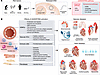
The cGAS/STING pathway and cardiovascular diseases.Figure 1

The cGAS/STING pathway and cardiovascular diseases. (A) Risk factors associated with cardiovascular disease — such as aging, sex differences, obesity, and smoking — contribute to the accumulation of intracellular nuclear DNA and mtDNA and extracellular cell-free DNA (cfDNA), which activate both canonical and noncanonical cGAS/STING signaling. (B) Activation of the cGAS/STING pathway modulates the function of vascular and cardiac cells, including vascular smooth muscle cells (VSMCs), vascular endothelial cells (ECs), immune cells, cardiomyocytes, and fibroblasts. This causes functional and structural changes of the blood vessels and heart, contributing to the pathophysiology of cardiovascular diseases, including atherosclerosis, hypertension, pulmonary hypertension, aortic aneurysm and dissection, myocardial infarction, myocardial ischemia/reperfusion injury, hypertensive heart disease, valvular heart disease, cardiomyopathy, myocarditis, and atrial fibrillation.
Overview of the cGAS/STING pathway

The cGAS/STING pathway links non-self- or self-dsDNA sensing to activation of proinflammatory transcription factors (Figure 1). cGAS, also known as MB21D1, is a member of the nucleotidyltransferase (NTase) enzyme family that detects cytosolic dsDNA in a sequence-independent manner (7–10). Upon binding to dsDNA, cGAS dimerizes and undergoes conformational changes that lead to the opening of a catalytic nucleotide binding pocket. Activated cGAS catalyzes the formation of the second messenger cyclic GMP-AMP (cGAMP) from ATP and GTP (10–14). The synthesized cGAMP binds to an active pocket site of STING, triggering its downstream signaling.

STING is a dimeric transmembrane adaptor protein localized on the ER, with its ligand-binding domain exposed to cytosol (10, 15–17). Upon binding to cGAMP, STING undergoes conformational changes to form a stable tetramer or higher-order oligomer that translocates from the ER to the Golgi apparatus, where STING recruits TANK-binding kinase 1 (TBK1) to promote the phosphorylation of TBK1 and STING (17, 18). Phosphorylated TBK1 promotes the phosphorylation and nuclear trafficking of downstream transcription factors, such as IFN regulatory factor 3 (IRF3) and STAT6 (7, 8, 17, 19). IRF3 induces expression of IFN-I and subsequent expression of IFN-stimulated genes (ISGs), while STAT6 induces chemokine expression (20, 21). STING can also activate NF-κB in both TBK1-dependent and -independent manners, which further promotes the expression of proinflammatory genes (10, 15, 16, 22).

DNA recognized by the cGAS/STING pathway

cGAS recognizes dsDNA in the cytosol from both extracellular and intracellular origins (Figure 1) (7). DNA fragments from microorganisms, viruses, dead cells (cell-free DNA), and extracellular vesicles can enter the cytosol by endocytosis, phagocytosis, or transmembrane diffusion (23–26). DNA fragments of intracellular origin include DNA released from mitochondria (mtDNA), chromatin fragments, and damaged DNA packaged into micronuclei in the nucleus (10). Although agnostic to the specific sequence, cGAS interacts with dsDNA in a nucleotide length–dependent manner (27–30). It has been reported that dsDNA fragments longer than 20 base pairs can bind cGAS but dsDNA fragments longer than 40 base pairs form a more stable complex for cGAS activation (29, 30).

cGAS/STING in the pathophysiology of vascular diseases

Atherosclerosis. Atherosclerosis is the thickening and hardening of the medium- and large-sized arteries due to a buildup of plaque in the subendothelium that consists of cholesterol, fatty acids, calcium, fibrin, blood cells, cellular waste products, and other substances in the blood. A large body of evidence now indicates that inflammation fundamentally contributes to the pathogenesis of atherosclerosis and accompanying thromboembolic events due to plaque rupture or erosion and that the cGAS/STING pathway plays important roles in producing that inflammation (Figure 1 and Tables 1 and 2). Upregulation of STING is observed in human atherosclerotic lesions (Table 1) (31, 32). In a mouse model of diabetes, genetic deletion and pharmacologic inhibition of STING ameliorate aortic endothelial dysfunction (33). Genetic disruption of IRF3 in Apoe–/– mice resulted in reduced lipid burden and macrophage infiltration in atherosclerotic plaques (34).

Table 1

Human evidence of cGAS/STING activation in cardiovascular diseases

Table 2

Emerging roles of the cGAS/STING pathway in atherosclerosis and myocardial infarction

In the arterial walls, the cGAS/STING pathway can be activated in various cell types, including vascular endothelial cells (ECs), vascular smooth muscle cells (VSMCs) and immune cells. Dong et al. reported that STING was upregulated in vascular ECs in Apoe–/– mice fed a high-fat diet, and EC-specific deletion of STING in high-fat diet–fed Apoe–/– mice resulted in a meaningful reduction in atherosclerotic plaques (32). Mechanistically, mtDNA, which is released into the cytoplasm of vascular ECs in response to low oscillatory shear stress, was suggested to activate the cGAS/STING pathway, leading to EC senescence and dysfunction and promoting atherogenesis. Oxidized LDL also induces mtDNA release in vascular ECs in Apoe–/– mice fed a Western diet, further contributing to STING activation and atherogenesis (35). Interestingly, activation of the cGAS/STING pathway by mtDNA was reported to contribute to NLRP3 inflammasome activation and subsequent pyroptosis in vascular ECs (36, 37). In vitro, treatment of human umbilical vein ECs with mtDNA or cGAMP increased inflammatory molecule expression and decreased endothelial NOS phosphorylation, partially through the cGAS/STING pathway (33).

Cigarette smoke extract can induce DNA damage in both the nucleus and mitochondria in human ECs, leading to IL-6 upregulation through activation of the cGAS/STING pathway (38). In addition, the cGAS/STING pathway was shown to activate the noncanonical effector PKR-like ER kinase (PERK), which causes vascular EC dysfunction through epigenetic alterations (39). Furthermore, Liu et al. demonstrated that IRF3 upregulation in vascular ECs contributes to plaque vulnerability through adhesion molecule induction in Apoe–/– mice (34). Collectively, these findings indicate the diverse contribution of the cGAS/STING pathway to vascular EC function. However, it is important to note that ECs are heterogenous, with different populations having differing roles in atherosclerosis (40), and the role of the cGAS/STING pathway in EC heterogeneity remains to be elucidated. In addition, the effect of the cGAS/STING pathway on vascular ECs in the peripheral arteries and small vessels needs to be clarified.

A role for the cGAS/STING pathway in atherosclerotic macrophages has also been reported. In Apoe–/– mice fed a Western diet, genetic deletion of STING ameliorated atherosclerosis, with decreased macrophage infiltration and proinflammatory cytokine expression in the aorta, whereas restoration of STING expression in bone marrow–derived cells in Sting–/–;Apoe–/– mice promoted atherogenesis with increased proinflammatory cytokine expression and TBK1 phosphorylation in the aorta (31). The release of mtDNA and subsequent activation of the cGAS/STING pathway have also been reported in oxidized LDL-treated macrophages (41, 42).

In VSMCs, the cGAS/STING pathway is implicated in senescence. Senescent VSMCs produce less collagen and more proinflammatory cytokines, which results in a thin fibrous cap and immune cell infiltration and promotes plaque vulnerability (43, 44). Bi et al. reported that, in Apoe–/– mice with chronic kidney disease, mtDNA released from damaged mitochondria activates the cGAS/STING pathway, resulting in IFN-I production, which causes VSMC senescence and phenotypic switching and promotes plaque vulnerability (43). Uryga et al. demonstrated that telomere-derived damaged DNA induces cellular senescence and proinflammatory cytokine expression in VSMCs through the cGAS/STING pathway, which leads to accelerated atherogenic changes after vascular injury (44).

Hypertension. Hypertension is the most prevalent CVD, in which systemic BP is persistently elevated. Mechanisms of hypertension include high-salt intake, activation of the renin-angiotensin-aldosterone system, increased sympathetic drive, and impaired nitric oxide bioavailability. Accumulating evidence implicates vascular inflammation in the initiation and maintenance of hypertension (45).

A recent study highlighted an important role of the cGAS/STING pathway in the development of hypertension (Figure 1 and Table 3) (46). Li et al. demonstrated that, in mice expressing a human risk variant of apolipoprotein L1 associated with hypertension, STING activation in vascular ECs was associated with hypertension, which promoted endothelin 1 production (46). EC-specific STING deletion markedly attenuated hypertension and subsequent vascular remodeling with suppressed endothelin 1 expression, demonstrating a causal role of STING activation in hypertension.

Table 3

Emerging roles of the cGAS/STING pathway in hypertension, hypertensive heart disease, and pulmonary hypertension

Chronobiological rhythms regulate BP and its variability (47, 48), and disruption of those rhythms has been linked to the pathophysiology of hypertension (47, 48). A recent study reported that disruption of chronobiological rhythm activates the cGAS/STING pathway and causes cell senescence in macrophages (49). Since innate immune cells, including macrophages, have been implicated in the pathophysiology of hypertension (50, 51), the cGAS/STING pathway might link disrupted chronobiological rhythms to hypertension. Further studies are needed to clarify the role of the cGAS/STING pathway in circadian control of BP and its variability.

PH. PH is a condition of high BP in the arteries of the lungs (52). Pham et al. observed STING upregulation in the lung of patients with PH (Table 1) (53, 54). In rodent models of PH, genetic disruption or pharmacologic inhibition of STING has been shown to improve pulmonary vascular remodeling and hypertension (Table 3) (54–58). Mechanistically, the cGAS/STING pathway induces VSMC senescence in the pulmonary arteries through NF-κB activation (54–57). Wu et al. showed that STING activates the NLRP3 inflammasome in macrophages to induce proliferation of VSMCs through proinflammatory cytokine production (59). In addition, the cGAS/STING pathway is implicated in immune cell dysfunction in PH. Pham et al. demonstrated that genetic deletion or pharmacologic inhibition of STING increases VEGF in CD11b+ and CD11c+ myeloid cells, which might contribute to protection against PH in an IFN signaling–independent manner (53). Interestingly, Pham et al. also reported that STING expression in myeloid cells might prevent severe PH by suppressing expression of PD-L1, suggesting diverse roles of STING in myeloid cells (54).

AAD. An aortic aneurysm is a dilatation of the aorta to greater than 1.5 times the original size due to structural weakening of the aortic wall. An aortic dissection is a tear between the intima and media of the aortic wall. AADs are lethal vascular conditions, characterized by progressive VSMC loss and extracellular matrix degradation, which can lead to aortic rupture or severe ischemia due to arterial occlusions. Persistent vascular inflammation has been implicated in the pathogenesis of AAD (60).

Recent studies have demonstrated that the cGAS/STING pathway plays important roles in the underlying mechanisms of AAD (Figure 1 and Table 4). Luo et al. reported marked upregulation of STING, TBK1, and IRF3 expression and phosphorylation in human sporadic AAD tissues (Table 1) (61). In a murine model of sporadic AAD induced by a combination of a high-fat diet and high-dose angiotensin II infusion, STING-deficient mice exhibited marked reductions in aortic enlargement, dissection, and rupture. Nuclear and mitochondrial DNA damage in VSMCs, and the subsequent leakage of DNA to the cytosol and extracellular space, activates STING in neighboring VSMCs and macrophages, leading to apoptotic and necroptotic cell death. Using single-cell transcriptome and transposase-accessible chromatin analyses, Chakraborty et al. revealed that activation of STING and IRF3 induces epigenetic modification and chromatin remodeling in VSMCs in the aorta of angiotensin II–infused mice, which resulted in VSMC phenotypic alterations through suppression of contractile genes and induction of proinflammatory genes (62). These epigenetic and phenotypic changes in VSMCs were prevented in Sting–/– mice.
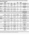
Table 4

Emerging roles of the cGAS/STING pathway in aortic aneurysm and dissection, valvular heart disease, cardiomyopathy, myocarditis, and atrial fibrillation

cGAS/STING in the pathophysiology of heart diseases

Heart diseases, including MI, hypertensive heart disease, valvular heart disease, cardiomyopathy, myocarditis, and AF all cause cardiac remodeling that can lead to heart failure (HF) (63, 64). A growing body of experimental and clinical evidence now demonstrates that inflammation plays a fundamental role in the pathophysiology of HF (65), although the underlying mechanisms remain largely unknown. Recent studies have provided evidence supporting involvement of the cGAS/STING pathway in various heart diseases and HF (17) (Figure 1 and Tables 1–4).

MI. MI is a disease characterized by myocardial cell death due to the interruption of blood flow in the coronary arteries to the corresponding region of the myocardium. Atherosclerosis is a main cause of MI. Following occlusion of the coronary arteries, intense sterile inflammation and immune cell infiltration, triggered by molecules released from damaged or dead cells, are initiated to digest and clear damaged cells and extracellular matrix (66). Subsequently, reparative pathways promote myofibroblast proliferation, neovascularization, resolution of inflammation, and scar formation for tissue repair. After the reparative phase, the heart continues to undergo structural and functional changes that lead to chamber dilatation and contractile dysfunction and exacerbate HF. Inflammation has been implicated not only in the reparative phase, but also in chronic cardiac remodeling after MI.

The cGAS/STING pathway has been implicated in the initiation of cardiac inflammation and the clearance of necrotic myocardium and tissue repair after MI (Figure 1). Upregulation of STING and IRF3 in the heart was observed in the reparative phase of murine hearts after MI (67, 68). King et al. found that genetic disruption of IRF3 or cGAS improved survival rate and cardiac remodeling after MI (67). Mechanistically, the cGAS/STING/IRF3 pathway is activated in macrophages that recognize dsDNA released from dying cells in the infarcted region. Subsequently, the signaling induces the production of IFN-I and ISGs, which impairs tissue repair and deteriorates cardiac remodeling after MI by promoting the expression of proinflammatory cytokines and chemokines and recruitment of leukocytes in the infarcted heart. Cao et al. also reported that the cGAS/STING pathway is essential for maintaining proinflammatory macrophages in the murine heart after MI through inducible NOS upregulation (68). Furthermore, Zhu et al. demonstrated that the cGAS/STING pathway was associated with neutrophil production and activation, although cGAS deficiency in neutrophils did not affect neutrophil extracellular trap (NET) formation after MI (69). Neutrophil-specific deletion of cGAS improved survival after MI in mice, although it did not affect post-MI cardiac remodeling.

Myocardial ischemia/reperfusion injury. Myocardial ischemia/reperfusion injury is myocardial damage caused by temporary interruption and subsequent restoration of blood flow to the heart (70). ROS, generated by the reactivated electron transport chain and other sources, play important roles in the pathogenesis of myocardial ischemia/reperfusion injury (70). ROS cause cardiomyocyte damage and cardiac dysfunction by inducing apoptosis, autophagy, and proinflammatory responses. Damage-associated molecules released from necrotic cardiac cells, including mitochondrial and nuclear DNA, also contribute to activation of proinflammatory responses (71).

Activation of the cGAS/STING pathway and accumulation of mtDNA in the cytosol have been reported in cardiac tissues of diabetic mice exposed to myocardial ischemia/reperfusion injury. Pharmacological inhibition of STING attenuated myocardial damage in this model (72). Zhang et al. reported that mitochondrial components, released from cardiomyocytes through extracellular vesicles, might be internalized by cardiac fibroblasts and promote myocardial fibrosis via fibroblast activation and proliferation (73). These findings suggest a potential role of the cGAS/STING pathway in the pathophysiology of myocardial ischemia/reperfusion injury, although further studies are necessary to establish its relevance in the human disease.

Hypertensive heart disease. Hypertensive heart disease is a condition in which cardiac structural and functional changes are attributable to systemic hypertension. Cardiac hypertrophy initially occurs as an adaptive response to pressure overload to maintain cardiac function (63). However, chronic stress responses cause contractile dysfunction and HF (64). Inflammation has been implicated in the pathophysiology of both adaptive hypertrophy and HF (74, 75).

The roles of the cGAS/STING pathway in hypertensive heart disease have been investigated by using a murine model of pressure overload-induced cardiac hypertrophy and HF. Upregulation of cGAS and STING has been reported in the murine heart subjected to pressure overload as well as in the human heart exhibiting cardiac hypertrophy (Table 1) (76–80). Hu et al. demonstrated that cGAS knockdown in cardiomyocytes improves survival rate and ameliorates cardiac hypertrophy, contractile dysfunction, interstitial fibrosis, immune cell infiltration, proinflammatory cytokine expression, and cardiomyocyte apoptosis during pressure overload (76). Zhang et al. reported that genetic deletion of STING resulted in improvement of cardiac hypertrophy, interstitial fibrosis, and contractile dysfunction with suppressed macrophage infiltration, IFN-I–mediated inflammation, and ER stress response in pressure-overloaded hearts (77). Guo et al. showed that upregulation of inducible NOS during pressure overload contributes to mtDNA release in cardiomyocytes, which promotes adverse remodeling through activation of the cGAS/STING/IRF3 pathway in mice (78). Brassington et al. reported that activation of the cGAS/STING pathway in cardiomyocytes might be involved in CD8+ T cell activation and macrophage recruitment, which induce cardiomyocyte apoptosis and interstitial fibrosis, respectively (81). Sanders et al. demonstrated that STING activation in cardiac ECs induces IL-6 secretion and HF during pressure overload in mice (82). Interestingly, transgenic mice with cardiomyocyte-specific overexpression of STING showed attenuated adverse remodeling and contractile dysfunction in response to pressure overload by inhibiting autophagy (79), while cardiomyocyte-specific activation of STING, using a constitutively active mutant, led to cardiac hypertrophy and HF in mice (80). How STING contributes to the adaptive and maladaptive mechanisms in hypertensive heart disease remains to be elucidated.

Valvular heart disease. Valvular heart disease is a structural or functional abnormality of cardiac valves that affects blood flow and mechanical stress on the heart. Progression of aortic valve calcification results in aortic stenosis (83). Hu et al. reported increased STING and its phosphorylation in calcified human valves compared with noncalcified valves (Table 1) (84). Pharmacological inhibition of STING suppressed calcium nodule formation and alkaline phosphatase activity in human aortic valve interstitial cells (HAVICs) treated with osteogenic medium, whereas treatment with a STING agonist promoted these changes. Genetic disruption or pharmacologic inhibition of STING also mitigated aortic valve calcification in mice. Mechanistically, mtDNA leakage through downregulation of calcium/calmodulin-dependent protein kinase 1 in activated HAVICs stimulates the cGAS/STING pathway, which leads to upregulation of alkaline phosphatase and runt-related transcription factor 2, the essential regulators of calcification, through TBK1 and NF-κB activation (84–86).

Cardiomyopathy. Cardiomyopathies are diseases of the heart muscle characterized by morphologically and functionally abnormal myocardium due to various causes, in the absence of coronary artery disease, hypertension, valvular disease, and congenital heart disease sufficient to cause the observed myocardial abnormality (87, 88). STING upregulation has been reported in human heart samples of patients with dilated and hypertrophic cardiomyopathy (HCM) (77), suggesting the involvement of the cGAS/STING pathway (Table 1).

Dilated cardiomyopathy is the most common form of cardiomyopathy, characterized by chamber dilation and dysfunction. Patients carrying a homozygous missense mutation in the nuclear lamina-associated inner nuclear membrane protein LEMD2 develop dilated cardiomyopathy (89). Using a knockin mouse model of the LEMD2 disease mutation and corresponding cell model (90), Chen et al. demonstrated that this mutation impairs nuclear envelope rupture repair capacity. The resulting accumulation of DNA in the cytoplasm activates cGAS/STING and promotes myocardial cellular senescence. Mutations in the LMNA gene, encoding the nuclear envelope protein lamin A/C, cause dilated cardiomyopathy (91). Cheedipudi et al. showed that genetic disruption of cGAS attenuated the dilated cardiomyopathy phenotype in Lmna-deficient mice (92). On the other hand, En et al. reported that the cGAS/STING pathway is not activated in adult mice with cardiomyocyte-specific Lmna deletion (93). Further investigation will be necessary to clarify the role of the cGAS/STING pathway in lamin-related dilated cardiomyopathy.

HCM is characterized by thickening of the heart muscle without pressure overload. The cGAS/STING/IFN pathway is implicated in the pathophysiology of sporadic HCM caused by a somatic loss-of-function mutation in NAP1L1, which encodes a member of the nucleosome assembly protein family (94). Lv et al. showed that in vivo overexpression of this variant in cardiomyocytes exacerbated angiotensin II–induced cardiac hypertrophy with activation of the cGAS/STING pathway in mice, while pharmacological inhibition of STING and IFN receptor reversed the cardiac phenotype (94). Mechanistically, this mutation causes destabilization of nucleosome formation and DNA leakage into the cytoplasm, which accelerates proinflammatory responses and cardiac hypertrophy.

Diabetic cardiomyopathy is characterized by lipid accumulation, mitochondrial dysfunction, inflammation, and fibrosis (95). Activation of the cGAS/STING pathway and the NLRP3 inflammasome have been observed in murine diabetic hearts (96–98). Yan et al. demonstrated that Sting knockdown in cardiomyocytes improved diabetes-induced cardiac hypertrophy and contractile dysfunction with suppressed pyroptosis and inflammatory responses (96). Mechanistically, palmitic acid–induced lipotoxicity activated the cGAS/STING pathway through mitochondrial oxidative damage and mtDNA release into the cytosol in cardiomyocytes, which promoted pyroptosis and proinflammatory cytokine production in an NLRP3 inflammasome-dependent manner, suggesting crosstalk between the cGAS/STING pathway and NLRP3 inflammasome activation (96–98).

Chronic kidney disease has been shown to contribute to the pathophysiology of cardiomyopathy (99). Han et al. reported the activation of the cGAS/STING pathway in murine hearts in a chronic kidney disease model, which was ameliorated by cardiomyocyte-specific genetic disruption of STING (100). Mitochondrial oxidative stress was implicated in activation of the cGAS/STING pathway in this setting.

Anthracycline chemotherapeutics for the treatment of cancer, such as doxorubicin, have been shown to cause cardiotoxicity with a poor prognosis (101, 102), and chronic doxorubicin cardiomyopathy has been associated with activation of the cGAS/STING pathway in murine hearts (103–105). Luo et al. demonstrated that EC-specific genetic deletion of STING prevented doxorubicin cardiomyopathy and EC dysfunction (103). Mechanistically, activation of the cGAS/STING pathway in cardiac ECs by doxorubicin caused mitochondrial dysfunction through a reduction of intracellular NAD levels. Another chemotherapeutic, cisplatin, has also been reported to have cardiotoxic effects (106) and shown to activate the cGAS/STING pathway in murine hearts (107). Wang et al. demonstrated that genetic disruption of STING attenuated cisplatin-induced cardiotoxicity in mice (107).

Immune checkpoint inhibitors are another group of cardiotoxic anticancer drugs (108). Cao et al. demonstrated that anti–PD-1 antibody activates the cGAS/STING pathway in macrophages with M1 polarization (109). Pharmacological inhibition of STING ameliorated cardiac injury induced by anti–PD-1 antibody in mice, although the mechanism remains unelucidated.

Myocarditis. Myocarditis is an inflammatory disease of the heart, characterized by inflammatory cell infiltration and myocardial injury, which can be caused by infections, immune systemic activation, and exposure to drugs (110). The cGAS/STING pathway has been implicated in the pathophysiology of various types of myocarditis (111–116). Qin et al. reported that the cGAS/STING pathway contributes to cardiac inflammation by coxsackievirus B3 (CVB3) infection (111). Genetic disruption of STING suppressed CVB3-induced proinflammatory cytokine expression in mice, which led to higher survival rates. CVB3-infected cardiomyocytes release mtDNA into the extracellular space, which activates STING signaling in cardiac macrophages. An important role of cGAS in macrophages was also reported in Chagas disease, which can cause myocarditis by Trypanosoma cruzi infection (112). Choudhuri et al. reported that extracellular vesicles containing oxidized DNA are released from infected cardiac cells, which activates macrophages through cGAS signaling (112). Li et al. demonstrated that STING is activated in a murine sepsis-induced cardiac injury model induced by LPS injection (113). In addition, genetic deletion of STING was shown to improve survival and cardiac function in this model, associated with suppressed cardiac inflammation and cardiomyocyte apoptosis and pyroptosis.

The expression of STING and single-stranded DNA accumulation in macrophages were also observed in human heart samples of patients with autoimmune myocarditis (114). Mice with genetic disruption of TREX1 or DNase II have been reported to exhibit autoimmune myocarditis with elevated expression of ISGs (115, 116). Gao et al. demonstrated that genetic ablation of cGAS in these murine models rescued the cardiac phenotypes (115). In another experimental autoimmune myocarditis model using subcutaneous injections of cardiac myosin heavy chain α peptides, Hua et al. showed that pharmacological inhibition of STING ameliorated myocarditis with suppressed proinflammatory responses in macrophages (114). Further studies will be necessary to clarify mechanistic insight more in detail.

AF. AF is the most common arrhythmia, characterized by irregular beating of the atrial chambers of the heart (117). Upregulation and activation of the cGAS/STING pathway have been reported in the atria of rodent models of diabetes and obesity (118, 119). Diabetic mice or obese rats show increased susceptibility to AF induced by rapid atrial pacing, but this phenotype was ameliorated by cardiomyocyte-specific knockdown of STING (118, 119).

STING activation is associated with atrial fibrosis, cardiomyocyte apoptosis, and abnormal calcium handling (118). In the atria of obese rats, expression levels of inositol 1,4,5-trisphosphate receptor type 1 (IP3R1) on the ER and voltage-dependent anion channel protein 1 (VDAC1) on the mitochondrial outer membrane are upregulated, resulting in excessive calcium transfer from the ER to the mitochondria (120). STING knockdown mitigated these structural and molecular changes. Interestingly, mitochondrial calcium overload mediated by IP3R1 and VDAC1 was reported to promote the leakage of mtDNA into the cytoplasm, leading to activation of cGAS (121). This finding suggests that the cGAS/STING pathway contributes to a vicious cycle in the pathophysiology of AF.

Targeting cGAS/STING for the treatment of CVDs

Pharmacological inhibitors of the cGAS/STING pathway have been developed, such as cGAS inhibitors, STING inhibitors, and TBK1 inhibitors (Table 5), and have been under clinical investigation in autoimmune disease and cancer (122, 123). VENT-03, an oral cGAS inhibitor, was shown to be safe and well-tolerated in healthy volunteers and is now in a phase IIa trial for the treatment of systemic lupus erythematosus (124). In cancer, nucleotide-based cGAMP analogs, which aim to exploit cGAS/STING-dependent antitumor immunity, have been tested, although trials have not yet shown therapeutic benefit (125). In infectious disease, modulation of cGAS/STING to either augment host defense or mitigate excessive inflammation has been under exploration (126). Collectively, these attempts provide important conceptual and translational frameworks for applying cGAS/STING-targeted therapies to CVDs. Here, we describe evidence of the therapeutic potential of these agents as demonstrated in animal models of CVDs.
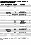
Table 5

Pharmacological inhibitors of the cGAS/STING pathway for the treatment of cardiovascular diseases

cGAS inhibitors. Three types of cGAS inhibitors have been developed: inhibitors of cGAS enzymatic activity, inhibitors of cGAS binding to dsDNA, and targeted modulators of cGAS by acetylation. RU.521 is a competitive inhibitor of cGAS that binds to the active site of cGAS and inhibits its interaction with ATP and GTP (127). RU.521 has been shown to be effective in the treatment of atherosclerosis, myocardial ischemia/reperfusion injury, and myocarditis in mice (36, 114, 128, 129). X6 and antimalarial drugs, which prevent the formation of the cGAS-dsDNA complex, were shown to have a beneficial effect in murine autoimmune myocarditis (116). Interestingly, the NSAID aspirin acetylates cGAS, which inhibits cGAS activation and improves autoimmune myocarditis in mice (130). Further studies are needed to clarify therapeutic potentials of these agents in human disease.

STING inhibitors. Nitrofuran derivatives, such as C-176, C-178, and H-151, effectively inhibit STING signaling by binding to Cys91, which results in nitroalkylation of STING’s palmitoylation site (131). These compounds have been shown to improve atherosclerosis, MI, myocardial ischemia/reperfusion injury, hypertensive heart disease, cardiomyopathy, and myocarditis in mice (31, 36, 43, 81, 100, 114, 128, 132, 133). Astin C and SN-011 competitively block interaction between STING and cyclic dinucleotides. These compounds showed protective effects against autoimmune myocarditis in mice (134, 135).

TBK1 inhibitors. Several kinase inhibitors, including K252a, dovitinib, and oxindole 91, have been shown to inhibit TBK1 by activity-based screens of compound libraries, although they are not TBK1-specific inhibitors (136). These compounds prevent autophosphorylation of TBK1 and IκB kinase ε (IKKε) at the Ser172 site through hydrogen bond network formation. Amlexanox is another TBK1 inhibitor that prevents its phosphorylation at the Ser366 site, and it also has an inhibitory effect on IKKε. Amlexanox is currently used for the treatment of recurrent aphthous ulcers and bronchial asthma. Several studies have shown that amlexanox mitigates atherosclerosis and AAD in mice (31, 61). Further investigation will be needed to determine the potential of TBK1 inhibitors in the treatment of CVDs.
Conclusions and remaining questions

The cGAS/STING pathway is a pivotal regulator of the innate immune system that functions through the sensing of both non-self- and self-DNA. Although indispensable for host defense, uncontrolled activation of the cGAS/STING pathway by self-DNA can drive chronic sterile inflammation and contribute to the development of a wide spectrum of diseases, including CVDs, which causes further cell and tissue damage to release self-DNA. Many questions remain regarding the contribution of cGAS/STING to CVD pathogenesis, and it will be important to address these in order to successfully translate therapies targeting the pathway.

Lifestyle factors, including smoking and obesity, have been reported to cause self-DNA release from damaged cells and tissues, which exacerbates inflammation via the cGAS/STING pathway (137). For example, Ueda et al. reported that in atherosclerotic patients, the serum level of circulating cell-free DNA was higher in smokers than in nonsmokers (38). It is possible that lifestyle modifications that can improve clinical outcomes of CVDs act in part by preventing the DNA release that activates the cGAS/STING pathway.

Aging is a risk factor for CVDs, and the effects may in part be mediated by cGAS/STING pathway activation. For example, levels of circulating mtDNA increase with age (138, 139), and nuclear integrity is impaired with age due to dysfunction of Yes-associated protein and transcriptional coactivator with PDZ-binding motif (140). Furthermore, levels of the cytosolic exonuclease TREX1 decrease in the kidney in aged rodents (141). These changes could result in cGAS/STING activation and drive senescence (142–144). Biological sex also affects CVD risk, and sex differences in protein expression profiles related to the cGAS/STING pathway have been reported (141, 145, 146). For example, activity of the cGAS/STING pathway is higher in the kidneys of male rats than in female rats, and this difference becomes more prominent with age (141). Such differences might contribute to the more pronounced age-related increase in the cumulative incidence of CVDs in men compared with women (147).

Many immune cell types contribute to the pathogenesis of CVDs, but the role of cGAS/STING in those populations is not fully elucidated. For example, granulocytes can form NETs to propagate inflammation and thrombosis in atherosclerotic plaques (148, 149), and the DNA in those NETs can be recognized by cGAS (150). However, whether the cGAS/STING pathway in these cells plays a role in the pathophysiology of CVDs needs further investigation.

In addition to the canonical cGAS/STING pathway that activates IRF3 and NF-κB, several noncanonical pathways have been reported. For example, STING/PERK/eIF2α signaling has been implicated in cell senescence (151), and in turn PERK may modulate the canonical pathway through binding to STING (39). The cGAS/STING pathway modulates autophagy, which contributes to innate immune response and metabolic homeostasis (152–155). Furthermore, STING has been reported to act as an ion channel that directly induces NLRP3 inflammasome activation (156). The importance of these noncanonical pathways to CVD pathophysiology remains to be elucidated.

Taken together, the cGAS/STING pathway is a promising therapeutic target for the treatment of CVDs. Indeed, as described above, in preclinical animal models, genetic and pharmacologic inhibition of the cGAS/STING pathway have protective effects against CVDs. However, nonspecific inhibition of this pathway might cause immune dysfunction and adverse effects, including impaired tissue repair. In addition, several studies have demonstrated crosstalk between the cGAS/STING and other signaling pathways (157), including TLR9 signaling (157–160), revealing complex and cell-type-specific relationships. Therefore, successful clinical translation of cGAS/STING-targeted therapies for CVD will require the development of strategies that enable spatiotemporal control of the cGAS/STING pathway.

Conflict of interest

The authors have declared that no conflict of interest exists.

Funding support
  • Japan Science and Technology Agency research grant (Fusion Oriented REsearch for disruptive Science and Technology Program JPMJFR245G) to YH.
  • Vehicle Racing Commemorative Foundation research grant to YH and MS.
  • Takeda Science Foundation research grant to YH.
  • SENSHIN Medical Research Foundation research grants to YH and DF.
  • Moriya Scholarship Foundation research grant to YH.
  • Koyanagi Foundation research grant to YH.
  • JSPS KAKENHI grants JP23K07501 and JP26K02416 to YH, JP24K02451 to DF, and JP25K02646 to MS.
  • Gout and uric acid foundation of Japan research grant to DF.
  • BIOTRONIK Japan research grant to DF.
Footnotes

Copyright: © 2026, Saitoh et al. This is an open access article published under the terms of the Creative Commons Attribution 4.0 International License.

Reference information: J Clin Invest. 2026;136(13):e204551. https://doi.org/10.1172/JCI204551.

References
  1. Global Burden of Cardiovascular Diseases Risks 2023 Collaborators. Global, regional, and national burden of cardiovascular diseases and risk factors in 204 countries and territories, 1990-2023. J Am Coll Cardiol. 2025;86(22):2167–2243.
    View this article via: CrossRef PubMed Google Scholar
  2. Mohebi R, et al. Cardiovascular disease projections in the United States based on the 2020 census estimates. J Am Coll Cardiol. 2022;80(6):565–578.
    View this article via: CrossRef PubMed Google Scholar
  3. West AP, Shadel GS. Mitochondrial DNA in innate immune responses and inflammatory pathology. Nat Rev Immunol. 2017;17(6):363–375.
    View this article via: CrossRef PubMed Google Scholar
  4. Ahn J, et al. STING manifests self DNA-dependent inflammatory disease. Proc Natl Acad Sci U S A. 2012;109(47):19386–19391.
    View this article via: CrossRef PubMed Google Scholar
  5. Bakhoum SF, et al. Chromosomal instability drives metastasis through a cytosolic DNA response. Nature. 2018;553(7689):467–472.
    View this article via: CrossRef PubMed Google Scholar
  6. Hartlova A, et al. DNA damage primes the type I interferon system via the cytosolic DNA sensor STING to promote anti-microbial innate immunity. Immunity. 2015;42(2):332–343.
    View this article via: CrossRef PubMed Google Scholar
  7. Sun L, et al. Cyclic GMP-AMP synthase is a cytosolic DNA sensor that activates the type I interferon pathway. Science. 2013;339(6121):786–791.
    View this article via: CrossRef PubMed Google Scholar
  8. Wu J, et al. Cyclic GMP-AMP is an endogenous second messenger in innate immune signaling by cytosolic DNA. Science. 2013;339(6121):826–830.
    View this article via: CrossRef PubMed Google Scholar
  9. Kuchta K, et al. Comprehensive classification of nucleotidyltransferase fold proteins: identification of novel families and their representatives in human. Nucleic Acids Res. 2009;37(22):7701–7714.
    View this article via: CrossRef PubMed Google Scholar
  10. Hopfner KP, Hornung V. Molecular mechanisms and cellular functions of cGAS-STING signalling. Nat Rev Mol Cell Biol. 2020;21(9):501–521.
    View this article via: CrossRef PubMed Google Scholar
  11. Zhang X, et al. Cyclic GMP-AMP containing mixed phosphodiester linkages is an endogenous high-affinity ligand for STING. Mol Cell. 2013;51(2):226–235.
    View this article via: CrossRef PubMed Google Scholar
  12. Ablasser A, et al. cGAS produces a 2’-5’-linked cyclic dinucleotide second messenger that activates STING. Nature. 2013;498(7454):380–384.
    View this article via: CrossRef PubMed Google Scholar
  13. Gao P, et al. Cyclic [G(2’,5’)pA(3’,5’)p] is the metazoan second messenger produced by DNA-activated cyclic GMP-AMP synthase. Cell. 2013;153(5):1094–1107.
    View this article via: CrossRef PubMed Google Scholar
  14. Diner EJ, et al. The innate immune DNA sensor cGAS produces a noncanonical cyclic dinucleotide that activates human STING. Cell Rep. 2013;3(5):1355–1361.
    View this article via: CrossRef PubMed Google Scholar
  15. Ishikawa H, Barber GN. STING is an endoplasmic reticulum adaptor that facilitates innate immune signalling. Nature. 2008;455(7213):674–678.
    View this article via: CrossRef PubMed Google Scholar
  16. Zhong B, et al. The adaptor protein MITA links virus-sensing receptors to IRF3 transcription factor activation. Immunity. 2008;29(4):538–550.
    View this article via: CrossRef PubMed Google Scholar
  17. Rouhi L, et al. The CGAS-STING1 pathway as a mediator of innate immune response in cardiovascular disease. JACC Asia. 2025;5(4):516–527.
    View this article via: CrossRef PubMed Google Scholar
  18. Zhang C, et al. Structural basis of STING binding with and phosphorylation by TBK1. Nature. 2019;567(7748):394–398.
    View this article via: CrossRef PubMed Google Scholar
  19. Tanaka Y, Chen ZJ. STING specifies IRF3 phosphorylation by TBK1 in the cytosolic DNA signaling pathway. Sci Signal. 2012;5(214):ra20.
    View this article via: CrossRef PubMed Google Scholar
  20. Chen H, et al. Activation of STAT6 by STING is critical for antiviral innate immunity. Cell. 2011;147(2):436–446.
    View this article via: CrossRef PubMed Google Scholar
  21. Tran DT, et al. Interferons and interferon-related pathways in heart disease. Front Cardiovasc Med. 2024;11:1357343.
    View this article via: CrossRef PubMed Google Scholar
  22. Martin M, et al. Analysis of drosophila STING reveals an evolutionarily conserved antimicrobial function. Cell Rep. 2018;23(12):3537–3550.
    View this article via: CrossRef PubMed Google Scholar
  23. Chamilos G, et al. Cytosolic sensing of extracellular self-DNA transported into monocytes by the antimicrobial peptide LL37. Blood. 2012;120(18):3699–3707.
    View this article via: CrossRef PubMed Google Scholar
  24. Klarquist J, et al. STING-mediated DNA sensing promotes antitumor and autoimmune responses to dying cells. J Immunol. 2014;193(12):6124–6134.
    View this article via: CrossRef PubMed Google Scholar
  25. Schoggins JW, et al. Pan-viral specificity of IFN-induced genes reveals new roles for cGAS in innate immunity. Nature. 2014;505(7485):691–695.
    View this article via: CrossRef PubMed Google Scholar
  26. Ziello JE, et al. Cellular endocytosis and gene delivery. Mol Med. 2010;16(5-6):222–229.
    View this article via: CrossRef PubMed Google Scholar
  27. Chen Q, et al. Regulation and function of the cGAS-STING pathway of cytosolic DNA sensing. Nat Immunol. 2016;17(10):1142–1149.
    View this article via: CrossRef PubMed Google Scholar
  28. Zhang X, et al. The cytosolic DNA sensor cGAS forms an oligomeric complex with DNA and undergoes switch-like conformational changes in the activation loop. Cell Rep. 2014;6(3):421–430.
    View this article via: CrossRef PubMed Google Scholar
  29. Barnett KC, et al. Phosphoinositide interactions position cGAS at the plasma membrane to ensure efficient distinction between self- and viral DNA. Cell. 2019;176(6):1432–1446.
    View this article via: CrossRef PubMed Google Scholar
  30. Luecke S, et al. cGAS is activated by DNA in a length-dependent manner. EMBO Rep. 2017;18(10):1707–1715.
    View this article via: CrossRef PubMed Google Scholar
  31. Pham PT, et al. STING, a cytosolic DNA sensor, plays a critical role in atherogenesis: a link between innate immunity and chronic inflammation caused by lifestyle-related diseases. Eur Heart J. 2021;42(42):4336–4348.
    View this article via: CrossRef PubMed Google Scholar
  32. Dong M, et al. Oscillatory shear stress promotes endothelial senescence and atherosclerosis via STING activation. Biochem Biophys Res Commun. 2024;715:149979.
    View this article via: CrossRef PubMed Google Scholar
  33. Pham PT, et al. Innate immune system regulated by stimulator of interferon genes, a cytosolic DNA sensor, regulates endothelial function. J Am Heart Assoc. 2023;12(22):e030084.
    View this article via: CrossRef PubMed Google Scholar
  34. Liu H, et al. Ablation of interferon regulatory factor 3 protects against atherosclerosis in apolipoprotein E-deficient mice. Hypertension. 2017;69(3):510–520.
    View this article via: CrossRef PubMed Google Scholar
  35. Xie S, et al. GSDME in endothelial cells: inducing vascular inflammation and atherosclerosis via mitochondrial damage and STING pathway Activation. Biomedicines. 2023;11(9):2579.
    View this article via: CrossRef PubMed Google Scholar
  36. An C, et al. IQGAP1 promotes mitochondrial damage and activation of the mtDNA sensor cGAS-STING pathway to induce endothelial cell pyroptosis leading to atherosclerosis. Int Immunopharmacol. 2023;123:110795.
    View this article via: CrossRef PubMed Google Scholar
  37. Liu W, Higashikuni Y. Self-DNA sensing in cigarette smoke-induced vascular inflammation: the role of mitochondrial DNA release in vascular endothelial cells. Hypertens Res. 2024;47(3):799–802.
    View this article via: CrossRef PubMed Google Scholar
  38. Ueda K, et al. Cigarette smoke induces mitochondrial DNA damage and activates cGAS-STING pathway: application to a biomarker for atherosclerosis. Clin Sci (Lond). 2023;137(2):163–180.
    View this article via: CrossRef PubMed Google Scholar
  39. Li X, et al. Non-canonical STING-PERK pathway dependent epigenetic regulation of vascular endothelial dysfunction via integrating IRF3 and NF-κB in inflammatory response. Acta Pharm Sin B. 2023;13(12):4765–4784.
    View this article via: CrossRef PubMed Google Scholar
  40. Becker LM, et al. Deciphering endothelial heterogeneity in health and disease at single-cell resolution: progress and perspectives. Cardiovasc Res. 2023;119(1):6–27.
    View this article via: CrossRef PubMed Google Scholar
  41. Huangfu N, et al. TDP43 exacerbates atherosclerosis progression by promoting inflammation and lipid uptake of macrophages. Front Cell Dev Biol. 2021;9:687169.
    View this article via: CrossRef PubMed Google Scholar
  42. Li W, et al. Tetrandrine alleviates atherosclerosis via inhibition of STING-TBK1 pathway and inflammation in macrophages. Int Immunopharmacol. 2023;119:110139.
    View this article via: CrossRef PubMed Google Scholar
  43. Bi X, et al. Mitochondrial damage-induced innate immune activation in vascular smooth muscle cells promotes chronic kidney disease-associated plaque vulnerability. Adv Sci (Weinh). 2021;8(5):2002738.
    View this article via: CrossRef PubMed Google Scholar
  44. Uryga AK, et al. Telomere damage promotes vascular smooth muscle cell senescence and immune cell recruitment after vessel injury. Commun Biol. 2021;4(1):611.
    View this article via: CrossRef PubMed Google Scholar
  45. Guzik TJ, et al. Immune and inflammatory mechanisms in hypertension. Nat Rev Cardiol. 2024;21(6):396–416.
    View this article via: CrossRef PubMed Google Scholar
  46. Li F, et al. Cell-specific inducible human APOL1 risk variant expression in mice causes hypertension and renal damage. Circulation. 2026;153(6):396–414.
    View this article via: CrossRef PubMed Google Scholar
  47. Crnko S, et al. Circadian rhythms and the molecular clock in cardiovascular biology and disease. Nat Rev Cardiol. 2019;16(7):437–447.
    View this article via: CrossRef PubMed Google Scholar
  48. Zeng Q, et al. Circadian effects on vascular immunopathologies. Circ Res. 2024;134(6):791–809.
    View this article via: CrossRef PubMed Google Scholar
  49. Li Y, et al. Deoxynivalenol-induced circadian CLOCK oscillation disruption promotes RAW264.7 macrophage immunosenescence by unleashing cGAS-STING signaling. Ecotoxicol Environ Saf. 2026;313:119983.
    View this article via: CrossRef PubMed Google Scholar
  50. Wenzel UO, et al. Immune mechanisms in arterial hypertension. Recent advances. Cell Tissue Res. 2021;385(2):393–404.
    View this article via: CrossRef PubMed Google Scholar
  51. Peter JK, et al. Macrophage depletion lowers blood pressure and reduces renal fibrosis progression in existing hypertension mice model. J Physiol Sci. 2025;75(3):100049.
    View this article via: CrossRef PubMed Google Scholar
  52. Maron BA. Revised definition of pulmonary hypertension and approach to management: a clinical primer. J Am Heart Assoc. 2023;12(8):e029024.
    View this article via: CrossRef PubMed Google Scholar
  53. Pham AT, et al. Non-interferon-dependent role of STING signaling in pulmonary hypertension. Arterioscler Thromb Vasc Biol. 2024;44(1):124–142.
    View this article via: CrossRef PubMed Google Scholar
  54. Pham AT, et al. Opposing roles for myeloid and smooth muscle cell STING in pulmonary hypertension. JCI Insight. 2025;10(13):e184792.
    View this article via: JCI Insight CrossRef PubMed Google Scholar
  55. Yan X, et al. CGRP attenuates pulmonary vascular remodeling by inhibiting the cGAS-STING-NFκB pathway in pulmonary arterial hypertension. Biochem Pharmacol. 2024;222:116093.
    View this article via: CrossRef PubMed Google Scholar
  56. Li J, et al. β-sitosterol alleviates pulmonary arterial hypertension by altering smooth muscle cell phenotype and DNA damage/cGAS/STING signaling. Phytomedicine. 2024;135:156030.
    View this article via: CrossRef PubMed Google Scholar
  57. Wang M, et al. Nuclear SIRT6 depletion activates LINE1-cGAS-STING pathway to induce PASMCs senescence in hypoxic pulmonary hypertension. Cell Signal. 2026;139:112351.
    View this article via: CrossRef PubMed Google Scholar
  58. Ding R, et al. Ginsenoside Rg1 ameliorates pulmonary hypertension by inhibiting cGAS/STING mediated cell senescence. Drug Des Devel Ther. 2025;19:6487–6504.
    View this article via: CrossRef PubMed Google Scholar
  59. Wu DD, et al. STING mediates SU5416/hypoxia-induced pulmonary arterial hypertension in rats by regulating macrophage NLRP3 inflammasome activation. Immunobiology. 2023;228(2):152345.
    View this article via: CrossRef PubMed Google Scholar
  60. Skotsimara G, et al. Aortic wall inflammation in the pathogenesis, diagnosis and treatment of aortic aneurysms. Inflammation. 2022;45(3):965–976.
    View this article via: CrossRef PubMed Google Scholar
  61. Luo W, et al. Critical role of cytosolic DNA and its sensing adaptor STING in aortic degeneration, dissection, and rupture. Circulation. 2020;141(1):42–66.
    View this article via: CrossRef PubMed Google Scholar
  62. Chakraborty A, et al. Epigenetic induction of smooth muscle cell phenotypic alterations in aortic aneurysms and dissections. Circulation. 2023;148(12):959–977.
    View this article via: CrossRef PubMed Google Scholar
  63. Frey N, Olson EN. Cardiac hypertrophy: the good, the bad, and the ugly. Annu Rev Physiol. 2003;65:45–79.
    View this article via: CrossRef PubMed Google Scholar
  64. Heineke J, Molkentin JD. Regulation of cardiac hypertrophy by intracellular signalling pathways. Nat Rev Mol Cell Biol. 2006;7(8):589–600.
    View this article via: CrossRef PubMed Google Scholar
  65. Mann DL. Innate immunity and the failing heart: the cytokine hypothesis revisited. Circ Res. 2015;116(7):1254–1268.
    View this article via: CrossRef PubMed Google Scholar
  66. Prabhu SD, Frangogiannis NG. The biological basis for cardiac repair after myocardial infarction: from inflammation to fibrosis. Circ Res. 2016;119(1):91–112.
    View this article via: CrossRef PubMed Google Scholar
  67. King KR, et al. IRF3 and type I interferons fuel a fatal response to myocardial infarction. Nat Med. 2017;23(12):1481–1487.
    View this article via: CrossRef PubMed Google Scholar
  68. Cao DJ, et al. Cytosolic DNA sensing promotes macrophage transformation and governs myocardial ischemic injury. Circulation. 2018;137(24):2613–2634.
    View this article via: CrossRef PubMed Google Scholar
  69. Zhu J, et al. The cGAS-STING pathway promotes acute ischemia-induced neutropoiesis and neutrophil priming in the bone marrow. Basic Res Cardiol. 2025;120(4):677–705.
    View this article via: CrossRef PubMed Google Scholar
  70. Hausenloy DJ, Yellon DM. Myocardial ischemia-reperfusion injury: a neglected therapeutic target. J Clin Invest. 2013;123(1):92–100.
    View this article via: JCI CrossRef PubMed Google Scholar
  71. Shah M, et al. The role of extracellular DNA and histones in ischaemia-reperfusion injury of the myocardium. Cardiovasc Drugs Ther. 2020;34(1):123–131.
    View this article via: CrossRef PubMed Google Scholar
  72. Xiong Y, et al. Decreased MFN2 activates the cGAS-STING pathway in diabetic myocardial ischaemia-reperfusion by triggering the release of mitochondrial DNA. Cell Commun Signal. 2023;21(1):192.
    View this article via: CrossRef PubMed Google Scholar
  73. Zhang C, et al. Intercellular mitochondrial component transfer triggers ischemic cardiac fibrosis. Sci Bull (Beijing). 2023;68(16):1784–1799.
    View this article via: CrossRef PubMed Google Scholar
  74. Higashikuni Y, et al. Toll-like receptor-2 mediates adaptive cardiac hypertrophy in response to pressure overload through interleukin-1β upregulation via nuclear factor κB activation. J Am Heart Assoc. 2013;2(6):e000267.
    View this article via: CrossRef PubMed Google Scholar
  75. Higashikuni Y, et al. NLRP3 inflammasome activation through heart-brain interaction initiates cardiac inflammation and hypertrophy during pressure overload. Circulation. 2023;147(4):338–355.
    View this article via: CrossRef PubMed Google Scholar
  76. Hu D, et al. Cytosolic DNA sensor cGAS plays an essential pathogenetic role in pressure overload-induced heart failure. Am J Physiol Heart Circ Physiol. 2020;318(6):1525–1537.
    View this article via: CrossRef PubMed Google Scholar
  77. Zhang Y, et al. STING is an essential regulator of heart inflammation and fibrosis in mice with pathological cardiac hypertrophy via endoplasmic reticulum (ER) stress. Biomed Pharmacother. 2020;125:110022.
    View this article via: CrossRef PubMed Google Scholar
  78. Guo Y, et al. iNOS aggravates pressure overload-induced cardiac dysfunction via activation of the cytosolic-mtDNA-mediated cGAS-STING pathway. Theranostics. 2023;13(12):4229–4246.
    View this article via: CrossRef PubMed Google Scholar
  79. Xiong R, et al. STING protects against cardiac dysfunction and remodelling by blocking autophagy. Cell Commun Signal. 2021;19(1):109.
    View this article via: CrossRef PubMed Google Scholar
  80. Wang L, et al. STING activation in cardiomyocytes drives hypertrophy-associated heart failure via NF-κB-mediated inflammatory response. Biochim Biophys Acta Mol Basis Dis. 2024;1870(3):166997.
    View this article via: CrossRef PubMed Google Scholar
  81. Brassington K, et al. Crosstalk between cytotoxic CD8+ T cells and stressed cardiomyocytes triggers development of interstitial cardiac fibrosis in hypertensive mouse hearts. Front Immunol. 2022;13:1040233.
    View this article via: CrossRef PubMed Google Scholar
  82. Sanders E, et al. Endothelial cell stimulator of interferon genes regulates IL-6 production and is required for pathologic cardiac hypertrophy and contractile dysfunction in experimental heart failure. Am J Pathol. 2025;195(10):1788–1807.
    View this article via: CrossRef PubMed Google Scholar
  83. Zhu Q, et al. New horizon in the treatment of calcific aortic stenosis: how far can we go? JACC Asia. 2024;4(11):882–884.
    View this article via: CrossRef PubMed Google Scholar
  84. Hu D, et al. CircZBTB44-Encoded Peptide ZBTB44-342aa Alleviates Aortic Valve Calcification Via cGAS-STING Inhibition. Circ Res. 2026;138(3):e326771.
    View this article via: CrossRef PubMed Google Scholar
  85. Gollmann-Tepekoylu C, et al. Toll-like receptor 3 mediates aortic stenosis through a conserved mechanism of calcification. Circulation. 2023;147(20):1518–1533.
    View this article via: CrossRef PubMed Google Scholar
  86. Yu B, et al. Lipoprotein(a) induces human aortic valve interstitial cell calcification. JACC Basic Transl Sci. 2017;2(4):358–371.
    View this article via: CrossRef PubMed Google Scholar
  87. Arbelo E, et al. 2023 ESC Guidelines for the management of cardiomyopathies. Eur Heart J. 2023;44(37):3503–3626.
    View this article via: CrossRef PubMed Google Scholar
  88. McKenna WJ, et al. Classification, epidemiology, and global burden of cardiomyopathies. Circ Res. 2017;121(7):722–730.
    View this article via: CrossRef PubMed Google Scholar
  89. Abdelfatah N, et al. Characterization of a unique form of arrhythmic cardiomyopathy caused by recessive mutation in LEMD2. JACC Basic Transl Sci. 2019;4(2):204–221.
    View this article via: CrossRef PubMed Google Scholar
  90. Chen R, et al. Mechanistic insights of the LEMD2 p.L13R mutation and its role in cardiomyopathy. Circ Res. 2023;132(2):43–58.
    View this article via: CrossRef Google Scholar
  91. De Vos WH, et al. Repetitive disruptions of the nuclear envelope invoke temporary loss of cellular compartmentalization in laminopathies. Hum Mol Genet. 2011;20(21):4175–4186.
    View this article via: CrossRef PubMed Google Scholar
  92. Cheedipudi SM, et al. Genetic Ablation of the DNA damage response pathway attenuates lamin-associated dilated cardiomyopathy in mice. JACC Basic Transl Sci. 2022;7(12):1232–1245.
    View this article via: CrossRef PubMed Google Scholar
  93. En A, et al. Pervasive nuclear envelope ruptures precede ECM signaling and disease onset without activating cGAS-STING in Lamin-cardiomyopathy mice. Cell Rep. 2024;43(6):114284.
    View this article via: CrossRef PubMed Google Scholar
  94. Lv C, et al. Somatic NAP1L1 p.D349E promotes cardiac hypertrophy through cGAS-STING-IFN signaling. Nat Commun. 2025;16(1):3140.
    View this article via: CrossRef PubMed Google Scholar
  95. Peterson LR, Gropler RJ. Metabolic and molecular imaging of the diabetic cardiomyopathy. Circ Res. 2020;126(11):1628–1645.
    View this article via: CrossRef PubMed Google Scholar
  96. Yan M, et al. Mitochondrial damage and activation of the cytosolic DNA sensor cGAS-STING pathway lead to cardiac pyroptosis and hypertrophy in diabetic cardiomyopathy mice. Cell Death Discov. 2022;8(1):258.
    View this article via: CrossRef PubMed Google Scholar
  97. Lu L, et al. Irisin improves diabetic cardiomyopathy-induced cardiac remodeling by regulating GSDMD-mediated pyroptosis through MITOL/STING signaling. Biomed Pharmacother. 2024;171:116007.
    View this article via: CrossRef PubMed Google Scholar
  98. Ma XM, et al. Lipotoxicity-induced mtDNA release promotes diabetic cardiomyopathy by activating the cGAS-STING pathway in obesity-related diabetes. Cell Biol Toxicol. 2023;39(1):277–299.
    View this article via: CrossRef PubMed Google Scholar
  99. Tonelli M, et al. Epidemiology and mechanisms of uremia-related cardiovascular disease. Circulation. 2016;133(5):518–536.
    View this article via: CrossRef PubMed Google Scholar
  100. Han W, et al. Targeting myocardial mitochondria-STING-polyamine axis prevents cardiac hypertrophy in chronic kidney disease. JACC Basic Transl Sci. 2022;7(8):820–840.
    View this article via: CrossRef PubMed Google Scholar
  101. Higashikuni Y, et al. Mitigation of doxorubicin cardiotoxicity with synergistic miRNA combinations identified using combinatorial genetics en masse (CombiGEM). JACC CardioOncol. 2025;7(4):396–410.
    View this article via: CrossRef PubMed Google Scholar
  102. Felker GM, et al. Underlying causes and long-term survival in patients with initially unexplained cardiomyopathy. N Engl J Med. 2000;342(15):1077–1084.
    View this article via: CrossRef PubMed Google Scholar
  103. Luo W, et al. Critical Role of the cGAS-STING Pathway in Doxorubicin-Induced Cardiotoxicity. Circ Res. 2023;132(11):223–242.
    View this article via: CrossRef PubMed Google Scholar
  104. Xiao Z, et al. GAS-STING signaling plays an essential pathogenetic role in Doxorubicin-Induced Cardiotoxicity. BMC Pharmacol Toxicol. 2023;24(1):19.
    View this article via: CrossRef PubMed Google Scholar
  105. Fang G, et al. Amentoflavone mitigates doxorubicin-induced cardiotoxicity by suppressing cardiomyocyte pyroptosis and inflammation through inhibition of the STING/NLRP3 signalling pathway. Phytomedicine. 2023;117:154922.
    View this article via: CrossRef PubMed Google Scholar
  106. El-Awady el SE, et al. Cisplatin-induced cardiotoxicity: Mechanisms and cardioprotective strategies. Eur J Pharmacol. 2011;650(1):335–341.
    View this article via: CrossRef PubMed Google Scholar
  107. Wang L, et al. Activation of STING pathway contributed to cisplatin-induced cardiac dysfunction via promoting the activation of TNF-α-AP-1 signal pathway. Front Pharmacol. 2021;12:711238.
    View this article via: CrossRef PubMed Google Scholar
  108. Ball S, et al. Cardiovascular toxicities of immune checkpoint inhibitors: JACC review topic of the week. J Am Coll Cardiol. 2019;74(13):1714–1727.
    View this article via: CrossRef PubMed Google Scholar
  109. Cao Z, et al. Immune checkpoint inhibitors mediate myocarditis by promoting macrophage polarization via cGAS/STING pathway. Cytokine. 2025;187:156873.
    View this article via: CrossRef PubMed Google Scholar
  110. Ammirati E, et al. Management of acute myocarditis and chronic inflammatory cardiomyopathy: an expert consensus document. Circ Heart Fail. 2020;13(11):e007405.
    View this article via: CrossRef PubMed Google Scholar
  111. Qin A, et al. Myocardial mitochondrial DNA drives macrophage inflammatory response through STING signaling in coxsackievirus B3-induced viral myocarditis. Cells. 2023;12(21):2555.
    View this article via: CrossRef PubMed Google Scholar
  112. Choudhuri S, Garg NJ. PARP1-cGAS-NF-κB pathway of proinflammatory macrophage activation by extracellular vesicles released during Trypanosoma cruzi infection and Chagas disease. PLoS Pathog. 2020;16(4):e1008474.
    View this article via: CrossRef PubMed Google Scholar
  113. Li N, et al. STING-IRF3 contributes to lipopolysaccharide-induced cardiac dysfunction, inflammation, apoptosis and pyroptosis by activating NLRP3. Redox Biol. 2019;24:101215.
    View this article via: CrossRef PubMed Google Scholar
  114. Hua X, et al. STING regulates the transformation of the proinflammatory macrophage phenotype by HIF1A into autoimmune myocarditis. Int Immunopharmacol. 2023;121:110523.
    View this article via: CrossRef PubMed Google Scholar
  115. Gao D, et al. Activation of cyclic GMP-AMP synthase by self-DNA causes autoimmune diseases. Proc Natl Acad Sci U S A. 2015;112(42):E5699–E5705.
    View this article via: CrossRef PubMed Google Scholar
  116. An J, et al. Inhibition of cyclic GMP-AMP synthase using a novel antimalarial drug derivative in Trex1-deficient mice. Arthritis Rheumatol. 2018;70(11):1807–1819.
    View this article via: CrossRef PubMed Google Scholar
  117. Kornej J, et al. Epidemiology of atrial fibrillation in the 21st century: novel methods and new insights. Circ Res. 2020;127(1):4–20.
    View this article via: CrossRef PubMed Google Scholar
  118. Cao Z, et al. Mitochondrial damage mediates STING activation driving obesity-mediated atrial fibrillation. Europace. 2025;27(4):euaf081.
    View this article via: CrossRef PubMed Google Scholar
  119. Meng S, et al. Impairment of mitochondrial quality control exacerbates diabetes-related atrial fibrillation by cGAS-STING signaling pathway and cardiomyocyte-macrophage crosstalk. Theranostics. 2026;16(4):1701–1719.
    View this article via: CrossRef PubMed Google Scholar
  120. Yuan M, et al. IP3R1/GRP75/VDAC1 complex mediates endoplasmic reticulum stress-mitochondrial oxidative stress in diabetic atrial remodeling. Redox Biol. 2022;52:102289.
    View this article via: CrossRef PubMed Google Scholar
  121. Li Y, et al. TGR5 supresses cGAS/STING pathway by inhibiting GRP75-mediated endoplasmic reticulum-mitochondrial coupling in diabetic retinopathy. Cell Death Dis. 2023;14(9):583.
    View this article via: CrossRef PubMed Google Scholar
  122. Decout A, et al. The cGAS-STING pathway as a therapeutic target in inflammatory diseases. Nat Rev Immunol. 2021;21(9):548–569.
    View this article via: CrossRef PubMed Google Scholar
  123. Samson N, Ablasser A. The cGAS-STING pathway and cancer. Nat Cancer. 2022;3(12):1452–1463.
    View this article via: CrossRef PubMed Google Scholar
  124. Mullard A. Biotechs step on cGAS for autoimmune diseases. Nat Rev Drug Discov. 2023;22(12):939–941.
    View this article via: CrossRef PubMed Google Scholar
  125. Qu J, et al. Potential therapeutic strategies for colitis and colon cancer: bidirectional targeting STING pathway. EBioMedicine. 2025;111:105491.
    View this article via: CrossRef PubMed Google Scholar
  126. Du Y, et al. Function and regulation of cGAS-STING signaling in infectious diseases. Front Immunol. 2023;14:1130423.
    View this article via: CrossRef PubMed Google Scholar
  127. Vincent J, et al. Small molecule inhibition of cGAS reduces interferon expression in primary macrophages from autoimmune mice. Nat Commun. 2017;8(1):750.
    View this article via: CrossRef PubMed Google Scholar
  128. Li JK, et al. Scutellarin ameliorates ischemia/reperfusion injury-induced cardiomyocyte apoptosis and cardiac dysfunction via inhibition of the cGAS-STING pathway. Exp Ther Med. 2023;25(4):155.
    View this article via: CrossRef PubMed Google Scholar
  129. Xu Q, et al. Small molecule inhibition of cyclic GMP-AMP synthase ameliorates sepsis-induced cardiac dysfunction in mice. Life Sci. 2020;260:118315.
    View this article via: CrossRef PubMed Google Scholar
  130. Dai J, et al. Acetylation blocks cGAS activity and inhibits self-DNA-induced autoimmunity. Cell. 2019;176(6):1447–1460.
    View this article via: CrossRef PubMed Google Scholar
  131. Haag SM, et al. Targeting STING with covalent small-molecule inhibitors. Nature. 2018;559(7713):269–273.
    View this article via: CrossRef PubMed Google Scholar
  132. Rech L, et al. Small molecule STING inhibition improves myocardial infarction remodeling. Life Sci. 2022;291:120263.
    View this article via: CrossRef PubMed Google Scholar
  133. Hu S, et al. The selective STING inhibitor H-151 preserves myocardial function and ameliorates cardiac fibrosis in murine myocardial infarction. Int Immunopharmacol. 2022;107:108658.
    View this article via: CrossRef PubMed Google Scholar
  134. Li S, et al. The cyclopeptide astin C specifically inhibits the innate immune CDN sensor STING. Cell Rep. 2018;25(12):3405–3421.
    View this article via: CrossRef PubMed Google Scholar
  135. Hong Z, et al. STING inhibitors target the cyclic dinucleotide binding pocket. Proc Natl Acad Sci U S A. 2021;118(24):e2105465118.
    View this article via: CrossRef PubMed Google Scholar
  136. Richters A, et al. Identification and further development of potent TBK1 inhibitors. ACS Chem Biol. 2015;10(1):289–298.
    View this article via: CrossRef PubMed Google Scholar
  137. An C, et al. The cGAS-STING pathway in cardiovascular diseases: from basic research to clinical perspectives. Cell Biosci. 2024;14(1):58.
    View this article via: CrossRef PubMed Google Scholar
  138. Padilla-Sanchez SD, et al. Circulating cell-free mitochondrial DNA levels correlate with body mass index and age. Biochim Biophys Acta Mol Basis Dis. 2020;1866(12):165963.
    View this article via: CrossRef PubMed Google Scholar
  139. Pinti M, et al. Circulating mitochondrial DNA increases with age and is a familiar trait: Implications for “inflamm-aging”. Eur J Immunol. 2014;44(5):1552–1562.
    View this article via: CrossRef PubMed Google Scholar
  140. Sladitschek-Martens HL, et al. YAP/TAZ activity in stromal cells prevents ageing by controlling cGAS-STING. Nature. 2022;607(7920):790–798.
    View this article via: CrossRef PubMed Google Scholar
  141. Khedr S, et al. Role of cGAS/STING pathway in aging and sexual dimorphism in diabetic kidney disease. JCI Insight. 2024;10(1):e174126.
    View this article via: JCI Insight CrossRef PubMed Google Scholar
  142. Takahashi A, et al. Downregulation of cytoplasmic DNases is implicated in cytoplasmic DNA accumulation and SASP in senescent cells. Nat Commun. 2018;9(1):1249.
    View this article via: CrossRef PubMed Google Scholar
  143. Cancado de Faria R, et al. A noncanonical cGAS-STING pathway drives cellular and organismal aging. Proc Natl Acad Sci U S A. 2025;122(28):e2424666122.
    View this article via: CrossRef PubMed Google Scholar
  144. Yu H, et al. Role of the cGAS-STING pathway in aging-related endothelial dysfunction. Aging Dis. 2022;13(6):1901–1918.
    View this article via: CrossRef PubMed Google Scholar
  145. Uppala R, et al. HERC6 regulates STING activity in a sex-biased manner through modulation of LATS2/VGLL3 Hippo signaling. iScience. 2024;27(2):108986.
    View this article via: CrossRef PubMed Google Scholar
  146. Elzinga SE, et al. Modeling the innate inflammatory cGAS/STING pathway: sexually dimorphic effects on microglia and cognition in obesity and prediabetes. Front Cell Neurosci. 2023;17:1167688.
    View this article via: CrossRef PubMed Google Scholar
  147. Freedman AA, et al. Sex differences in age of onset of premature cardiovascular disease and subtypes: the coronary artery risk development in young adults study. J Am Heart Assoc. 2026;15(3):e044922.
    View this article via: CrossRef PubMed Google Scholar
  148. Libby P. The changing landscape of atherosclerosis. Nature. 2021;592(7855):524–533.
    View this article via: CrossRef PubMed Google Scholar
  149. Hidalgo A, et al. Neutrophil extracellular traps: from physiology to pathology. Cardiovasc Res. 2022;118(13):2737–2753.
    View this article via: CrossRef PubMed Google Scholar
  150. Apel F, et al. The cytosolic DNA sensor cGAS recognizes neutrophil extracellular traps. Sci Signal. 2021;14(673):eaax7942.
    View this article via: CrossRef PubMed Google Scholar
  151. Zhang D, et al. A non-canonical cGAS-STING-PERK pathway facilitates the translational program critical for senescence and organ fibrosis. Nat Cell Biol. 2022;24(5):766–782.
    View this article via: CrossRef PubMed Google Scholar
  152. Gui X, et al. Autophagy induction via STING trafficking is a primordial function of the cGAS pathway. Nature. 2019;567(7747):262–266.
    View this article via: CrossRef PubMed Google Scholar
  153. Saitoh T, et al. Atg9a controls dsDNA-driven dynamic translocation of STING and the innate immune response. Proc Natl Acad Sci U S A. 2009;106(49):20842–20846.
    View this article via: CrossRef PubMed Google Scholar
  154. Zheng Z, et al. Noncanonical autophagy is a new strategy to inhibit HSV-1 through STING1 activation. Autophagy. 2023;19(12):3096–3112.
    View this article via: CrossRef PubMed Google Scholar
  155. Rong Y, et al. STING controls energy stress-induced autophagy and energy metabolism via STX17. J Cell Biol. 2022;221(7):e202202060.
    View this article via: CrossRef PubMed Google Scholar
  156. Liu B, et al. Human STING is a proton channel. Science. 2023;381(6657):508–514.
    View this article via: CrossRef PubMed Google Scholar
  157. Yu X, et al. Cross-regulation of two type I interferon signaling pathways in plasmacytoid dendritic cells controls anti-malaria immunity and host mortality. Immunity. 2016;45(5):1093–1107.
    View this article via: CrossRef PubMed Google Scholar
  158. Deb P, et al. Triggering of the cGAS-STING pathway in human plasmacytoid dendritic cells inhibits TLR9-mediated IFN production. J Immunol. 2020;205(1):223–236.
    View this article via: CrossRef PubMed Google Scholar
  159. Temizoz B, et al. TLR9 and STING agonists synergistically induce innate and adaptive type-II IFN. Eur J Immunol. 2015;45(4):1159–1169.
    View this article via: CrossRef PubMed Google Scholar
  160. Wang W, et al. STING promotes NLRP3 localization in ER and facilitates NLRP3 deubiquitination to activate the inflammasome upon HSV-1 infection. PLoS Pathog. 2020;16(3):e1008335.
    View this article via: CrossRef PubMed Google Scholar
Version history
  • Version 1 (July 1, 2026): Electronic publication

Article tools

  • View PDF
  • Download citation information
  • Send a comment
  • Terms of use
  • Standard abbreviations
  • Need help? Email the journal

Review Series

The cGAS-STING pathway: DNA sensing in health and disease

  • Interstitial lung disease and the STING pathway
    Prasad Palani Velu et al.
  • Therapeutic targeting of the cGAS-STING pathway in human disease
    Akanksha S. Mahajan et al.
  • Molecular mechanisms regulating cGAS/STING activation in health and disease
    Min-Guk Cho et al.
  • Autoinflammatory syndromes of STING and TREX1 dysfunction
    Debby J. Park et al.
  • Expanding roles of cGAS-STING signaling in neuroinflammation
    Weixi Feng et al.
  • Emerging roles of the cGAS/STING pathway in cardiovascular diseases
    Wataru Saitoh et al.

Metrics

  • Article usage
  • Citations to this article

Go to

  • Top
  • Abstract
  • Introduction
  • Overview of the cGAS/STING pathway
  • DNA recognized by the cGAS/STING pathway
  • cGAS/STING in the pathophysiology of vascular diseases
  • cGAS/STING in the pathophysiology of heart diseases
  • Targeting cGAS/STING for the treatment of CVDs
  • Conclusions and remaining questions
  • Conflict of interest
  • Funding support
  • Footnotes
  • References
  • Version history
Advertisement
Advertisement

Copyright © 2026 American Society for Clinical Investigation
ISSN: 0021-9738 (print), 1558-8238 (online)

Sign up for email alerts