
Advertisement
Research ArticleCell biologyOncology
Open Access |  10.1172/JCI199747
METTL8 orchestrates TFH cell differentiation through m³C-mediated TCF1 regulation in infection and cancer models
Jiaxi Song,1 Dan Cui,2 Jiabao Wang,1 Xuewei Qi,1 Jiaxin Ma,1 Haoyu Yu,1 Qing Liu,1 Jing Yang,1 Tengfei Hu,3 Xu Han,3 Handong Ma,1 Rentao Yu,4 Ying Dai,5 Xiaona Zhang,6,7 Qunfa Cao,6,7 Xiaoyuan Song,3 Bofeng Li,1 Lilin Ye,8,9 Hua Wang,5,10 Qizhao Huang,8 Jia Li,2,11,12 and Fengyin Li1
1State Key Laboratory of Immune Response and Immunotherapy, School of Basic Medical Sciences, Division of Life Sciences and Medicine, University of Science and Technology of China, Hefei, China.
2Guangzhou National Laboratory, Guangzhou, China.
3Hefei National Research Center for Physical Sciences at the Microscale, Ministry of Education Key Laboratory of Cellular Dynamics, Chinese Academy of Sciences Key Laboratory of Brain Function and Disease, School of Life Sciences, Division of Life Sciences and Medicine, University of Science and Technology of China, Hefei, China.
4Department of Dermatology, The First Affiliated Hospital of Chongqing Medical University, Chongqing, China.
5Department of Oncology, The First Affiliated Hospital of Anhui Medical University, Hefei, China.
6National Engineering Center for Biochip at Shanghai, Shanghai, China.
7Shanghai Biochip Corporation, Shanghai, China.
8Institute of Immunological Innovation and Translation, Chongqing Medical University, Chongqing, China.
9Changping Laboratory, Beijing, China.
10Key Laboratory of Anti-Inflammatory and Immune Medicine, Ministry of Education, Hefei, China.
11State Key Laboratory of Respiratory Disease, National Clinical Research Center for Respiratory Disease, National Center for Respiratory Medicine, Joint International Research Laboratory of Respiratory Health, Guangdong Basic Research Center of Excellence for Respiratory Medicine, Guangzhou Institute of Respiratory Health, the First Affiliated Hospital of Guangzhou Medical University, Guangzhou, China.
12China-Portugal Artificial Intelligence and Public Health Technologies Joint Laboratory, Guangdong-Hong Kong-Macao Joint Laboratory of Respiratory Infectious Diseases, Guangdong Provincial Key Laboratory of Respiratory Disease Research, Guangzhou Medical University. Guangzhou, China.
Address for correspondence. Fengyin Li, 443 Huangshan Road, University of Science and Technology of China, Anhui, China. Phone: 86.0551.63600939; Email: fyli88@ustc.edu.cn. Or: Jia Li, 151 Yanjiang West Road, The First Affiliated Hospital of Guangzhou Medical University, Guangdong, China. Phone: 86.19878341601; Email: jiali@gzhmu.edu.cn. Or: Qizhao Huang, 1 Yixueyuan Road, Chongqing Medical University, Chongqing, China. Phone: 86.13648303833; Email: huangqizhaotcell@cqmu.edu.cn. Or: Hua Wang, 81 Meishan Road, Anhui Medical University, Anhui, China. Phone: 86.0551. 65161057; Email: wanghua@ahmu.edu.cn.
Authorship note: JS and DC are co–first authors.
Find articles by Song, J. in: PubMed | Google Scholar
1State Key Laboratory of Immune Response and Immunotherapy, School of Basic Medical Sciences, Division of Life Sciences and Medicine, University of Science and Technology of China, Hefei, China.
2Guangzhou National Laboratory, Guangzhou, China.
3Hefei National Research Center for Physical Sciences at the Microscale, Ministry of Education Key Laboratory of Cellular Dynamics, Chinese Academy of Sciences Key Laboratory of Brain Function and Disease, School of Life Sciences, Division of Life Sciences and Medicine, University of Science and Technology of China, Hefei, China.
4Department of Dermatology, The First Affiliated Hospital of Chongqing Medical University, Chongqing, China.
5Department of Oncology, The First Affiliated Hospital of Anhui Medical University, Hefei, China.
6National Engineering Center for Biochip at Shanghai, Shanghai, China.
7Shanghai Biochip Corporation, Shanghai, China.
8Institute of Immunological Innovation and Translation, Chongqing Medical University, Chongqing, China.
9Changping Laboratory, Beijing, China.
10Key Laboratory of Anti-Inflammatory and Immune Medicine, Ministry of Education, Hefei, China.
11State Key Laboratory of Respiratory Disease, National Clinical Research Center for Respiratory Disease, National Center for Respiratory Medicine, Joint International Research Laboratory of Respiratory Health, Guangdong Basic Research Center of Excellence for Respiratory Medicine, Guangzhou Institute of Respiratory Health, the First Affiliated Hospital of Guangzhou Medical University, Guangzhou, China.
12China-Portugal Artificial Intelligence and Public Health Technologies Joint Laboratory, Guangdong-Hong Kong-Macao Joint Laboratory of Respiratory Infectious Diseases, Guangdong Provincial Key Laboratory of Respiratory Disease Research, Guangzhou Medical University. Guangzhou, China.
Address for correspondence. Fengyin Li, 443 Huangshan Road, University of Science and Technology of China, Anhui, China. Phone: 86.0551.63600939; Email: fyli88@ustc.edu.cn. Or: Jia Li, 151 Yanjiang West Road, The First Affiliated Hospital of Guangzhou Medical University, Guangdong, China. Phone: 86.19878341601; Email: jiali@gzhmu.edu.cn. Or: Qizhao Huang, 1 Yixueyuan Road, Chongqing Medical University, Chongqing, China. Phone: 86.13648303833; Email: huangqizhaotcell@cqmu.edu.cn. Or: Hua Wang, 81 Meishan Road, Anhui Medical University, Anhui, China. Phone: 86.0551. 65161057; Email: wanghua@ahmu.edu.cn.
Authorship note: JS and DC are co–first authors.
Find articles by Cui, D. in: PubMed | Google Scholar
1State Key Laboratory of Immune Response and Immunotherapy, School of Basic Medical Sciences, Division of Life Sciences and Medicine, University of Science and Technology of China, Hefei, China.
2Guangzhou National Laboratory, Guangzhou, China.
3Hefei National Research Center for Physical Sciences at the Microscale, Ministry of Education Key Laboratory of Cellular Dynamics, Chinese Academy of Sciences Key Laboratory of Brain Function and Disease, School of Life Sciences, Division of Life Sciences and Medicine, University of Science and Technology of China, Hefei, China.
4Department of Dermatology, The First Affiliated Hospital of Chongqing Medical University, Chongqing, China.
5Department of Oncology, The First Affiliated Hospital of Anhui Medical University, Hefei, China.
6National Engineering Center for Biochip at Shanghai, Shanghai, China.
7Shanghai Biochip Corporation, Shanghai, China.
8Institute of Immunological Innovation and Translation, Chongqing Medical University, Chongqing, China.
9Changping Laboratory, Beijing, China.
10Key Laboratory of Anti-Inflammatory and Immune Medicine, Ministry of Education, Hefei, China.
11State Key Laboratory of Respiratory Disease, National Clinical Research Center for Respiratory Disease, National Center for Respiratory Medicine, Joint International Research Laboratory of Respiratory Health, Guangdong Basic Research Center of Excellence for Respiratory Medicine, Guangzhou Institute of Respiratory Health, the First Affiliated Hospital of Guangzhou Medical University, Guangzhou, China.
12China-Portugal Artificial Intelligence and Public Health Technologies Joint Laboratory, Guangdong-Hong Kong-Macao Joint Laboratory of Respiratory Infectious Diseases, Guangdong Provincial Key Laboratory of Respiratory Disease Research, Guangzhou Medical University. Guangzhou, China.
Address for correspondence. Fengyin Li, 443 Huangshan Road, University of Science and Technology of China, Anhui, China. Phone: 86.0551.63600939; Email: fyli88@ustc.edu.cn. Or: Jia Li, 151 Yanjiang West Road, The First Affiliated Hospital of Guangzhou Medical University, Guangdong, China. Phone: 86.19878341601; Email: jiali@gzhmu.edu.cn. Or: Qizhao Huang, 1 Yixueyuan Road, Chongqing Medical University, Chongqing, China. Phone: 86.13648303833; Email: huangqizhaotcell@cqmu.edu.cn. Or: Hua Wang, 81 Meishan Road, Anhui Medical University, Anhui, China. Phone: 86.0551. 65161057; Email: wanghua@ahmu.edu.cn.
Authorship note: JS and DC are co–first authors.
Find articles by Wang, J. in: PubMed | Google Scholar
1State Key Laboratory of Immune Response and Immunotherapy, School of Basic Medical Sciences, Division of Life Sciences and Medicine, University of Science and Technology of China, Hefei, China.
2Guangzhou National Laboratory, Guangzhou, China.
3Hefei National Research Center for Physical Sciences at the Microscale, Ministry of Education Key Laboratory of Cellular Dynamics, Chinese Academy of Sciences Key Laboratory of Brain Function and Disease, School of Life Sciences, Division of Life Sciences and Medicine, University of Science and Technology of China, Hefei, China.
4Department of Dermatology, The First Affiliated Hospital of Chongqing Medical University, Chongqing, China.
5Department of Oncology, The First Affiliated Hospital of Anhui Medical University, Hefei, China.
6National Engineering Center for Biochip at Shanghai, Shanghai, China.
7Shanghai Biochip Corporation, Shanghai, China.
8Institute of Immunological Innovation and Translation, Chongqing Medical University, Chongqing, China.
9Changping Laboratory, Beijing, China.
10Key Laboratory of Anti-Inflammatory and Immune Medicine, Ministry of Education, Hefei, China.
11State Key Laboratory of Respiratory Disease, National Clinical Research Center for Respiratory Disease, National Center for Respiratory Medicine, Joint International Research Laboratory of Respiratory Health, Guangdong Basic Research Center of Excellence for Respiratory Medicine, Guangzhou Institute of Respiratory Health, the First Affiliated Hospital of Guangzhou Medical University, Guangzhou, China.
12China-Portugal Artificial Intelligence and Public Health Technologies Joint Laboratory, Guangdong-Hong Kong-Macao Joint Laboratory of Respiratory Infectious Diseases, Guangdong Provincial Key Laboratory of Respiratory Disease Research, Guangzhou Medical University. Guangzhou, China.
Address for correspondence. Fengyin Li, 443 Huangshan Road, University of Science and Technology of China, Anhui, China. Phone: 86.0551.63600939; Email: fyli88@ustc.edu.cn. Or: Jia Li, 151 Yanjiang West Road, The First Affiliated Hospital of Guangzhou Medical University, Guangdong, China. Phone: 86.19878341601; Email: jiali@gzhmu.edu.cn. Or: Qizhao Huang, 1 Yixueyuan Road, Chongqing Medical University, Chongqing, China. Phone: 86.13648303833; Email: huangqizhaotcell@cqmu.edu.cn. Or: Hua Wang, 81 Meishan Road, Anhui Medical University, Anhui, China. Phone: 86.0551. 65161057; Email: wanghua@ahmu.edu.cn.
Authorship note: JS and DC are co–first authors.
Find articles by Qi, X. in: PubMed | Google Scholar
1State Key Laboratory of Immune Response and Immunotherapy, School of Basic Medical Sciences, Division of Life Sciences and Medicine, University of Science and Technology of China, Hefei, China.
2Guangzhou National Laboratory, Guangzhou, China.
3Hefei National Research Center for Physical Sciences at the Microscale, Ministry of Education Key Laboratory of Cellular Dynamics, Chinese Academy of Sciences Key Laboratory of Brain Function and Disease, School of Life Sciences, Division of Life Sciences and Medicine, University of Science and Technology of China, Hefei, China.
4Department of Dermatology, The First Affiliated Hospital of Chongqing Medical University, Chongqing, China.
5Department of Oncology, The First Affiliated Hospital of Anhui Medical University, Hefei, China.
6National Engineering Center for Biochip at Shanghai, Shanghai, China.
7Shanghai Biochip Corporation, Shanghai, China.
8Institute of Immunological Innovation and Translation, Chongqing Medical University, Chongqing, China.
9Changping Laboratory, Beijing, China.
10Key Laboratory of Anti-Inflammatory and Immune Medicine, Ministry of Education, Hefei, China.
11State Key Laboratory of Respiratory Disease, National Clinical Research Center for Respiratory Disease, National Center for Respiratory Medicine, Joint International Research Laboratory of Respiratory Health, Guangdong Basic Research Center of Excellence for Respiratory Medicine, Guangzhou Institute of Respiratory Health, the First Affiliated Hospital of Guangzhou Medical University, Guangzhou, China.
12China-Portugal Artificial Intelligence and Public Health Technologies Joint Laboratory, Guangdong-Hong Kong-Macao Joint Laboratory of Respiratory Infectious Diseases, Guangdong Provincial Key Laboratory of Respiratory Disease Research, Guangzhou Medical University. Guangzhou, China.
Address for correspondence. Fengyin Li, 443 Huangshan Road, University of Science and Technology of China, Anhui, China. Phone: 86.0551.63600939; Email: fyli88@ustc.edu.cn. Or: Jia Li, 151 Yanjiang West Road, The First Affiliated Hospital of Guangzhou Medical University, Guangdong, China. Phone: 86.19878341601; Email: jiali@gzhmu.edu.cn. Or: Qizhao Huang, 1 Yixueyuan Road, Chongqing Medical University, Chongqing, China. Phone: 86.13648303833; Email: huangqizhaotcell@cqmu.edu.cn. Or: Hua Wang, 81 Meishan Road, Anhui Medical University, Anhui, China. Phone: 86.0551. 65161057; Email: wanghua@ahmu.edu.cn.
Authorship note: JS and DC are co–first authors.
Find articles by Ma, J. in: PubMed | Google Scholar
1State Key Laboratory of Immune Response and Immunotherapy, School of Basic Medical Sciences, Division of Life Sciences and Medicine, University of Science and Technology of China, Hefei, China.
2Guangzhou National Laboratory, Guangzhou, China.
3Hefei National Research Center for Physical Sciences at the Microscale, Ministry of Education Key Laboratory of Cellular Dynamics, Chinese Academy of Sciences Key Laboratory of Brain Function and Disease, School of Life Sciences, Division of Life Sciences and Medicine, University of Science and Technology of China, Hefei, China.
4Department of Dermatology, The First Affiliated Hospital of Chongqing Medical University, Chongqing, China.
5Department of Oncology, The First Affiliated Hospital of Anhui Medical University, Hefei, China.
6National Engineering Center for Biochip at Shanghai, Shanghai, China.
7Shanghai Biochip Corporation, Shanghai, China.
8Institute of Immunological Innovation and Translation, Chongqing Medical University, Chongqing, China.
9Changping Laboratory, Beijing, China.
10Key Laboratory of Anti-Inflammatory and Immune Medicine, Ministry of Education, Hefei, China.
11State Key Laboratory of Respiratory Disease, National Clinical Research Center for Respiratory Disease, National Center for Respiratory Medicine, Joint International Research Laboratory of Respiratory Health, Guangdong Basic Research Center of Excellence for Respiratory Medicine, Guangzhou Institute of Respiratory Health, the First Affiliated Hospital of Guangzhou Medical University, Guangzhou, China.
12China-Portugal Artificial Intelligence and Public Health Technologies Joint Laboratory, Guangdong-Hong Kong-Macao Joint Laboratory of Respiratory Infectious Diseases, Guangdong Provincial Key Laboratory of Respiratory Disease Research, Guangzhou Medical University. Guangzhou, China.
Address for correspondence. Fengyin Li, 443 Huangshan Road, University of Science and Technology of China, Anhui, China. Phone: 86.0551.63600939; Email: fyli88@ustc.edu.cn. Or: Jia Li, 151 Yanjiang West Road, The First Affiliated Hospital of Guangzhou Medical University, Guangdong, China. Phone: 86.19878341601; Email: jiali@gzhmu.edu.cn. Or: Qizhao Huang, 1 Yixueyuan Road, Chongqing Medical University, Chongqing, China. Phone: 86.13648303833; Email: huangqizhaotcell@cqmu.edu.cn. Or: Hua Wang, 81 Meishan Road, Anhui Medical University, Anhui, China. Phone: 86.0551. 65161057; Email: wanghua@ahmu.edu.cn.
Authorship note: JS and DC are co–first authors.
Find articles by Yu, H. in: PubMed | Google Scholar
1State Key Laboratory of Immune Response and Immunotherapy, School of Basic Medical Sciences, Division of Life Sciences and Medicine, University of Science and Technology of China, Hefei, China.
2Guangzhou National Laboratory, Guangzhou, China.
3Hefei National Research Center for Physical Sciences at the Microscale, Ministry of Education Key Laboratory of Cellular Dynamics, Chinese Academy of Sciences Key Laboratory of Brain Function and Disease, School of Life Sciences, Division of Life Sciences and Medicine, University of Science and Technology of China, Hefei, China.
4Department of Dermatology, The First Affiliated Hospital of Chongqing Medical University, Chongqing, China.
5Department of Oncology, The First Affiliated Hospital of Anhui Medical University, Hefei, China.
6National Engineering Center for Biochip at Shanghai, Shanghai, China.
7Shanghai Biochip Corporation, Shanghai, China.
8Institute of Immunological Innovation and Translation, Chongqing Medical University, Chongqing, China.
9Changping Laboratory, Beijing, China.
10Key Laboratory of Anti-Inflammatory and Immune Medicine, Ministry of Education, Hefei, China.
11State Key Laboratory of Respiratory Disease, National Clinical Research Center for Respiratory Disease, National Center for Respiratory Medicine, Joint International Research Laboratory of Respiratory Health, Guangdong Basic Research Center of Excellence for Respiratory Medicine, Guangzhou Institute of Respiratory Health, the First Affiliated Hospital of Guangzhou Medical University, Guangzhou, China.
12China-Portugal Artificial Intelligence and Public Health Technologies Joint Laboratory, Guangdong-Hong Kong-Macao Joint Laboratory of Respiratory Infectious Diseases, Guangdong Provincial Key Laboratory of Respiratory Disease Research, Guangzhou Medical University. Guangzhou, China.
Address for correspondence. Fengyin Li, 443 Huangshan Road, University of Science and Technology of China, Anhui, China. Phone: 86.0551.63600939; Email: fyli88@ustc.edu.cn. Or: Jia Li, 151 Yanjiang West Road, The First Affiliated Hospital of Guangzhou Medical University, Guangdong, China. Phone: 86.19878341601; Email: jiali@gzhmu.edu.cn. Or: Qizhao Huang, 1 Yixueyuan Road, Chongqing Medical University, Chongqing, China. Phone: 86.13648303833; Email: huangqizhaotcell@cqmu.edu.cn. Or: Hua Wang, 81 Meishan Road, Anhui Medical University, Anhui, China. Phone: 86.0551. 65161057; Email: wanghua@ahmu.edu.cn.
Authorship note: JS and DC are co–first authors.
Find articles by Liu, Q. in: PubMed | Google Scholar
1State Key Laboratory of Immune Response and Immunotherapy, School of Basic Medical Sciences, Division of Life Sciences and Medicine, University of Science and Technology of China, Hefei, China.
2Guangzhou National Laboratory, Guangzhou, China.
3Hefei National Research Center for Physical Sciences at the Microscale, Ministry of Education Key Laboratory of Cellular Dynamics, Chinese Academy of Sciences Key Laboratory of Brain Function and Disease, School of Life Sciences, Division of Life Sciences and Medicine, University of Science and Technology of China, Hefei, China.
4Department of Dermatology, The First Affiliated Hospital of Chongqing Medical University, Chongqing, China.
5Department of Oncology, The First Affiliated Hospital of Anhui Medical University, Hefei, China.
6National Engineering Center for Biochip at Shanghai, Shanghai, China.
7Shanghai Biochip Corporation, Shanghai, China.
8Institute of Immunological Innovation and Translation, Chongqing Medical University, Chongqing, China.
9Changping Laboratory, Beijing, China.
10Key Laboratory of Anti-Inflammatory and Immune Medicine, Ministry of Education, Hefei, China.
11State Key Laboratory of Respiratory Disease, National Clinical Research Center for Respiratory Disease, National Center for Respiratory Medicine, Joint International Research Laboratory of Respiratory Health, Guangdong Basic Research Center of Excellence for Respiratory Medicine, Guangzhou Institute of Respiratory Health, the First Affiliated Hospital of Guangzhou Medical University, Guangzhou, China.
12China-Portugal Artificial Intelligence and Public Health Technologies Joint Laboratory, Guangdong-Hong Kong-Macao Joint Laboratory of Respiratory Infectious Diseases, Guangdong Provincial Key Laboratory of Respiratory Disease Research, Guangzhou Medical University. Guangzhou, China.
Address for correspondence. Fengyin Li, 443 Huangshan Road, University of Science and Technology of China, Anhui, China. Phone: 86.0551.63600939; Email: fyli88@ustc.edu.cn. Or: Jia Li, 151 Yanjiang West Road, The First Affiliated Hospital of Guangzhou Medical University, Guangdong, China. Phone: 86.19878341601; Email: jiali@gzhmu.edu.cn. Or: Qizhao Huang, 1 Yixueyuan Road, Chongqing Medical University, Chongqing, China. Phone: 86.13648303833; Email: huangqizhaotcell@cqmu.edu.cn. Or: Hua Wang, 81 Meishan Road, Anhui Medical University, Anhui, China. Phone: 86.0551. 65161057; Email: wanghua@ahmu.edu.cn.
Authorship note: JS and DC are co–first authors.
Find articles by Yang, J. in: PubMed | Google Scholar
1State Key Laboratory of Immune Response and Immunotherapy, School of Basic Medical Sciences, Division of Life Sciences and Medicine, University of Science and Technology of China, Hefei, China.
2Guangzhou National Laboratory, Guangzhou, China.
3Hefei National Research Center for Physical Sciences at the Microscale, Ministry of Education Key Laboratory of Cellular Dynamics, Chinese Academy of Sciences Key Laboratory of Brain Function and Disease, School of Life Sciences, Division of Life Sciences and Medicine, University of Science and Technology of China, Hefei, China.
4Department of Dermatology, The First Affiliated Hospital of Chongqing Medical University, Chongqing, China.
5Department of Oncology, The First Affiliated Hospital of Anhui Medical University, Hefei, China.
6National Engineering Center for Biochip at Shanghai, Shanghai, China.
7Shanghai Biochip Corporation, Shanghai, China.
8Institute of Immunological Innovation and Translation, Chongqing Medical University, Chongqing, China.
9Changping Laboratory, Beijing, China.
10Key Laboratory of Anti-Inflammatory and Immune Medicine, Ministry of Education, Hefei, China.
11State Key Laboratory of Respiratory Disease, National Clinical Research Center for Respiratory Disease, National Center for Respiratory Medicine, Joint International Research Laboratory of Respiratory Health, Guangdong Basic Research Center of Excellence for Respiratory Medicine, Guangzhou Institute of Respiratory Health, the First Affiliated Hospital of Guangzhou Medical University, Guangzhou, China.
12China-Portugal Artificial Intelligence and Public Health Technologies Joint Laboratory, Guangdong-Hong Kong-Macao Joint Laboratory of Respiratory Infectious Diseases, Guangdong Provincial Key Laboratory of Respiratory Disease Research, Guangzhou Medical University. Guangzhou, China.
Address for correspondence. Fengyin Li, 443 Huangshan Road, University of Science and Technology of China, Anhui, China. Phone: 86.0551.63600939; Email: fyli88@ustc.edu.cn. Or: Jia Li, 151 Yanjiang West Road, The First Affiliated Hospital of Guangzhou Medical University, Guangdong, China. Phone: 86.19878341601; Email: jiali@gzhmu.edu.cn. Or: Qizhao Huang, 1 Yixueyuan Road, Chongqing Medical University, Chongqing, China. Phone: 86.13648303833; Email: huangqizhaotcell@cqmu.edu.cn. Or: Hua Wang, 81 Meishan Road, Anhui Medical University, Anhui, China. Phone: 86.0551. 65161057; Email: wanghua@ahmu.edu.cn.
Authorship note: JS and DC are co–first authors.
Find articles by Hu, T. in: PubMed | Google Scholar
1State Key Laboratory of Immune Response and Immunotherapy, School of Basic Medical Sciences, Division of Life Sciences and Medicine, University of Science and Technology of China, Hefei, China.
2Guangzhou National Laboratory, Guangzhou, China.
3Hefei National Research Center for Physical Sciences at the Microscale, Ministry of Education Key Laboratory of Cellular Dynamics, Chinese Academy of Sciences Key Laboratory of Brain Function and Disease, School of Life Sciences, Division of Life Sciences and Medicine, University of Science and Technology of China, Hefei, China.
4Department of Dermatology, The First Affiliated Hospital of Chongqing Medical University, Chongqing, China.
5Department of Oncology, The First Affiliated Hospital of Anhui Medical University, Hefei, China.
6National Engineering Center for Biochip at Shanghai, Shanghai, China.
7Shanghai Biochip Corporation, Shanghai, China.
8Institute of Immunological Innovation and Translation, Chongqing Medical University, Chongqing, China.
9Changping Laboratory, Beijing, China.
10Key Laboratory of Anti-Inflammatory and Immune Medicine, Ministry of Education, Hefei, China.
11State Key Laboratory of Respiratory Disease, National Clinical Research Center for Respiratory Disease, National Center for Respiratory Medicine, Joint International Research Laboratory of Respiratory Health, Guangdong Basic Research Center of Excellence for Respiratory Medicine, Guangzhou Institute of Respiratory Health, the First Affiliated Hospital of Guangzhou Medical University, Guangzhou, China.
12China-Portugal Artificial Intelligence and Public Health Technologies Joint Laboratory, Guangdong-Hong Kong-Macao Joint Laboratory of Respiratory Infectious Diseases, Guangdong Provincial Key Laboratory of Respiratory Disease Research, Guangzhou Medical University. Guangzhou, China.
Address for correspondence. Fengyin Li, 443 Huangshan Road, University of Science and Technology of China, Anhui, China. Phone: 86.0551.63600939; Email: fyli88@ustc.edu.cn. Or: Jia Li, 151 Yanjiang West Road, The First Affiliated Hospital of Guangzhou Medical University, Guangdong, China. Phone: 86.19878341601; Email: jiali@gzhmu.edu.cn. Or: Qizhao Huang, 1 Yixueyuan Road, Chongqing Medical University, Chongqing, China. Phone: 86.13648303833; Email: huangqizhaotcell@cqmu.edu.cn. Or: Hua Wang, 81 Meishan Road, Anhui Medical University, Anhui, China. Phone: 86.0551. 65161057; Email: wanghua@ahmu.edu.cn.
Authorship note: JS and DC are co–first authors.
Find articles by Han, X. in: PubMed | Google Scholar
1State Key Laboratory of Immune Response and Immunotherapy, School of Basic Medical Sciences, Division of Life Sciences and Medicine, University of Science and Technology of China, Hefei, China.
2Guangzhou National Laboratory, Guangzhou, China.
3Hefei National Research Center for Physical Sciences at the Microscale, Ministry of Education Key Laboratory of Cellular Dynamics, Chinese Academy of Sciences Key Laboratory of Brain Function and Disease, School of Life Sciences, Division of Life Sciences and Medicine, University of Science and Technology of China, Hefei, China.
4Department of Dermatology, The First Affiliated Hospital of Chongqing Medical University, Chongqing, China.
5Department of Oncology, The First Affiliated Hospital of Anhui Medical University, Hefei, China.
6National Engineering Center for Biochip at Shanghai, Shanghai, China.
7Shanghai Biochip Corporation, Shanghai, China.
8Institute of Immunological Innovation and Translation, Chongqing Medical University, Chongqing, China.
9Changping Laboratory, Beijing, China.
10Key Laboratory of Anti-Inflammatory and Immune Medicine, Ministry of Education, Hefei, China.
11State Key Laboratory of Respiratory Disease, National Clinical Research Center for Respiratory Disease, National Center for Respiratory Medicine, Joint International Research Laboratory of Respiratory Health, Guangdong Basic Research Center of Excellence for Respiratory Medicine, Guangzhou Institute of Respiratory Health, the First Affiliated Hospital of Guangzhou Medical University, Guangzhou, China.
12China-Portugal Artificial Intelligence and Public Health Technologies Joint Laboratory, Guangdong-Hong Kong-Macao Joint Laboratory of Respiratory Infectious Diseases, Guangdong Provincial Key Laboratory of Respiratory Disease Research, Guangzhou Medical University. Guangzhou, China.
Address for correspondence. Fengyin Li, 443 Huangshan Road, University of Science and Technology of China, Anhui, China. Phone: 86.0551.63600939; Email: fyli88@ustc.edu.cn. Or: Jia Li, 151 Yanjiang West Road, The First Affiliated Hospital of Guangzhou Medical University, Guangdong, China. Phone: 86.19878341601; Email: jiali@gzhmu.edu.cn. Or: Qizhao Huang, 1 Yixueyuan Road, Chongqing Medical University, Chongqing, China. Phone: 86.13648303833; Email: huangqizhaotcell@cqmu.edu.cn. Or: Hua Wang, 81 Meishan Road, Anhui Medical University, Anhui, China. Phone: 86.0551. 65161057; Email: wanghua@ahmu.edu.cn.
Authorship note: JS and DC are co–first authors.
Find articles by Ma, H. in: PubMed | Google Scholar
1State Key Laboratory of Immune Response and Immunotherapy, School of Basic Medical Sciences, Division of Life Sciences and Medicine, University of Science and Technology of China, Hefei, China.
2Guangzhou National Laboratory, Guangzhou, China.
3Hefei National Research Center for Physical Sciences at the Microscale, Ministry of Education Key Laboratory of Cellular Dynamics, Chinese Academy of Sciences Key Laboratory of Brain Function and Disease, School of Life Sciences, Division of Life Sciences and Medicine, University of Science and Technology of China, Hefei, China.
4Department of Dermatology, The First Affiliated Hospital of Chongqing Medical University, Chongqing, China.
5Department of Oncology, The First Affiliated Hospital of Anhui Medical University, Hefei, China.
6National Engineering Center for Biochip at Shanghai, Shanghai, China.
7Shanghai Biochip Corporation, Shanghai, China.
8Institute of Immunological Innovation and Translation, Chongqing Medical University, Chongqing, China.
9Changping Laboratory, Beijing, China.
10Key Laboratory of Anti-Inflammatory and Immune Medicine, Ministry of Education, Hefei, China.
11State Key Laboratory of Respiratory Disease, National Clinical Research Center for Respiratory Disease, National Center for Respiratory Medicine, Joint International Research Laboratory of Respiratory Health, Guangdong Basic Research Center of Excellence for Respiratory Medicine, Guangzhou Institute of Respiratory Health, the First Affiliated Hospital of Guangzhou Medical University, Guangzhou, China.
12China-Portugal Artificial Intelligence and Public Health Technologies Joint Laboratory, Guangdong-Hong Kong-Macao Joint Laboratory of Respiratory Infectious Diseases, Guangdong Provincial Key Laboratory of Respiratory Disease Research, Guangzhou Medical University. Guangzhou, China.
Address for correspondence. Fengyin Li, 443 Huangshan Road, University of Science and Technology of China, Anhui, China. Phone: 86.0551.63600939; Email: fyli88@ustc.edu.cn. Or: Jia Li, 151 Yanjiang West Road, The First Affiliated Hospital of Guangzhou Medical University, Guangdong, China. Phone: 86.19878341601; Email: jiali@gzhmu.edu.cn. Or: Qizhao Huang, 1 Yixueyuan Road, Chongqing Medical University, Chongqing, China. Phone: 86.13648303833; Email: huangqizhaotcell@cqmu.edu.cn. Or: Hua Wang, 81 Meishan Road, Anhui Medical University, Anhui, China. Phone: 86.0551. 65161057; Email: wanghua@ahmu.edu.cn.
Authorship note: JS and DC are co–first authors.
Find articles by Yu, R. in: PubMed | Google Scholar
1State Key Laboratory of Immune Response and Immunotherapy, School of Basic Medical Sciences, Division of Life Sciences and Medicine, University of Science and Technology of China, Hefei, China.
2Guangzhou National Laboratory, Guangzhou, China.
3Hefei National Research Center for Physical Sciences at the Microscale, Ministry of Education Key Laboratory of Cellular Dynamics, Chinese Academy of Sciences Key Laboratory of Brain Function and Disease, School of Life Sciences, Division of Life Sciences and Medicine, University of Science and Technology of China, Hefei, China.
4Department of Dermatology, The First Affiliated Hospital of Chongqing Medical University, Chongqing, China.
5Department of Oncology, The First Affiliated Hospital of Anhui Medical University, Hefei, China.
6National Engineering Center for Biochip at Shanghai, Shanghai, China.
7Shanghai Biochip Corporation, Shanghai, China.
8Institute of Immunological Innovation and Translation, Chongqing Medical University, Chongqing, China.
9Changping Laboratory, Beijing, China.
10Key Laboratory of Anti-Inflammatory and Immune Medicine, Ministry of Education, Hefei, China.
11State Key Laboratory of Respiratory Disease, National Clinical Research Center for Respiratory Disease, National Center for Respiratory Medicine, Joint International Research Laboratory of Respiratory Health, Guangdong Basic Research Center of Excellence for Respiratory Medicine, Guangzhou Institute of Respiratory Health, the First Affiliated Hospital of Guangzhou Medical University, Guangzhou, China.
12China-Portugal Artificial Intelligence and Public Health Technologies Joint Laboratory, Guangdong-Hong Kong-Macao Joint Laboratory of Respiratory Infectious Diseases, Guangdong Provincial Key Laboratory of Respiratory Disease Research, Guangzhou Medical University. Guangzhou, China.
Address for correspondence. Fengyin Li, 443 Huangshan Road, University of Science and Technology of China, Anhui, China. Phone: 86.0551.63600939; Email: fyli88@ustc.edu.cn. Or: Jia Li, 151 Yanjiang West Road, The First Affiliated Hospital of Guangzhou Medical University, Guangdong, China. Phone: 86.19878341601; Email: jiali@gzhmu.edu.cn. Or: Qizhao Huang, 1 Yixueyuan Road, Chongqing Medical University, Chongqing, China. Phone: 86.13648303833; Email: huangqizhaotcell@cqmu.edu.cn. Or: Hua Wang, 81 Meishan Road, Anhui Medical University, Anhui, China. Phone: 86.0551. 65161057; Email: wanghua@ahmu.edu.cn.
Authorship note: JS and DC are co–first authors.
Find articles by
Dai, Y.
in:
PubMed
|
Google Scholar
|

1State Key Laboratory of Immune Response and Immunotherapy, School of Basic Medical Sciences, Division of Life Sciences and Medicine, University of Science and Technology of China, Hefei, China.
2Guangzhou National Laboratory, Guangzhou, China.
3Hefei National Research Center for Physical Sciences at the Microscale, Ministry of Education Key Laboratory of Cellular Dynamics, Chinese Academy of Sciences Key Laboratory of Brain Function and Disease, School of Life Sciences, Division of Life Sciences and Medicine, University of Science and Technology of China, Hefei, China.
4Department of Dermatology, The First Affiliated Hospital of Chongqing Medical University, Chongqing, China.
5Department of Oncology, The First Affiliated Hospital of Anhui Medical University, Hefei, China.
6National Engineering Center for Biochip at Shanghai, Shanghai, China.
7Shanghai Biochip Corporation, Shanghai, China.
8Institute of Immunological Innovation and Translation, Chongqing Medical University, Chongqing, China.
9Changping Laboratory, Beijing, China.
10Key Laboratory of Anti-Inflammatory and Immune Medicine, Ministry of Education, Hefei, China.
11State Key Laboratory of Respiratory Disease, National Clinical Research Center for Respiratory Disease, National Center for Respiratory Medicine, Joint International Research Laboratory of Respiratory Health, Guangdong Basic Research Center of Excellence for Respiratory Medicine, Guangzhou Institute of Respiratory Health, the First Affiliated Hospital of Guangzhou Medical University, Guangzhou, China.
12China-Portugal Artificial Intelligence and Public Health Technologies Joint Laboratory, Guangdong-Hong Kong-Macao Joint Laboratory of Respiratory Infectious Diseases, Guangdong Provincial Key Laboratory of Respiratory Disease Research, Guangzhou Medical University. Guangzhou, China.
Address for correspondence. Fengyin Li, 443 Huangshan Road, University of Science and Technology of China, Anhui, China. Phone: 86.0551.63600939; Email: fyli88@ustc.edu.cn. Or: Jia Li, 151 Yanjiang West Road, The First Affiliated Hospital of Guangzhou Medical University, Guangdong, China. Phone: 86.19878341601; Email: jiali@gzhmu.edu.cn. Or: Qizhao Huang, 1 Yixueyuan Road, Chongqing Medical University, Chongqing, China. Phone: 86.13648303833; Email: huangqizhaotcell@cqmu.edu.cn. Or: Hua Wang, 81 Meishan Road, Anhui Medical University, Anhui, China. Phone: 86.0551. 65161057; Email: wanghua@ahmu.edu.cn.
Authorship note: JS and DC are co–first authors.
Find articles by Zhang, X. in: PubMed | Google Scholar
1State Key Laboratory of Immune Response and Immunotherapy, School of Basic Medical Sciences, Division of Life Sciences and Medicine, University of Science and Technology of China, Hefei, China.
2Guangzhou National Laboratory, Guangzhou, China.
3Hefei National Research Center for Physical Sciences at the Microscale, Ministry of Education Key Laboratory of Cellular Dynamics, Chinese Academy of Sciences Key Laboratory of Brain Function and Disease, School of Life Sciences, Division of Life Sciences and Medicine, University of Science and Technology of China, Hefei, China.
4Department of Dermatology, The First Affiliated Hospital of Chongqing Medical University, Chongqing, China.
5Department of Oncology, The First Affiliated Hospital of Anhui Medical University, Hefei, China.
6National Engineering Center for Biochip at Shanghai, Shanghai, China.
7Shanghai Biochip Corporation, Shanghai, China.
8Institute of Immunological Innovation and Translation, Chongqing Medical University, Chongqing, China.
9Changping Laboratory, Beijing, China.
10Key Laboratory of Anti-Inflammatory and Immune Medicine, Ministry of Education, Hefei, China.
11State Key Laboratory of Respiratory Disease, National Clinical Research Center for Respiratory Disease, National Center for Respiratory Medicine, Joint International Research Laboratory of Respiratory Health, Guangdong Basic Research Center of Excellence for Respiratory Medicine, Guangzhou Institute of Respiratory Health, the First Affiliated Hospital of Guangzhou Medical University, Guangzhou, China.
12China-Portugal Artificial Intelligence and Public Health Technologies Joint Laboratory, Guangdong-Hong Kong-Macao Joint Laboratory of Respiratory Infectious Diseases, Guangdong Provincial Key Laboratory of Respiratory Disease Research, Guangzhou Medical University. Guangzhou, China.
Address for correspondence. Fengyin Li, 443 Huangshan Road, University of Science and Technology of China, Anhui, China. Phone: 86.0551.63600939; Email: fyli88@ustc.edu.cn. Or: Jia Li, 151 Yanjiang West Road, The First Affiliated Hospital of Guangzhou Medical University, Guangdong, China. Phone: 86.19878341601; Email: jiali@gzhmu.edu.cn. Or: Qizhao Huang, 1 Yixueyuan Road, Chongqing Medical University, Chongqing, China. Phone: 86.13648303833; Email: huangqizhaotcell@cqmu.edu.cn. Or: Hua Wang, 81 Meishan Road, Anhui Medical University, Anhui, China. Phone: 86.0551. 65161057; Email: wanghua@ahmu.edu.cn.
Authorship note: JS and DC are co–first authors.
Find articles by Cao, Q. in: PubMed | Google Scholar
1State Key Laboratory of Immune Response and Immunotherapy, School of Basic Medical Sciences, Division of Life Sciences and Medicine, University of Science and Technology of China, Hefei, China.
2Guangzhou National Laboratory, Guangzhou, China.
3Hefei National Research Center for Physical Sciences at the Microscale, Ministry of Education Key Laboratory of Cellular Dynamics, Chinese Academy of Sciences Key Laboratory of Brain Function and Disease, School of Life Sciences, Division of Life Sciences and Medicine, University of Science and Technology of China, Hefei, China.
4Department of Dermatology, The First Affiliated Hospital of Chongqing Medical University, Chongqing, China.
5Department of Oncology, The First Affiliated Hospital of Anhui Medical University, Hefei, China.
6National Engineering Center for Biochip at Shanghai, Shanghai, China.
7Shanghai Biochip Corporation, Shanghai, China.
8Institute of Immunological Innovation and Translation, Chongqing Medical University, Chongqing, China.
9Changping Laboratory, Beijing, China.
10Key Laboratory of Anti-Inflammatory and Immune Medicine, Ministry of Education, Hefei, China.
11State Key Laboratory of Respiratory Disease, National Clinical Research Center for Respiratory Disease, National Center for Respiratory Medicine, Joint International Research Laboratory of Respiratory Health, Guangdong Basic Research Center of Excellence for Respiratory Medicine, Guangzhou Institute of Respiratory Health, the First Affiliated Hospital of Guangzhou Medical University, Guangzhou, China.
12China-Portugal Artificial Intelligence and Public Health Technologies Joint Laboratory, Guangdong-Hong Kong-Macao Joint Laboratory of Respiratory Infectious Diseases, Guangdong Provincial Key Laboratory of Respiratory Disease Research, Guangzhou Medical University. Guangzhou, China.
Address for correspondence. Fengyin Li, 443 Huangshan Road, University of Science and Technology of China, Anhui, China. Phone: 86.0551.63600939; Email: fyli88@ustc.edu.cn. Or: Jia Li, 151 Yanjiang West Road, The First Affiliated Hospital of Guangzhou Medical University, Guangdong, China. Phone: 86.19878341601; Email: jiali@gzhmu.edu.cn. Or: Qizhao Huang, 1 Yixueyuan Road, Chongqing Medical University, Chongqing, China. Phone: 86.13648303833; Email: huangqizhaotcell@cqmu.edu.cn. Or: Hua Wang, 81 Meishan Road, Anhui Medical University, Anhui, China. Phone: 86.0551. 65161057; Email: wanghua@ahmu.edu.cn.
Authorship note: JS and DC are co–first authors.
Find articles by Song, X. in: PubMed | Google Scholar
1State Key Laboratory of Immune Response and Immunotherapy, School of Basic Medical Sciences, Division of Life Sciences and Medicine, University of Science and Technology of China, Hefei, China.
2Guangzhou National Laboratory, Guangzhou, China.
3Hefei National Research Center for Physical Sciences at the Microscale, Ministry of Education Key Laboratory of Cellular Dynamics, Chinese Academy of Sciences Key Laboratory of Brain Function and Disease, School of Life Sciences, Division of Life Sciences and Medicine, University of Science and Technology of China, Hefei, China.
4Department of Dermatology, The First Affiliated Hospital of Chongqing Medical University, Chongqing, China.
5Department of Oncology, The First Affiliated Hospital of Anhui Medical University, Hefei, China.
6National Engineering Center for Biochip at Shanghai, Shanghai, China.
7Shanghai Biochip Corporation, Shanghai, China.
8Institute of Immunological Innovation and Translation, Chongqing Medical University, Chongqing, China.
9Changping Laboratory, Beijing, China.
10Key Laboratory of Anti-Inflammatory and Immune Medicine, Ministry of Education, Hefei, China.
11State Key Laboratory of Respiratory Disease, National Clinical Research Center for Respiratory Disease, National Center for Respiratory Medicine, Joint International Research Laboratory of Respiratory Health, Guangdong Basic Research Center of Excellence for Respiratory Medicine, Guangzhou Institute of Respiratory Health, the First Affiliated Hospital of Guangzhou Medical University, Guangzhou, China.
12China-Portugal Artificial Intelligence and Public Health Technologies Joint Laboratory, Guangdong-Hong Kong-Macao Joint Laboratory of Respiratory Infectious Diseases, Guangdong Provincial Key Laboratory of Respiratory Disease Research, Guangzhou Medical University. Guangzhou, China.
Address for correspondence. Fengyin Li, 443 Huangshan Road, University of Science and Technology of China, Anhui, China. Phone: 86.0551.63600939; Email: fyli88@ustc.edu.cn. Or: Jia Li, 151 Yanjiang West Road, The First Affiliated Hospital of Guangzhou Medical University, Guangdong, China. Phone: 86.19878341601; Email: jiali@gzhmu.edu.cn. Or: Qizhao Huang, 1 Yixueyuan Road, Chongqing Medical University, Chongqing, China. Phone: 86.13648303833; Email: huangqizhaotcell@cqmu.edu.cn. Or: Hua Wang, 81 Meishan Road, Anhui Medical University, Anhui, China. Phone: 86.0551. 65161057; Email: wanghua@ahmu.edu.cn.
Authorship note: JS and DC are co–first authors.
Find articles by Li, B. in: PubMed | Google Scholar
1State Key Laboratory of Immune Response and Immunotherapy, School of Basic Medical Sciences, Division of Life Sciences and Medicine, University of Science and Technology of China, Hefei, China.
2Guangzhou National Laboratory, Guangzhou, China.
3Hefei National Research Center for Physical Sciences at the Microscale, Ministry of Education Key Laboratory of Cellular Dynamics, Chinese Academy of Sciences Key Laboratory of Brain Function and Disease, School of Life Sciences, Division of Life Sciences and Medicine, University of Science and Technology of China, Hefei, China.
4Department of Dermatology, The First Affiliated Hospital of Chongqing Medical University, Chongqing, China.
5Department of Oncology, The First Affiliated Hospital of Anhui Medical University, Hefei, China.
6National Engineering Center for Biochip at Shanghai, Shanghai, China.
7Shanghai Biochip Corporation, Shanghai, China.
8Institute of Immunological Innovation and Translation, Chongqing Medical University, Chongqing, China.
9Changping Laboratory, Beijing, China.
10Key Laboratory of Anti-Inflammatory and Immune Medicine, Ministry of Education, Hefei, China.
11State Key Laboratory of Respiratory Disease, National Clinical Research Center for Respiratory Disease, National Center for Respiratory Medicine, Joint International Research Laboratory of Respiratory Health, Guangdong Basic Research Center of Excellence for Respiratory Medicine, Guangzhou Institute of Respiratory Health, the First Affiliated Hospital of Guangzhou Medical University, Guangzhou, China.
12China-Portugal Artificial Intelligence and Public Health Technologies Joint Laboratory, Guangdong-Hong Kong-Macao Joint Laboratory of Respiratory Infectious Diseases, Guangdong Provincial Key Laboratory of Respiratory Disease Research, Guangzhou Medical University. Guangzhou, China.
Address for correspondence. Fengyin Li, 443 Huangshan Road, University of Science and Technology of China, Anhui, China. Phone: 86.0551.63600939; Email: fyli88@ustc.edu.cn. Or: Jia Li, 151 Yanjiang West Road, The First Affiliated Hospital of Guangzhou Medical University, Guangdong, China. Phone: 86.19878341601; Email: jiali@gzhmu.edu.cn. Or: Qizhao Huang, 1 Yixueyuan Road, Chongqing Medical University, Chongqing, China. Phone: 86.13648303833; Email: huangqizhaotcell@cqmu.edu.cn. Or: Hua Wang, 81 Meishan Road, Anhui Medical University, Anhui, China. Phone: 86.0551. 65161057; Email: wanghua@ahmu.edu.cn.
Authorship note: JS and DC are co–first authors.
Find articles by Ye, L. in: PubMed | Google Scholar
1State Key Laboratory of Immune Response and Immunotherapy, School of Basic Medical Sciences, Division of Life Sciences and Medicine, University of Science and Technology of China, Hefei, China.
2Guangzhou National Laboratory, Guangzhou, China.
3Hefei National Research Center for Physical Sciences at the Microscale, Ministry of Education Key Laboratory of Cellular Dynamics, Chinese Academy of Sciences Key Laboratory of Brain Function and Disease, School of Life Sciences, Division of Life Sciences and Medicine, University of Science and Technology of China, Hefei, China.
4Department of Dermatology, The First Affiliated Hospital of Chongqing Medical University, Chongqing, China.
5Department of Oncology, The First Affiliated Hospital of Anhui Medical University, Hefei, China.
6National Engineering Center for Biochip at Shanghai, Shanghai, China.
7Shanghai Biochip Corporation, Shanghai, China.
8Institute of Immunological Innovation and Translation, Chongqing Medical University, Chongqing, China.
9Changping Laboratory, Beijing, China.
10Key Laboratory of Anti-Inflammatory and Immune Medicine, Ministry of Education, Hefei, China.
11State Key Laboratory of Respiratory Disease, National Clinical Research Center for Respiratory Disease, National Center for Respiratory Medicine, Joint International Research Laboratory of Respiratory Health, Guangdong Basic Research Center of Excellence for Respiratory Medicine, Guangzhou Institute of Respiratory Health, the First Affiliated Hospital of Guangzhou Medical University, Guangzhou, China.
12China-Portugal Artificial Intelligence and Public Health Technologies Joint Laboratory, Guangdong-Hong Kong-Macao Joint Laboratory of Respiratory Infectious Diseases, Guangdong Provincial Key Laboratory of Respiratory Disease Research, Guangzhou Medical University. Guangzhou, China.
Address for correspondence. Fengyin Li, 443 Huangshan Road, University of Science and Technology of China, Anhui, China. Phone: 86.0551.63600939; Email: fyli88@ustc.edu.cn. Or: Jia Li, 151 Yanjiang West Road, The First Affiliated Hospital of Guangzhou Medical University, Guangdong, China. Phone: 86.19878341601; Email: jiali@gzhmu.edu.cn. Or: Qizhao Huang, 1 Yixueyuan Road, Chongqing Medical University, Chongqing, China. Phone: 86.13648303833; Email: huangqizhaotcell@cqmu.edu.cn. Or: Hua Wang, 81 Meishan Road, Anhui Medical University, Anhui, China. Phone: 86.0551. 65161057; Email: wanghua@ahmu.edu.cn.
Authorship note: JS and DC are co–first authors.
Find articles by Wang, H. in: PubMed | Google Scholar
1State Key Laboratory of Immune Response and Immunotherapy, School of Basic Medical Sciences, Division of Life Sciences and Medicine, University of Science and Technology of China, Hefei, China.
2Guangzhou National Laboratory, Guangzhou, China.
3Hefei National Research Center for Physical Sciences at the Microscale, Ministry of Education Key Laboratory of Cellular Dynamics, Chinese Academy of Sciences Key Laboratory of Brain Function and Disease, School of Life Sciences, Division of Life Sciences and Medicine, University of Science and Technology of China, Hefei, China.
4Department of Dermatology, The First Affiliated Hospital of Chongqing Medical University, Chongqing, China.
5Department of Oncology, The First Affiliated Hospital of Anhui Medical University, Hefei, China.
6National Engineering Center for Biochip at Shanghai, Shanghai, China.
7Shanghai Biochip Corporation, Shanghai, China.
8Institute of Immunological Innovation and Translation, Chongqing Medical University, Chongqing, China.
9Changping Laboratory, Beijing, China.
10Key Laboratory of Anti-Inflammatory and Immune Medicine, Ministry of Education, Hefei, China.
11State Key Laboratory of Respiratory Disease, National Clinical Research Center for Respiratory Disease, National Center for Respiratory Medicine, Joint International Research Laboratory of Respiratory Health, Guangdong Basic Research Center of Excellence for Respiratory Medicine, Guangzhou Institute of Respiratory Health, the First Affiliated Hospital of Guangzhou Medical University, Guangzhou, China.
12China-Portugal Artificial Intelligence and Public Health Technologies Joint Laboratory, Guangdong-Hong Kong-Macao Joint Laboratory of Respiratory Infectious Diseases, Guangdong Provincial Key Laboratory of Respiratory Disease Research, Guangzhou Medical University. Guangzhou, China.
Address for correspondence. Fengyin Li, 443 Huangshan Road, University of Science and Technology of China, Anhui, China. Phone: 86.0551.63600939; Email: fyli88@ustc.edu.cn. Or: Jia Li, 151 Yanjiang West Road, The First Affiliated Hospital of Guangzhou Medical University, Guangdong, China. Phone: 86.19878341601; Email: jiali@gzhmu.edu.cn. Or: Qizhao Huang, 1 Yixueyuan Road, Chongqing Medical University, Chongqing, China. Phone: 86.13648303833; Email: huangqizhaotcell@cqmu.edu.cn. Or: Hua Wang, 81 Meishan Road, Anhui Medical University, Anhui, China. Phone: 86.0551. 65161057; Email: wanghua@ahmu.edu.cn.
Authorship note: JS and DC are co–first authors.
Find articles by
Huang, Q.
in:
PubMed
|
Google Scholar
|
1State Key Laboratory of Immune Response and Immunotherapy, School of Basic Medical Sciences, Division of Life Sciences and Medicine, University of Science and Technology of China, Hefei, China.
2Guangzhou National Laboratory, Guangzhou, China.
3Hefei National Research Center for Physical Sciences at the Microscale, Ministry of Education Key Laboratory of Cellular Dynamics, Chinese Academy of Sciences Key Laboratory of Brain Function and Disease, School of Life Sciences, Division of Life Sciences and Medicine, University of Science and Technology of China, Hefei, China.
4Department of Dermatology, The First Affiliated Hospital of Chongqing Medical University, Chongqing, China.
5Department of Oncology, The First Affiliated Hospital of Anhui Medical University, Hefei, China.
6National Engineering Center for Biochip at Shanghai, Shanghai, China.
7Shanghai Biochip Corporation, Shanghai, China.
8Institute of Immunological Innovation and Translation, Chongqing Medical University, Chongqing, China.
9Changping Laboratory, Beijing, China.
10Key Laboratory of Anti-Inflammatory and Immune Medicine, Ministry of Education, Hefei, China.
11State Key Laboratory of Respiratory Disease, National Clinical Research Center for Respiratory Disease, National Center for Respiratory Medicine, Joint International Research Laboratory of Respiratory Health, Guangdong Basic Research Center of Excellence for Respiratory Medicine, Guangzhou Institute of Respiratory Health, the First Affiliated Hospital of Guangzhou Medical University, Guangzhou, China.
12China-Portugal Artificial Intelligence and Public Health Technologies Joint Laboratory, Guangdong-Hong Kong-Macao Joint Laboratory of Respiratory Infectious Diseases, Guangdong Provincial Key Laboratory of Respiratory Disease Research, Guangzhou Medical University. Guangzhou, China.
Address for correspondence. Fengyin Li, 443 Huangshan Road, University of Science and Technology of China, Anhui, China. Phone: 86.0551.63600939; Email: fyli88@ustc.edu.cn. Or: Jia Li, 151 Yanjiang West Road, The First Affiliated Hospital of Guangzhou Medical University, Guangdong, China. Phone: 86.19878341601; Email: jiali@gzhmu.edu.cn. Or: Qizhao Huang, 1 Yixueyuan Road, Chongqing Medical University, Chongqing, China. Phone: 86.13648303833; Email: huangqizhaotcell@cqmu.edu.cn. Or: Hua Wang, 81 Meishan Road, Anhui Medical University, Anhui, China. Phone: 86.0551. 65161057; Email: wanghua@ahmu.edu.cn.
Authorship note: JS and DC are co–first authors.
Find articles by Li, J. in: PubMed | Google Scholar
1State Key Laboratory of Immune Response and Immunotherapy, School of Basic Medical Sciences, Division of Life Sciences and Medicine, University of Science and Technology of China, Hefei, China.
2Guangzhou National Laboratory, Guangzhou, China.
3Hefei National Research Center for Physical Sciences at the Microscale, Ministry of Education Key Laboratory of Cellular Dynamics, Chinese Academy of Sciences Key Laboratory of Brain Function and Disease, School of Life Sciences, Division of Life Sciences and Medicine, University of Science and Technology of China, Hefei, China.
4Department of Dermatology, The First Affiliated Hospital of Chongqing Medical University, Chongqing, China.
5Department of Oncology, The First Affiliated Hospital of Anhui Medical University, Hefei, China.
6National Engineering Center for Biochip at Shanghai, Shanghai, China.
7Shanghai Biochip Corporation, Shanghai, China.
8Institute of Immunological Innovation and Translation, Chongqing Medical University, Chongqing, China.
9Changping Laboratory, Beijing, China.
10Key Laboratory of Anti-Inflammatory and Immune Medicine, Ministry of Education, Hefei, China.
11State Key Laboratory of Respiratory Disease, National Clinical Research Center for Respiratory Disease, National Center for Respiratory Medicine, Joint International Research Laboratory of Respiratory Health, Guangdong Basic Research Center of Excellence for Respiratory Medicine, Guangzhou Institute of Respiratory Health, the First Affiliated Hospital of Guangzhou Medical University, Guangzhou, China.
12China-Portugal Artificial Intelligence and Public Health Technologies Joint Laboratory, Guangdong-Hong Kong-Macao Joint Laboratory of Respiratory Infectious Diseases, Guangdong Provincial Key Laboratory of Respiratory Disease Research, Guangzhou Medical University. Guangzhou, China.
Address for correspondence. Fengyin Li, 443 Huangshan Road, University of Science and Technology of China, Anhui, China. Phone: 86.0551.63600939; Email: fyli88@ustc.edu.cn. Or: Jia Li, 151 Yanjiang West Road, The First Affiliated Hospital of Guangzhou Medical University, Guangdong, China. Phone: 86.19878341601; Email: jiali@gzhmu.edu.cn. Or: Qizhao Huang, 1 Yixueyuan Road, Chongqing Medical University, Chongqing, China. Phone: 86.13648303833; Email: huangqizhaotcell@cqmu.edu.cn. Or: Hua Wang, 81 Meishan Road, Anhui Medical University, Anhui, China. Phone: 86.0551. 65161057; Email: wanghua@ahmu.edu.cn.
Authorship note: JS and DC are co–first authors.
Find articles by Li, F. in: PubMed | Google Scholar
Published November 16, 2026 - More info
J Clin Invest. 2026;136(22):e199747. https://doi.org/10.1172/JCI199747.
© 2026 Song et al. This work is licensed under the Creative Commons Attribution 4.0 International License. To view a copy of this license, visit http://creativecommons.org/licenses/by/4.0/.
Received: September 2, 2025; Accepted: September 22, 2026

-
Results
METTL8 is highly expressed in CXCL13hi CD4+ T cells. Although ICIs have achieved therapeutic success in various malignancies, their administration drives marked expansion of CD4+ T cells and can elicit autoimmune‑like tissue manifestations, with the skin being one of the most frequently affected sites. To characterize the CD4+ T cell populations induced by anti–PD‑1 therapy, we collected skin samples from patients with tumors who had developed dermatitis after anti–PD-1 treatment and performed spatial transcriptome sequencing (Supplemental Figure 1A; supplemental material available online with this article; https://doi.org/10.1172/JCI199747DS1). The data revealed CD4 expression in the skin samples (Figure 1A and Supplemental Figure 1B). As CXCL13 is expressed in CD4+ T cells within autoimmune-affected organs (20), we detected CXCL13 expression in many CD4+ regions of the skin samples (Figure 1A and Supplemental Figure 1B) (21). PDCD1, the characteristic marker of GC TFH cells, was also detected in tissue regions enriched for CD4 and CXCL13 expression (Figure 1A and Supplemental Figure 1B), suggesting the presence of TFH-like CD4+ cells in these areas. To further explore CXCL13 expression in the TFH cells generated by anti–PD-1 therapy, we treated B16F10 tumor–bearing mice with anti–PD-1 antibody and tested TFH cells in their spleens and tumor-draining lymph nodes (TdLNs) (Figure 1B). Anti–PD-1 treatment significantly suppressed tumor growth (Supplemental Figure 1, C and D). Compared with IgG-treated controls, anti–PD-1–treated mice exhibited markedly increased frequencies of GC TFH cells in both spleens and TdLNs (Figure 1C and Supplemental Figure 1E). Notably, anti–PD-1 treatment significantly increased the absolute number of CXCL13hi GC TFH cells and modestly increased their frequency (Figure 1D and Supplemental Figure 1F), and CXCL13 expression levels were substantially elevated in TFH cells following anti–PD-1 treatment (Figure 1E and Supplemental Figure 1G). Collectively, these results indicate that anti–PD-1 treatment expands the population of CXCL13hi TFH cells.
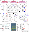 Figure 1
METTL8 is expressed in CXCL13hi CD4+ T cells. (A) Spatial expression of CD4, CXCL13, and PDCD1 in skin lesions from patients with anti–PD-1 treated tumors. (B) Schematic diagram of mouse model: B16F10 cells were subcutaneously injected into B6 mice and intraperitoneally injected with anti–PD-1 or IgG every 2 days from days 8 to 12, then analyzed at day 14. (C) Representative plots (left) and quantification (right) of PD-1hiCXCR5hi and BCL6+CXCR5+ GC TFH cells (gated on CD44hiCD62Llo CD4+ T cells) in the TdLNs of non-tumor, IgG‑, or anti–PD‑1–treated tumor mice (n = 5/group). (D) Frequency and absolute number of CXCL13hi cells (gated on BCL6+CXCR5+ GC TFH cells) in TdLNs of B16F10 tumor–bearing mice treated with IgG or anti–PD‑1 (n = 8–9/group). (E) MFI of CXCL13 in BCL6+CXCR5+ TFH cells in the TdLNs of B16F10 tumor–bearing mice treated with IgG or anti–PD‑1 (n = 7/group). (F) Mettl8-tdTomato-Flag mice were infected with LCMV-Armstrong and analyzed at 8 dpi. Splenic CD44hiCD62Llo CD4+ cells were gated; CXCL13 expression is shown for PD-1hiCXCR5hi GC TFH, PD-1loCXCR5+ TFH, and CXCR5− non-TFH subsets (n = 3/group). (G) Bubble plot of selected TFH/TH1-related differentially expressed genes between CXCL13hi and CXCL13lo clusters. (H) Heatmap of top 50 differentially expressed genes between CD4+ T cells from patients with SS versus healthy controls (scRNA-seq). (I) Cross-comparison of gene expression changes in CD4+ T cells from patients with disease versus healthy controls and in TFH versus TH1 cells. Selected genes shared between the 2 comparisons are highlighted. Mouse data represent 2–3 independent experiments; mean ± SEM. P values were calculated using 1-way ANOVA (C and F) and 2-tailed Student’s t test (D and E). *P < 0.05; ***P < 0.001; ****P < 0.0001.
Given that cancer immunotherapy elicits autoimmune responses characterized by expansion of CXCL13hi GC TFH cells, which share similar features with spontaneous autoimmune diseases, and that GC TFH cells in the classic LCMV mouse infection model also express high levels of CXCL13 (Figure 1F), we sought to identify key epitranscriptomic regulators that commonly govern these cells. Therefore, we compared CD4+ T cells from the salivary glands of patients with Sjögren’s syndrome (SS), a spontaneous autoimmune disease, with those from healthy individuals treated as controls and performed integrative analysis with differentially expressed genes between TFH and TH1 cells from the classical LCMV mouse infection model.
By reanalyzing published scRNA-seq data from salivary glands in healthy individuals and patients with SS (22), we identified 4 CD4+ T cell clusters, including proliferating CD4+ T cells, Treg cells, CXCL13hi, and CXCL13lo CD4+ T cells (Supplemental Figure 1I). To depict the characteristics of the latter 2 clusters, we analyzed the differentially expressed genes between these cells and found that CXCL13hi CD4+ T cells highly expressed TFH-featured genes, such as CXCR5, PDCD1, BCL6, and TCF7, while CXCL13lo CD4+ T cells were prone to TH1 cells (Figure 1G). Further analysis of differentially expressed genes revealed that these highly expressed TFH-associated genes were also enriched in the CD4+ T cells of patients with SS compared with healthy individuals (Figure 1H), suggesting that these CXCL13hi TFH cells increased in affected organs of the autoimmune disease. These results further support the presence of CXCL13hi TFH-like cells with B cell helper features in autoimmune-affected tissues.
To identify factors potentially involved in TFH cell development, we performed a cross-analysis of genes highly expressed in TFH cells from the canonical LCMV-infected mouse model (11) and those upregulated in CXCL13hi CD4+ T cells from patients with SS. The overlapping gene set predominantly comprised known TFH-associated genes, including CXCR5, TCF7, and PDCD1. Notably, METTL8, which encodes an m³C methyltransferase and has not previously been implicated in TFH cell biology, was also present among the common genes (Figure 1I). These findings suggest that METTL8 may serve as a shared regulator of CXCL13hi TFH cells across distinct pathological and physiological contexts. Further analysis revealed that METTL8 was highly expressed in CD4+ T cells of patients with SS compared with those of healthy individuals, which is consistent with the expression of CXCL13 (Figure 1I). Thus, we hypothesize that METTL8 contributes to CXCL13hi TFH cell specification.
To determine whether METTL8 is expressed in TFH cells following ICI treatment, we first analyzed transcriptomic data from skin samples and observed high METTL8 expression in the CD4+CXCL13+PD-1+ regions (Figure 2A). We then examined METTL8 expression in the CD4+BCL6+ TFH cells within skin samples from patients with anti–PD-1–treated tumors. Compared with untreated controls, anti–PD-1–treated patients exhibited increased CD4+ T cell infiltration in the skin. Notably, these infiltrating CD4+ T cells expressed both BCL6 and METTL8, whereas such cells showed little or no detectable staining for these markers in untreated controls (Figure 2B and Supplemental Figure 2A). Collectively, these findings indicate that METTL8 is expressed in TFH cells upon anti–PD-1 therapy.
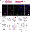 Figure 2
METTL8 is commonly expressed in CXCL13hi CD4+ T cells among diverse pathological and experimental settings. (A) The distribution and expression of METTL8 in the skin of patients with anti–PD-1–treated tumors by spatial transcriptome sequencing. (B) The expression of CD4, Bcl6, and METTL8 in the skin from anti–PD-1–treated or untreated tumor patients by immunofluorescence. Scale bars: 100 μm. (C) Schematic of the mouse model: B16F10 cells were subcutaneously injected into Mettl8-tdTomato-Flag mice and intraperitoneally injected with anti–PD-1 or IgG every 2 days from days 7 to 11, then analyzed at day 13. (D) The MFI of Mettl8-tdTomato reporter in PD-1hiCXCR5hi GC TFH cells in the spleens of anti–PD-1– or IgG-treated B16F10 tumor–bearing mice (n = 6/group). (E) Schematic of the mouse model: Mettl8-tdTomato-Flag mice were intraperitoneally injected with 2 × 10⁵ PFU LCMV-Armstrong and analyzed at 8 dpi. (F) Representative plots and quantification illustrating the expression of Mettl8-tdTomato gated on PD-1hiCXCR5hi GC TFH cells, PD-1loCXCR5+ TFH cells, and CXCR5− non-TFH cells (n = 5/group). (G) Representative histogram and quantification of Mettl8-tdTomato MFI in CXCL13hi and CXCL13lo CD4+ T cell subsets (n = 3/group). (H) Violin plots displaying the expression of CXCL13 and METTL8 within healthy controls and patients with SS. (I) Bubble plots displaying the expression of METTL8 within CXCL13hi and CXCL13lo clusters from scRNA-seq analysis. Mouse data represent 2–3 independent experiments; mean ± SEM. P values were calculated using a 2-tailed Student’s t test. **P < 0.01; ***P < 0.001; ****P < 0.0001.
To further validate this observation, we inoculated B16F10 cells into Mettl8-tdTomato-Flag reporter mice and treated them with either IgG or anti–PD-1 antibody (Figure 2C). Analysis of PD-1hiCXCR5hi GC TFH cells in the spleens and TdLNs revealed that METTL8 expression levels were higher in anti–PD-1–treated mice than in IgG-treated controls (Figure 2D and Supplemental Figure 2B). Taken together, these results support the conclusion that METTL8 is expressed in TFH cells following treatment in both human and mouse samples.
To further investigate whether METTL8 is expressed preferentially in TFH cells, we infected Mettl8-tdTomato-Flag mice with LCMV-Armstrong and analyzed them at 8 days after infection (Figure 2E). We confirmed that Mettl8-tdTomato was highly expressed in PD-1hiCXCR5hi GC TFH cells compared with PD-1loCXCR5+ TFH cells or CXCR5− non-TFH cells (Figure 2F). Considering these consistent expression trends of CXCL13 and METTL8 in both the human disease and the mouse model, we conducted further analysis that revealed that the expression of METTL8 was much higher in CXCL13hi CD4+ T cells than in CXCL13lo CD4+ T cells (Figure 2G). These results indicate that METTL8 is preferentially expressed in CXCL13hi GC TFH cells.
Further analysis revealed that METTL8 expression was significantly higher in CD4+ T cells of SS patients than in those from healthy controls, a pattern consistent with CXCL13 expression (Figure 2H). In addition, METTL8 expression was markedly elevated in CXCL13hi clusters compared with CXCL13lo clusters (Figure 2I). These findings suggest that METTL8 may contribute to the specification of CXCL13hi TFH cells.
METTL8 promotes TFH cell differentiation. To further explore whether METTL8 promotes TFH cell generation by tumor immunotherapy, we subcutaneously inoculated B16F10 or MC38 cells into Mettl8fl/fl Cd4cre mice and littermate controls and then treated mice with anti–PD-1 antibody or IgG (Figure 3A). The data showed that Mettl8 deficiency did not affect the frequency and absolute number of CD4+ T cells in either spleens or TdLNs (Supplemental Figure 3, A and B). CD4+ T cell activation was also not detectably affected by Mettl8 deficiency (Supplemental Figure 3, C and D). However, PD-1hiCXCR5hi and BCL6+CXCR5+ GC TFH cells were decreased in Mettl8-deficient mice compared with littermate controls after anti–PD-1 treatment in both spleens and TdLNs (Figure 3B and Supplemental Figure 3, E and F). Thus, these data indicate that METTL8 promotes TFH cell accumulation after anti–PD-1 treatment.
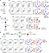 Figure 3
METTL8 promotes TFH cell differentiation. (A) Schematic diagram of the mouse model: 2 × 10⁵ B16F10 cells were subcutaneously injected into Mettl8fl/fl Cd4cre and littermate control mice and intraperitoneally injected with anti–PD-1 or IgG every 2 days from days 7 to 11. Mice were analyzed at day 13. (B) Representative plots (left) and quantification (right) of PD-1hiCXCR5hi and BCL6+CXCR5+ GC TFH cells gated on CD44hiCD62Llo CD4+ T cells in the TdLNs of B16F10 tumor–bearing Mettl8fl/fl Cd4cre and littermate control mice treated with IgG or anti–PD‑1 antibody (n = 5–8/group). (C) Schematic of adoptive transfer: CD45.1+ Mettl8−/− or Ctrl Smarta CD4+ T cells were transferred into CD45.2+ WT recipients, followed by LCMV-Armstrong (LCMV-Arm) 24 h later, and analyzed at 8 dpi. (D) Representative plots (left) and quantification (right) of PD-1hiCXCR5hi and BCL6+CXCR5+ GC TFH cells in CD45.1+ Smarta CD4+ T cells in spleens of WT recipients (n = 8/group). (E) Schematic of the cotransfer experiment. CD45.2+ Mettl8−/− or Ctrl Smarta CD4+ T cells were primed in vivo for 24 h and transduced with EV-GFP or Mettl8-GFP retrovirus. GFP+ and GFP− cells were sorted and cotransferred at a 1:1 ratio into CD45.1+ WT recipients, followed by LCMV-Armstrong infection 24 h later and analyzed at 8 dpi. (F) Representative plots (left) and quantification (right) of PD-1hiCXCR5hi and BCL6+CXCR5+ GC TFH cells gated on CD45.2+ Smarta CD4+ T cells in the spleens of mice in E (n = 6/group). Cell counts were normalized to the corresponding WT GFP– group within each experiment. Data represent 2–3 independent experiments; mean ± SEM. P values were calculated using 1-way ANOVA (B and F) and 2-tailed Student’s t test (D). *P < 0.05; **P < 0.01; ***P < 0.001; ****P < 0.0001.
To further validate the effect of METTL8 on TFH cells, we used the canonical LCMV-infected mouse model. Mettl8fl/fl Cd4cre mice were crossed with a Smarta TCR transgenic strain expressing an MHC II–restricted TCR specifically recognizing the LCMV gp61 epitope. WT and Mettl8−/− Smarta CD4+ T cells were adoptively transferred into congenic recipients, followed by infection with LCMV-Armstrong (Figure 3C). In response to infection, Mettl8−/− Smarta CD4+ T cells exhibited a marked decrease in PD-1hiCXCR5hi and BCL6+CXCR5+ GC TFH cells (Figure 3D). Furthermore, LCMV infection showed no difference between the frequencies and absolute numbers of CD44hiCD62lo activated CD4+ T cells of Mettl8fl/fl Cd4cre mice and littermate controls (Supplemental Figure 4A), but GC TFH cells were impaired by Mettl8 deficiency (Supplemental Figure 4, B and C). In addition, we observed a reduction in SLAMloCXCR5+ TFH cells among monoclonal and polyclonal CD4+ T cells, accompanied by increased frequencies and unchanged counts of SLAMhiCXCR5− TH1 cells (Supplemental Figure 4, D and E). The PD-1loCXCR5+ TFH cells were also reduced by Mettl8 deficiency, whereas the number of CXCR5− non-TFH cells was not affected, although the frequency increased (Supplemental Figure 4F). Notably, the expression of caspase and Ki67 in TFH cells was unaffected by Mettl8 deficiency, suggesting that METTL8 does not affect apoptosis or proliferation of TFH cells (Supplemental Figure 4G). Therefore, these results indicate that Mettl8 deficiency impairs TFH cell differentiation.
To further substantiate the requirement of METTL8 in TFH cell differentiation, we enforced METTL8 expression in Mettl8−/− or control (Ctrl) Smarta CD4+ T cells using pMSCV-IRES-GFP retroviruses. GFP+ and GFP− cells were sorted and cotransferred into recipient mice at a 1:1 ratio (Figure 3E and Supplemental Figure 4H). Mice were infected with LCMV-Armstrong, and at the 8 day postinfection (dpi) time point, the ratio of GFP+ to GFP− cells remained unaltered (Supplemental Figure 4I), indicating comparable survival and proliferation of transduced and nontransduced cells. Relative to empty vector–GFP (EV-GFP) control, forced expression of METTL8 in Ctrl Smarta cells significantly increased PD-1hiCXCR5hi and BCL6+CXCR5+ GC TFH cells (Figure 3F). Importantly, ectopic METTL8 expression in Mettl8−/− Smarta cells significantly rescued the reduction in GC TFH cells caused by Mettl8 deficiency (Figure 3F). Moreover, SLAMloCXCR5+ TFH cells were restored, and the frequency of SLAMhiCXCR5− TH1 cells was decreased by METTL8 overexpression in Mettl8−/− Smarta cells (Supplemental Figure 4J). These data further support a requirement for METTL8 in TFH cell differentiation.
METTL8 promotes TFH cell function. To assess the impact of Mettl8-deficient TFH cells on tumor progression, we monitored tumor growth and survival in MC38 or B16F10 tumor–bearing Mettl8fl/fl Cd4cre mice and littermate controls. Antitumor responses, with or without anti–PD-1 treatment, were comparable between genotypes in these models (Figure 4, A and B, and Supplemental Figure 5, A and B). Given the essential role of TFH cells in supporting B cell responses, we next examined the GC response in TdLNs. Upon anti–PD-1 treatment, Mettl8 deficiency led to a reduction in GC B cells and plasma cells within TdLNs (Figure 4C and Supplemental Figure 5C). Collectively, these data indicate that Mettl8 deficiency impairs the GC response in TdLNs following anti–PD-1 therapy, without affecting tumor progression.
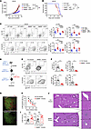 Figure 4
METTL8 promotes TFH cell function. (A and B) Tumor growth (A) and survival (B) of MC38-bearing Mettl8fl/fl Cd4cre and littermate controls, treated with IgG or anti–PD-1 (n = 8/group). (C) Representative plots (left) and quantification (right) of GL-7+Fas+ GC B cells and CD138+IgDlo plasma cells (gated on B220+CD19+ cells) in the TdLNs of the above groups (n = 4–8/group). (D) Schematic of the adoptive transfer model: CD45.1+ Mettl8−/− or Ctrl Smarta CD4+ T cells were transferred into CD45.2+ Bcl6fl/fl Cd4cre recipients, followed by LCMV-Armstrong 24 h later, and analyzed at 14 dpi. (E and F) Representative plots (E) and quantification (F) of GC B cells and plasma cells in the spleens of the mice in D (n = 7–8/group). (G) Immunofluorescence of spleens from LCMV-Armstrong–infected Mettl8fl/fl Cd4cre mice and littermate controls at 8 dpi, indicating B cell follicle (green) and GC (red). Scale bars: 100 μm. (H) LCMV-specific IgG in serum of mice in D by ELISA (n = 5/group). (I) Serum samples were collected from Mettl8−/− or Ctrl Smarta cell–transferred mice at 4 dpi with LCMV‑Armstrong. Viral titers of the sera were quantified by qPCR using 2 independent primer pairs targeting the LCMV glycoprotein (GP) and nucleoprotein (NP) genes (n = 9–10/group). (J) Liver samples were collected from Mettl8−/− or Ctrl Smarta cell–transferred mice at 8 dpi with LCMV‑Armstrong. H&E staining showing inflammatory cell infiltration (arrows) in indicated groups. Scale bars: 200 μm (left), 50 μm (right). Data represent 2–3 independent experiments; mean ± SEM. P values were calculated using 2-way ANOVA (A), log-rank test (B), 1-way ANOVA (C), and 2-tailed Student’s t test (F, H, and I), *P < 0.05; **P < 0.01; ***P < 0.001; ****P < 0.0001.
To examine the functional requirement of METTL8 in the infection model, we transferred Mettl8−/− or Ctrl Smarta CD4+ T cells to Bcl6fl/fl Cd4cre mice, which were deficient in endogenous GC TFH cells (Figure 4D) (23). Mettl8-deficient PD-1hiCXCR5hi and BCL6+CXCR5+ GC TFH cells were dramatically reduced at 14 dpi (Supplemental Figure 5, D and E). Accordingly, recipients of Mettl8−/− Smarta cells exhibited reduced Fas+GL7+ GC B cells and CD138+IgDlo plasma cells (Figure 4, E and F). Similarly, direct LCMV-Armstrong infection of Mettl8fl/fl Cd4cre mice resulted in decreased GC B cells and plasma cells at 8 dpi compared with littermate controls (Supplemental Figure 5, F and G), and GC formation was impaired (Figure 4G). Consistently, LCMV-specific IgG in the serum of Bcl6fl/fl Cd4cre recipients was significantly reduced by Mettl8 deficiency (Figure 4H). Furthermore, Mettl8 deficiency led to elevated viral loads (Figure 4I). Although LCMV-Armstrong infection normally induces only mild hepatitis in mice (24), representative H&E images showed more prominent inflammatory infiltrates in portal tracts and intralobular regions (Figure 4J). Histological examination further revealed that follicle-associated regions were diminished in the spleens of Mettl8−/− Smarta cell–transferred mice (Supplemental Figure 5H). These data support an important role for METTL8 in GC responses.
METTL8 promotes TFH cell accumulation and maintenance. As TH1 and TFH fate determination occurs promptly following the activation of CD4+ T cells, we investigated the potential involvement of METTL8 in the TFH fate decision process. Initially, we assessed METTL8 expression in activated CD4+ T cells in vitro, observing heightened expression with prolonged stimulation (Supplemental Figure 6A). To further explore METTL8 expression, we utilized Mettl8-tdTomato Smarta CD4+ T cells labeled with cell-trace violet and adoptively transferred them into congenic recipients, followed by LCMV-Armstrong infection (Supplemental Figure 6B). Harvesting cells at the 40 and 60 h postinfection (hpi) time point, corresponding to the initiation of the first division at 36 hpi and the fifth division at 60 hpi (25) for Smarta CD4+ T cells, we examined METTL8 expression in each division from the first to the fifth (Supplemental Figure 6C). The data revealed elevated METTL8 expression during CD4+ T cell activation and the first division, maintaining a sustained high level despite slight decreases during subsequent divisions (Figure 5A). These results indicate that METTL8 expression is increased by CD4+ T cell activation during the early TH1-TFH fate bifurcation stage, prompting us to investigate whether METTL8 plays a role during this period.
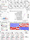 Figure 5
Mettl8 deficiency impairs TFH differentiation due to altered transcriptional profiles. (A) Representative plots (top) and quantification (bottom) of MFI of tdTomato in each division (Div) of CD45.2+ Smarta CD4+ T cells. All the MFI of tdTomato in divisions 1–5 are compared with that in division 0 (n = 4–10/group). (B) Representative plots (left) and quantification (right) of the frequency of CXCR5+SLAMlo TFH and CXCR5−SLAMhi TH1 cells in the spleens of Mettl8−/− or Ctrl Smarta cell–transferred mice at 4–7 dpi with LCMV‑Armstrong (n = 3–4/group). (C–F) RNA-seq was conducted on PD-1hiCXCR5hi GC TFH cells sorted from spleens of LCMV-Armstrong–infected Mettl8fl/fl Cd4cre (KO) mice and littermate controls (WT) at 8 dpi. In C, Volcano plot illustrates DEGs between GC TFH cells from KO and WT mice. Lines indicate a 2-fold difference between GC TFH cells from KO and WT mice (edgeR, FDR < 0.05). In D, KEGG analysis of DEGs obtained from the R package named clusterProfiler. In E, GSEA shows the enrichment of TFH versus non-TFH cell-associated downregulated (top) and upregulated (bottom) genes in the transcriptome of KO versus WT GC TFH cells. In F, heatmap displaying DEGs related to TFH and TH1 cells. (G) Representative histograms (top) and cumulative data (bottom) showing the expression of the indicated molecules gated on PD-1hiCXCR5hi GC TFH cells (n = 4–8/group). For mouse experiments, data represent 2–3 independent experiments; mean ± SEM. P values were calculated using a 2-tailed Student’s t test. *P < 0.05; **P < 0.01; ***P < 0.001; ****P < 0.0001.
To explore the role of METTL8 in TFH lineage specification, we utilized Mettl8fl/fl Cd4cre mice and littermate controls. Following adoptive transfer of cell-trace violet–labeled Mettl8−/− or Ctrl Smarta CD4+ T cells and LCMV-Armstrong infection, both groups underwent at least 5 divisions, with the majority of cells in the fourth and fifth divisions at 60 hpi, indicating comparable division rates (Supplemental Figure 6D). These findings suggest that the loss of METTL8 does not disrupt CD4+ T cell activation and early division. Despite this, the reduction of TFH cells upon Mettl8 deficiency led us to infer that METTL8 may contribute to TFH versus TH1 lineage bifurcation.
As previously reported, the combination of CD25 and CXCR5 was employed to distinguish nascent TH1 and TFH cells in activated CD4+ T cells (25). In the fifth division, Mettl8−/− CD25loCXCR5+/lo nascent TFH cells were detected at a lower frequency compared with their WT counterparts, accompanied by an opposite change in the frequency of CD25hiCXCR5− nascent TH1 cells (Supplemental Figure 6E). Additionally, CXCR5 expression was slightly but significantly reduced in Mettl8-deficient nascent TFH cells, whereas it remained unchanged in nascent TH1 cells (Supplemental Figure 6E). These results suggest that METTL8 exerts modest effects on early TFH specification. We therefore examined TFH cells from days 4 to 7 after infection and found that the reduction in TFH cells caused by Mettl8 deficiency became progressively more pronounced over time (Figure 5B and Supplemental Figure 6F). Consequently, our findings indicate that METTL8 has a limited impact on early TFH differentiation but plays a substantial role in TFH accumulation and maintenance.
To unravel the underlying mechanism governing TFH cell development mediated by METTL8, we conducted RNA sequencing on sorted PD-1hiCXCR5hi GC TFH cells obtained from LCMV-Armstrong–infected Mettl8fl/fl Cd4cre mice and their littermate controls at 8 dpi. Our analysis identified 1,597 downregulated genes and 1,469 upregulated genes in Mettl8-deficient GC TFH cells, each exhibiting a fold change > 2 (Figure 5C). Notably, these differentially expressed genes showed enrichment in pathways related to TFH cell differentiation (Figure 5D). GSEA further revealed that genes downregulated in TFH cells compared with non-TFH cells were enriched in Mettl8-deficient GC TFH cells, while genes upregulated in TFH cells versus non-TFH cells were enriched in WT GC TFH cells (Figure 5E). Among the identified upregulated and downregulated genes, those encoding transcription factors crucial for TFH cell differentiation, such as Bcl6, Maf, Tcf7, Cebpa, Tox, Id3, Pou2af1, Sostdc1, and Ascl2, were consistently downregulated in Mettl8-deficient GC TFH cells (Figure 5F and Supplemental Figure 6G). Additionally, the expression of genes defining GC TFH cell characteristics, including Cxcr5, Pdcd1, Icos, and Il21, exhibited a decrease in Mettl8-deficient mice (Figure 5F and Supplemental Figure 6G). Consistent with our RNA-seq findings, flow cytometric analysis confirmed a significant reduction in the expression of TFH cell surface markers CXCR5 and ICOS, as well as transcription factors BCL6 and TCF1 (Figure 5G). Conversely, genes upregulated in Mettl8-deficient GC TFH cells tended to exhibit TH1 signatures, including Prdm1, Tbx21, Id2, and Il2ra (Figure 5F and Supplemental Figure 6G). The increased expression of T-bet was validated by flow cytometry (Figure 5G). In summary, these results indicate that METTL8 biases activated CD4+ T cells toward the TFH program rather than the TH1 program.
METTL8 facilitates m3C-associated regulation of Tcf7 mRNA and cooperates with TCF1-associated chromatin programs. To elucidate the mechanisms by which METTL8-mediated m3C methylation modulates the TFH transcriptional program, we conducted AlkAniline-seq (26) to identify distinct m3C modification loci between PD-1hiCXCR5hi GC TFH cells isolated from LCMV-Armstrong–infected Mettl8fl/fl Cd4cre mice and littermate controls. The global mapping of the m3C landscape revealed abundant m3C peaks within the coding sequence of mRNA (Supplemental Figure 7, A and B). Comparative analysis of m3C modification loci in Mettl8-deficient and WT GC TFH cells identified 4,491 different sites, with 1,425 in WT TFH cells and 3,066 in Mettl8-deficient TFH cells (Supplemental Figure 7C). Significantly enriched RNA motifs flanking m³C modification loci from WT GC TFH cells are presented (Supplemental Figure 7D). These data revealed AlkAniline-seq for mapping m3C modifications in TFH cells.
Given that METTL8 is a mitochondrially localized enzyme that catalyzes m3C modification on tRNAs and regulates mitochondrial protein translation and respiration (16–18), we first performed tRNA m³C-seq to examine whether Mettl8 deficiency affects tRNA modification in TFH cells. However, we observed no significant differences in tRNA m³C modification between WT and Mettl8-deficient TFH cells (Supplemental Figure 7, F and G). To further assess mitochondrial respiratory function, we performed Seahorse metabolic flux analysis in WT and Mettl8-deficient TFH cells. No statistically significant differences were detected in the measured bulk oxygen consumption rate (OCR) and extracellular acidification rate (ECAR) parameters under the tested conditions (Supplemental Figure 7H). Collectively, these results suggest that METTL8 does not appreciably affect tRNA m³C modification or global mitochondrial metabolism in TFH cells, prompting us to investigate its potential mRNA-targeted functions.
To further investigate METTL8 binding sites on mRNA, RNA-immunoprecipitation and sequencing (RIP-seq) was performed on PD-1hiCXCR5hi GC TFH cells isolated from LCMV-Armstrong–infected mice. A combined analysis of RNA-seq, m3C-seq, and RIP-seq revealed 94 genes commonly targeted by m3C and METTL8, with differential RNA expression between Mettl8-deficient and WT GC TFH cells (Supplemental Figure 7E). To define a direct relationship between METTL8 binding and differential m³C modification, we required that a METTL8 binding site must be located within 500 bp of a differential m3C modification site on the same transcript (27, 28), rather than merely co-occurring anywhere on the same transcript. To evaluate the biological significance of the observed overlap, we performed a Fisher’s exact test based on the entire set of expressed genes to compare the observed number of overlapping genes against the expected value under a random distribution. Our analysis demonstrated that the overlap is statistically significant (P < 0.01, OR > 2.5), indicating that the intersecting genes are unlikely to arise by chance. Notably, Tcf7 showed overlapping m3C-seq and RIP-seq signals (Figure 6A). To validate the regulation of METTL8 on Tcf7, a pMigR1-GFP-Tcf7 vector was constructed, and retrovirus was generated to enforce TCF1 expression in Smarta cells. Strikingly, TCF1 overexpression in Mettl8−/− Smarta cells significantly restored the PD-1hiCXCR5hi GC TFH cell frequency (Figure 6, B and C). Correspondingly, the frequency of PD-1loCXCR5+ TFH cells was increased, and that of CXCR5− non-TFH cells was decreased by TCF1 overexpression in Mettl8−/− Smarta cells compared with Mettl8−/− Smarta cells transfected with pMigR1 EV (Supplemental Figure 7I). To test whether METTL8 regulates Tcf7 mRNA stability, an RNA decay assay was performed using actinomycin D and showed that Tcf7 mRNA declined faster in Mettl8-deficient cells than in control cells (Figure 6D), indicating that METTL8 enhances Tcf7 mRNA stability.
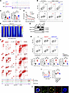 Figure 6
METTL8 promotes m3C modification of Tcf7 mRNA and chromatin accessibility in TFH cells. (A) m³C-seq and METTL8-Flag RIP-seq signals mapped to the Tcf7 transcript. (B and C) Representative plots (B) and quantification (C) of PD-1hiCXCR5hi and BCL6+CXCR5+ GC TFH populations gated on Smarta GFP+CD4+ T cells from the spleens of different recipient mice adoptively transferred with EV or Tcf7 retrovirus-introduced Smarta cells. Mice were infected with LCMV-Armstrong and analyzed at 8 dpi (n = 9–13/group). (D) RNA decay assay showing remaining Tcf7 mRNA detected by RT-qPCR, normalized to t = 0 (n = 2/group). (E) ATAC‑seq heatmaps of local chromatin changes between WT and Mettl8−/− PD-1hiCXCR5hi GC TFH cells. (F) Diamond plots of chromatin interactions in TH1, WT, and Mettl8−/− (KO) TFH cells at the Adar, Il6ra, Ccdc153, and Cxcr5 gene loci (top), with CUT&Tag and ATAC-seq tracks and gene structures on the bottom. Vertical lines with circles denote chromatin loop anchors. (G) Coimmunoprecipitation of TCF1 with METTL8-Flag in HEK293T cells. (H) Representative plots (top) and quantification (bottom) of PD-1hiCXCR5hi and BCL6+CXCR5+ GC TFH cells gated on CD45.2+ Smarta CD4+ T cells in the spleens of recipient mice transferred with Smarta cells transduced with EV, Mettl8‑WT, or Mettl8‑D220A retrovirus. Mice were infected with LCMV‑Armstrong and analyzed at 8 dpi (n = 6–8/group). (I) TCF1 MFI in CXCR5+ CD4+ TFH cells from the spleens (n = 7–8/group). (J) Immunofluorescence staining showing METTL8 localization in activated CD4+ T cells. Scale bars: 2 μm. Data represent 2–3 independent experiments; mean ± SEM. P values were calculated using 1-way ANOVA (C, H, and I) and 2-way ANOVA (D). *P < 0.05; **P < 0.01; ***P < 0.001; ****P < 0.0001.
Considering the genomic coordination and histone acetylation modification ability of TCF1 (29–31), we performed ATAC-seq (assay for transposase-accessible chromatin with high-throughput sequencing) on Mettl8−/− and control PD-1hiCXCR5hi GC TFH cells and observed reduced chromatin accessibility in Mettl8-deficient GC TFH cells (Figure 6E). The decrease in chromatin accessibility mainly occurred at the gene transcription initiation site (Supplemental Figure 7J). To investigate the cooperation of METTL8 with TCF1 in organizing chromatin architecture (32) in GC TFH cells, we integrated Hi-C (high-resolution chromosome-conformation capture), TCF1 CUT&Tag, and ATAC-seq data from Mettl8−/− and control GC TFH cells with METTL8 CUT&Tag data from Mettl8-tdTomato GC TFH cells. We also performed Hi‑C on TH1 cells to identify TFH‑specific loops affected by METTL8. The analysis identified locus-specific chromatin loops at TCF1 target genes, including Il6ra and Cxcr5, where METTL8 and TCF1 CUT&Tag signals overlapped. These regions also showed H3K27ac enrichment and increased chromatin accessibility, consistent with active regulatory elements at key GC TFH genes (Figure 6F). Notably, these TFH‑specific loops were largely absent in TH1 cells, indicating that the 3D genome organization at these loci is lineage restricted (Figure 6F).
In Mettl8−/− GC TFH cells, TCF1 binding at these loci was substantially reduced, accompanied by disruption of chromatin looping and decreased chromatin accessibility. These findings suggest that METTL8 promotes TCF1-dependent chromatin organization and maintains an accessible regulatory landscape at genes essential for GC TFH cell identity and function (Figure 6F). Together, these results suggest that METTL8 cooperates with TCF1-associated regulatory programs to promote TFH-associated gene expression and chromatin states at TFH-related loci.
To validate the interaction between METTL8 and TCF1, coimmunoprecipitation was conducted using HEK293T cells cotransfected with pMigR1-Mettl8-Flag and pMigR1-Tcf7 plasmids. The results demonstrated that TCF1 coimmunoprecipitated with METTL8 (Figure 6G). Thus, METTL8 can interact with TCF1 protein, supporting a potential mechanism by which METTL8 influences TCF1-associated chromatin accessibility and 3D genome organization.
To determine whether the phenotypic changes in CD4+ T cells are caused by the catalytic activity of METTL8, we used a retroviral transduction system to introduce a point mutation (D220A) at the conserved catalytic residue aspartic acid 220 (27, 28) of METTL8 in Smarta cells. These cells were then adoptively transferred into recipient mice followed by LCMV-Armstrong infection. On day 8 after infection, the D220A mutation abolished the TFH-promoting effect of METTL8 overexpression (Figure 6H) and failed to restore TCF1 expression (Figure 6I). These results suggest that catalytic activity of METTL8 is required for the TFH-promoting function.
Considering that distinct METTL8 isoforms generated by alternative splicing exhibit differential subcellular localization, residing either in the mitochondria or the nucleolus, we investigated METTL8 distribution in CD4+ T cells. Immunofluorescence analysis revealed that METTL8 was present in both the mitochondria and the nucleus (Figure 6J and Supplemental Figure 8C), confirming its dual subcellular localization in this cell type.
Ginkgolic acid suppresses TFH cell responses through a METTL8-associated mechanism. A recent study reported that the nuclear localization of METTL8 was regulated by SUMOylation, which could be completely disrupted by ginkgolic acid (GA), a well-known SUMOylation inhibitor (33). To investigate whether GA affects TFH cell differentiation, we treated LCMV-Armstrong–infected Mettl8-tdTomato-Flag mice with GA (Figure 7A). The data revealed that METTL8 expression reflected by the MFI of tdTomato was significantly decreased in PD-1hiCXCR5hi GC TFH cells upon GA treatment (Figure 7B), suggesting that GA may disrupt METTL8 SUMOylation or METTL8-dependent activity.
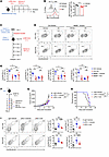 Figure 7
GA treatment reduces TFH cell responses without detectably reducing anti–PD-1 antitumor activity. (A) Schematic diagram of GA-treated infection model: Mettl8-tdTomato-Flag mice were infected with LCMV-Armstrong on day 0; treated with GA or vehicle on days 1, 3, 5, and 7; and analyzed on day 8. (B) Representative histograms and quantification of Mettl8-tdTomato reporter MFI in splenic PD-1hiCXCR5hi GC TFH cells (n = 3–4/group). (C) Schematic of the GA-treated adoptive transfer model: CD45.2+ Ctrl or Mettl8–/– Smarta CD4+ T cells were transferred into CD45.1+ WT recipients, followed by LCMV-Armstrong 24 h later, and treated with GA or vehicle every 2 days from days 1 to 7 after infection. Spleen cells were analyzed at 8 dpi. (D and E) Representative plots (D) and quantification (E) of PD-1hiCXCR5hi and BCL6+CXCR5+ GC TFH cells among donor Smarta cells (n = 5/group). (F) Schematic of the GA or anti–PD-1 antibody treatment. Mice were subcutaneously injected with B16F10 cells, followed by GA and anti–PD-1 treatment alternately from 7 to 12 days after tumor inoculation. (G and H) Tumor growth (G) and survival curve (H) of mice in F (n = 7–8/group). (I) Representative plots (left) and quantification (right) of PD-1hiCXCR5hi and BCL6+CXCR5+ GC TFH cells (gated on CD44hiCD62Llo CD4+ T cells) in TdLNs of IgG- or anti–PD-1–treated B16F10 tumor–bearing mice, with or without GA (n = 4–5/group). Data represent 2–3 independent experiments; mean ± SEM. P values were calculated using a 2-tailed Student’s t test (B), 1-way ANOVA (E and I), 2-way ANOVA (G), and log-rank test (H). *P < 0.05; **P < 0.01; ***P < 0.001.
To further validate that GA‑mediated suppression of GC TFH cells is associated with METTL8, we treated Mettl8−/− Smarta cell–transferred mice (KO mice) with GA following LCMV infection (Figure 7C). In these KO mice, GA treatment did not alter total lymphocyte or Smarta cell numbers (Supplemental Figure 9, A and B). However, in WT recipients, GA led to a significant decrease in PD-1hiCXCR5hi and BCL6+CXCR5+ GC TFH cells (Figure 7, D and E), along with a reduction in PD-1loCXCR5+ and SLAMloCXCR5+ TFH cells, while the number of CXCR5− non-TFH cells remained unchanged (Supplemental Figure 9, C and D). Similar results were obtained in the direct LCMV infection model (Supplemental Figure 9, E−G). These data indicate that GA inhibits TFH cell differentiation.
Notably, in Mettl8‑deficient mice, the frequencies and absolute numbers of PD-1hiCXCR5hi and BCL6+CXCR5+ GC TFH cells were comparable between GA-treated and untreated groups (Figure 7, D and E), indicating that GA exerts little or no additional effect on GC TFH differentiation in the absence of METTL8. Taken together, these data suggest that GA‑mediated suppression of TFH differentiation is associated with METTL8.
Considering that ICI therapy can trigger autoimmune responses and that neoantigen-driven B cell and TFH cell interactions enhance CD8+ T cell antitumor immunity (34), we further investigated whether GA treatment could adversely affect ICI therapy. Thus, we administered GA in combination with anti–PD-1 antibody in B16F10 and MC38 subcutaneous tumor models (Figure 7F). The addition of GA did not result in a statistically detectable reduction in the antitumor effect of anti–PD-1 (Figure 7, G and H), suggesting that GA does not compromise the antitumor activity of PD-1 blockade. Furthermore, we tested TFH cell development and function following GA treatment and found that GA impaired TFH differentiation and GC response after anti–PD-1 therapy in both B16F10 (Figure 7I and Supplemental Figure 10) and MC38 (Supplemental Figure 11) tumor models. Therefore, GA treatment inhibits TFH response in anti–PD-1 immunotherapy.
In summary, these results identify METTL8 as a key regulator of TFH differentiation via Tcf7 mRNA stabilization and 3D genomic remodeling. Furthermore, genetic loss of METTL8 impairs TFH cell development and function. GA treatment suppresses TFH cell responses in a METTL8‑associated manner.


Copyright © 2026 American Society for Clinical Investigation
ISSN: 0021-9738 (print), 1558-8238 (online)