
Research Article

Abstract
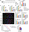
Autosomal dominant polycystic kidney disease (ADPKD) accounts for 5%–10% of prevalent end-stage kidney failure (ESKD). ADPKD cysts result from a loss of sufficient functional expression of PKD1/Polycystin-1 (PC1) in approximately 80% of families. Kidney disease severity correlates with the extent to which PC1 dosage is reduced below a critical level, and evidence suggests therapeutic benefit from increasing PC1 expression in these conditions. Upstream open reading frame (uORF) translation can reduce translation of a protein’s coding sequence. Ribosome profiling data and bioinformatic predictions suggested the presence of conserved PKD1 uORFs, so we sought to explore their biological role. We generated luciferase reporters and two humanized PKD1 5’ UTR mouse models with or without single nucleotide edits removing uORF start codons (ΔuORF) to define active uORFs and test their impact on PC1 translation. PKD1 uORF start codons can robustly initiate translation, and ΔuORF conveys a 2–4 fold increase in PC1 protein expression and resultant prevention of kidney cysts in Dnajb11 as well as in Pkd1 missense models. PKD1 uORF1-blocking steric antisense oligonucleotides (ASOs) substantially increase PC1 expression in vitro. PKD1 uORFs play an important role in the low basal expression of WT PKD1, and their inhibition represents an opportunity to therapeutically increase PC1 translation in polycystic kidney and liver disease resulting from reduced dosage of PC1.
Authors
Zhigui Li, Zi Guo, Soyoung Cho, Rishi Bhardwaj, Ke Dong, Sorin Fedeles, Whitney Besse
Abstract
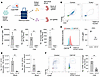
Amyotrophic lateral sclerosis (ALS) is a neurodegenerative disease characterized by progressive motor neuron loss, skeletal muscle atrophy, paralysis, and eventually death. Mitochondrial dysfunction plays a pivotal role in ALS pathogenesis, although the precise pathogenic mechanisms remain elusive, and effective therapeutic strategies are extremely limited. In this study, we developed a small-molecule inhibitor, UA-30, which directly targets RalA, and explored its potential for the treatment of ALS. We found that when administered via oral gavage for 6 weeks following the onset of motor deficit, UA-30 extended lifespan and improved motor function of SOD1G93A mice, a model of ALS. UA-30 ameliorated motor neuron loss, neuroinflammation, fibrosis, and mitochondrial dysfunction, as evidenced by energy recovery, decreased oxidative stress, and enhanced mitophagy. Mechanistically, UA-30 inhibited RalA activity and thereby modulated ERK/FOXO3a signaling, which inhibited FOXO3a degradation via the ubiquitin-proteasome pathway; enhanced FOXO3a stability; and upregulated the expression of mitophagy-related genes in this ALS mouse model. The beneficial effects of UA-30 in ALS were abolished by overexpression of the constitutively active form of RalA (RalAG23V) or Mdivi-1 treatment. These findings support RalA inhibition as a therapeutic strategy for enhancing mitophagy and mitigating ALS-like pathology and support UA-30 as an orally active candidate for further preclinical development.
Authors
Bingge Zhang, Ye He, Ting Su, Xiaomei Li, Xiufen Zhang, Ruijuan Liu, Xiao Han, Ruiming Zhang, Chao Yang, Xinlei Liu, Qinghua Hou, Zaijun Zhang, Yongmei Xie, Gongping Liu, Xifei Yang
Abstract
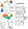
Loss-of-function mutation in the human gene dipeptidyl peptidase 9 (DPP9) causes Hatipoglu syndrome leading to severe inflammasomopathy. A key feature of the disease is pancytopenia, and patients require bone marrow transplantation, but the mechanism of cell loss is unclear, since Dpp9-mutant mice have normal hematopoiesis, suggesting that a distinct mechanism of disease occurs in humans. Here, we present a model of human DPP9 deficiency leveraging reverse genetics in the MISTRG6 humanized mice. We found that CRISPR editing of human CD34+ hematopoietic stem and progenitor cells (HSPCs) led to very efficient and persistent gene deletion in vivo. Human DPP9 deletion recapitulated cytopenia in peripheral blood and in the bone marrow, and cell loss was cell intrinsic. However, DPP9 deletion led to few transcriptional changes suggesting posttranscriptional regulation in human HSPCs. Mechanistically, DPP9 deficiency led to activation of the CARD8 inflammasome, resulting in HSPC pyroptosis, whereas NLRP1 was dispensable for cell death. Thus, our results reveal a unique human mechanism of disease and offer therapeutic insight for this inflammasomopathy.
Authors
Tianli Xiao, J. Richard Brewer, Maximillian Carlino, Ailin Han, Yamato J. Takabe, Chia-Yi Lee, Fengrui Zhang, Mi Chen, Holly Nicole Blackburn, Amin H. Nassar, Qiankun Wang, Kristen Brennand, Liang Shan, Esen Sefik, Diane S. Krause, Richard A. Flavell

Abstract
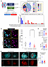
Aging occurs heterogeneously across organs, leading to progressive tissue dysfunction. Cellular senescence is a stress response triggered by age-associated insults, yet the mechanisms regulating senescence and organ aging remain incompletely understood. Here, we defined a role for lysine-specific demethylase 1 (LSD1) in DNA damage–mediated senescence and organ aging. LSD1 was upregulated in aged organs and senescent cells. In response to natural aging or ionizing radiation–induced DNA damage, LSD1 interacted with and demethylated ATM at lysine 3,016, as confirmed using a newly generated ATM-K3016me antibody. This modification sustained ATM phosphorylation, amplified DNA damage signaling, and delayed checkpoint recovery, promoting senescence and organ aging. Inhibition of LSD1 accelerated ATM dephosphorylation via WIP1, enhanced DNA repair, reduced senescence and DNA damage, and prevented irradiation-induced hair graying. Elimination of senescent cells with senolytics reduced LSD1 protein in aged organs, indicating a feedback loop between LSD1 and senescence. Mechanistically, LSD1 underwent autophagosome-lysosome degradation through interaction with LC3 and Beclin1, and autophagy impairment during DNA damage contributed to LSD1 accumulation in senescent cells. This study revealed LSD1 as a key regulator of DNA damage–induced senescence and organ aging and suggested that targeting LSD1 may attenuate senescence, delay organ aging, and prevent hair graying.
Authors
Yingying Zhang, Chen Yu, Xiaoqin Zhang, Linda Xiaoyan Li, Alice Shasha Cheng, Xiaogang Li
Abstract
Effector CD8+ T cells are key drivers of type 1 diabetes (T1D) pathogenesis, yet questions remain regarding the molecular defects leading to altered cytotoxicity, peripheral tissue phenotype, and receptor specificity. We analyzed human pancreatic lymph nodes (pLNs) using mass cytometry and single-cell RNA-seq (scRNA-seq) with combined T cell receptor (TCR) profiling. Cytometric analysis revealed enrichment of T stem cell memory–like (TSCM-like) cells (CD8+CD45RA+CD27+CD28+CCR7+CXCR3+) in T1D pLNs. scRNA-seq indicated an elevated inflammatory cytokine gene signature (IFITM3, LTB) along with regulators of terminal differentiation (BCL6, BCL3), coupled with downregulation of exhaustion-associated genes (DUSP2, NR4A2, TSC22D3) in CD8+ T cells in T1D pLNs. Immune response enrichment analysis (IREA) indicated IL-15 signaling as a significant driver of these phenotypes. Integrated TCR and transcriptomics analysis revealed a cluster of diverse naive-like CD8+ T cell clones in T1D pLNs. Comparison of pLNs and pancreatic tissue slice isolates indicated sharing of effector CD8+ T cells, with enhanced terminal effector signatures within the pancreas relative to paired pLNs. Multiplex imaging revealed differential localization of T cell factor 1 (TCF1)- and thymocyte selection-associated high mobility group box protein (TOX)-expressing T cells in the pancreas, with islet-proximal TCF1+TOX+ cells displaying a mixture of activation and exhaustion-associated phenotypes. Thus, we provide multimodal cellular profiles enriched in T1D tissues for consideration in therapeutic targeting.
Authors
Leeana D. Peters, Howard R. Seay, Justin A. Smith, Amanda L. Posgai, Reed L. Berkowitz, Clive H. Wasserfall, Mark A. Atkinson, Rhonda Bacher, Maigan A. Brusko, Todd M. Brusko

Abstract
Trigeminal neuralgia (TN) is a severe orofacial pain disorder accompanied by anxiety, yet its central mechanisms remain elusive. Analysis of human functional MRI data identified the parafascicular nucleus (PF) as a candidate region. Using a TN mouse model, we uncovered 2 spatially and functionally distinct PF neuronal ensembles that separately encoded sensory and affective dimensions of pain. One population received inhibitory input from GABAergic neurons in the oral spinal trigeminal nucleus (Sp5O) and mediated nociception. The second population, driven by a glutamatergic Sp5O-lateral parabrachial nucleus (lPBN)-PF pathway, encoded pain-related anxiety. The engagement of the anxiety-encoding ensemble lagged behind that of the pain-encoding ensemble, with a shorter delay in females. Single-nucleus RNA sequencing identified Col25a1 and Syn2 as markers of the anxiety-encoding ensemble. Notably, this population, localized in the medial PF, formed a reciprocal lPBN-mPF-lPBN excitatory feedback loop that sustained affective pain. These findings positioned PF as a key node linking pain and emotion in TN.
Authors
Yitian Lu, Yangyang Yi, Jiao Liu, Hao Zhi, Qing Chang, Zihao Huang, Yumeng Chen, Han L. Tan, Yiheng Tu, Yun Wang, Cheng Cen

Abstract
Chronic primary pain conditions (CPPCs), such as fibromyalgia and vestibulodynia, affect over 100 million Americans, predominantly women, and pose a substantial healthcare challenge. CPPCs arise from genetic and environmental factors that enhance catecholamine tone, potentially through miRNA dysregulation following catecholamine activation of β-adrenergic receptors. Here, we identified miR-133a-3p as a biomarker of CPPC status and investigated its functions using in vivo and in vitro approaches. Plasma levels of miR-133a-3p were consistently downregulated in humans with ≥ 1 CPPC and in rat and mouse models of primary pain. Our data suggest that miR-133a-3p is packaged in extracellular vesicles that are secreted by adipocytes and trafficked to the spinal cord. Activation of adrenergic receptors on white adipocytes resulted in downregulation of miR-133a-3p, negatively regulating pain-related genes in the spinal cord, such as MAP3K3, which is critical for sensory neuron activation. Adipose-specific overexpression of miR-133a-3p in a mouse model of primary pain reversed mechanical hypersensitivity in both sexes. These findings implicate miR-133a-3p dysregulation in primary pain across conditions and species and establish its role in multisite mechanical hypersensitivity. Furthermore, miR-133a-3p overexpression shows therapeutic potential for the millions of individuals with CPPCs.
Authors
Nathaniel P. Hernandez, Jiegen Chen, Yiling Qian, Xin Zhang, Yaomin Wang, Brittney P. Ciszek, Xianglong Gao, Marguerita E. Klein, Yun-Ling Pai, Mohamad Karaky, Carolina Beraldo Meloto, Francesca Montagna, Matt Kanke, Clair Crewe, Luda Diatchenko, Praveen Sethupathy, Andrea G. Nackley
Abstract
A subset of people living with HIV (PLWH) can produce broadly neutralizing antibodies (bNAbs) against HIV, but the lymph node (LN) dynamics that promote the generation of these Abs are poorly understood. Here, we explored LN-associated histological, immunological, and virological determinants of bNAb generation in a cohort of antiretroviral therapy–naive PLWH. We found that participants who produce bNAbs, termed “neutralizers” (Ns), have a better-preserved LN-associated B cell follicle architecture than do PLWH who do not. The former was associated with a substantially higher in situ prevalence of B-cell lymphoma 6 (Bcl-6hi) follicular helper CD4+ T cells (Tfh), expressing a molecular program that favors their differentiation and stemness, and substantially reduced IL-10 follicular suppressor CD4+ T cells. Furthermore, our data reveal possible molecular targets mediating Tfh–B cell interactions in Ns. Together, we identify germinal center cellular and molecular signatures that could contribute to the development of bNAbs in PLWH.
Authors
Eirini Moysi, Ashish A. Sharma, Sijy O’Dell, Spiros Georgakis, Perla Mariana Del Rio Estrada, Ghneim Khader, Alonso Arana, Fernanda Torres-Ruiz, Mauricio González Navarro, Yara Andrea Luna Villalobos, Santiago Avila Rios, Gustavo Reyes-Teran, Margaret H. Beddall, Sung Hee Ko, Frida Belinky, Michail Orfanakis, Laurence de Leval, Ana B. Enriquez, Clarisa M. Buckner, Susan Moir, Helen Lindsay, Raphael Gottardo, Nicole Doria-Rose, Eli A. Boritz, John R. Mascola, Rafick-Pierre Sekaly, Richard A. Koup, Constantinos Petrovas

Abstract
Epilepsy affects approximately 50 million people worldwide, yet more than half of individuals with a presumed genetic cause still lack a molecular diagnosis despite the identification of over 1,000 monogenic epilepsy genes. This diagnostic gap is unlikely to be resolved by improved variant detection alone, suggesting that variants affecting the same biological pathway may combine to cause disease. By studying epilepsy-associated actin regulatory genes, we identified a conserved actin/mitochondria/glutamate (AMG) pathway. We demonstrate that reduced actin polymerization promoted DRP1-mediated mitochondrial fission, increased ROS levels, and enhanced glutamatergic transmission, leading to seizures. The glial innate immune pathway, a recently recognized contributor to epilepsy, is activated when the AMG pathway is affected. Reducing mitochondrial fission with the mitochondria division inhibitor (Mdivi-1), or suppressing ROS with N-acetyl-l-cysteine amide (NACA), significantly alleviated seizures. Importantly, digenic heterozygous loss-of-function variants in AMG pathway genes combined to cause seizures, and individuals with epilepsy of unknown etiology showed an increased burden of such variants when compared with the controls. Modeling patient-specific digenic combinations in Drosophila confirmed that many combinations promote seizure susceptibility. Together, these findings establish the AMG pathway as a mechanistic framework for identifying digenic etiologies in epilepsy and highlight potential therapeutic targets.
Authors
Shenzhao Lu, Mengqi Ma, Shabab B. Hannan, Mingxi Deng, Hu Chen, Zhijian Yu, Lindsey D. Goodman, Haein Kim, Yun Zhao, Sandeep Kumar Dubey, Wen-Wen Lin, Xueyang Pan, Debdeep Dutta, Vishnu Anand Cuddapah, Jill A. Rosenfeld, Xi Luo, Zhandong Liu, Joshua M. Shulman, Hugo J. Bellen

Abstract
Opioids are essential analgesics for managing severe pain but can paradoxically increase pain sensitivity (hyperalgesia) and diminish analgesic efficacy (tolerance). Hyperactivity of NMDA-type glutamate receptors (NMDARs) at primary afferent terminals in the spinal cord contributes to both phenomena; however, the underlying signaling mechanisms remain unclear. Here, we report that morphine administration in rats promoted the translocation of monomeric BRAF, an oncogenic kinase, from the dorsal root ganglion (DRG) to spinal cord synaptosomes, leading to increased MEK-ERK phosphorylation at nociceptor central terminals. BRAF physically interacted with NMDARs in both rat and human spinal cords. Inhibition of BRAF activity with vemurafenib reversed morphine-induced NMDAR phosphorylation and synaptic localization of α2δ-1–bound NMDARs. Vemurafenib also abolished morphine-induced presynaptic NMDAR hyperactivity in spinal dorsal horn neurons. Correspondingly, conditional Braf knockout in DRG neurons normalized morphine-enhanced NMDAR phosphorylation, synaptic trafficking of α2δ-1–bound NMDARs, and NMDAR hyperactivity in the spinal cord. Furthermore, pharmacological inhibition of BRAF or MEK, or Braf deletion in DRG neurons, enhanced morphine analgesia while mitigating morphine-induced hyperalgesia and tolerance. These findings identify BRAF overactivity at nociceptor central terminals as a key mediator of opioid-induced NMDAR hyperactivity. Clinically approved BRAF inhibitors could be repurposed to enhance opioid analgesia while minimizing adverse effects.
Authors
Daozhong Jin, Hong Chen, Yuying Huang, Shao-Rui Chen, Hui-Lin Pan
No posts were found with this tag.


Copyright © 2026 American Society for Clinical Investigation
ISSN: 0021-9738 (print), 1558-8238 (online)