
Oncology

Abstract
γ-Secretase is a transmembrane protease complex that cleaves multiple type I transmembrane proteins, including amyloid precursor protein and neurogenic locus notch homolog protein (NOTCH). Although numerous γ-secretase inhibitors and modulators targeting Notch-dependent cancers have been developed in recent decades, their clinical translation has been hampered by low substrate specificity and on-target gut toxicity. Using a proteomics-based screening approach, we identified dedicator of cytokinesis protein 2 (DOCK2) as an interactor of the γ-secretase subunit nicastrin (NCSTN). We further demonstrate that DOCK2 regulates mannosylation of NCSTN N-glycans, which in turn modulates γ-secretase activity toward NOTCH receptors. Both genetic depletion of DOCK2 and pharmacological inhibition of NCSTN mannosylation with kifunensine attenuated Notch-dependent leukemia progression in vivo. Collectively, these findings uncover a regulatory mechanism underlying substrate-specific activation of γ-secretase and suggest a promising therapeutic strategy for Notch-related diseases.
Authors
Hua Jiang, Weixiang Bian, Yanjun Cao, Zhuo Zhang, Yijia Chen, Yue Sui, Hongqiang Qin, Xu Li

Abstract
Hypoxia-inducible factor 1 (HIF-1) orchestrates the transcriptional regulation of thousands of genes involved in breast cancer (BC) progression. Here, we identified protein phosphatase 2A (PP2A) methylesterase 1 (PPME1) as a critical HIF-1 target gene that drives oncogenic signaling under hypoxic conditions. In BC cells, HIF-1–dependent PPME1 expression caused inhibition of the PP2A catalytic subunit (PP2Ac), thereby diminishing PP2A activity, which led to AKT activation, phosphorylation of β-catenin, and its nuclear translocation. Nuclear β-catenin cooperates with HIF-1 to promote BC stem cell specification by activating transcription of the NANOG and KLF4 genes, which encode pluripotency factors, and to drive immune evasion by activating transcription of VEGFA, which recruits and polarizes immunosuppressive tumor-associated macrophages and ISG20, which represses STAT1/IRF1-dependent expression of CXCL10, thereby impairing CD8+ T cell recruitment. In vivo, PPME1 knockdown altered the tumor immune microenvironment, enhanced antitumor immunity, and synergized with anti–CTLA-4 immunotherapy to enable complete tumor eradication. These findings establish PPME1 as a critical regulator linking hypoxia signaling, stemness, and immune evasion and highlight its potential as a BC therapeutic target in combination with immune checkpoint blockade.
Authors
Yajing Lyu, Varen Talwar, Yongkang Yang, Si-Sim Kang, Shuyi Li, Shaima Salman, Daiana Drehmer, Yufeng Wang, Chelsey Chen, Vijay Ramu, Sujin Kim, Dylan Park, Tina Yi-Ting Huang, Emmanuel Datan, Dominic Dordai, Jonathan P. Schneck, Gregg L. Semenza
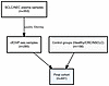
Plasma nucleosome profiling reports on tumor burden and molecular subtypes in small cell lung cancer
Abstract
BACKGROUND Small-cell lung cancer (SCLC) is an aggressive malignancy with a poor prognosis and marked transcriptional heterogeneity that may drive distinct therapeutic vulnerabilities. Clinical translation of molecular subtyping has been limited by restricted access to tumor biopsies, particularly at relapse.METHODS We applied chromatin immunoprecipitation of cell-free nucleosomes carrying active histone modifications followed by sequencing (cfChIP-seq) to 441 plasma samples from individuals with advanced SCLC, other neuroendocrine carcinomas, or non-SCLC cancers, as well as from healthy controls. Plasma cfChIP-seq profiles were integrated with matched tumor transcriptomes from 73 samples, including 41 time-matched pairs.RESULTS cfChIP-seq captured the epigenetic and transcriptional landscape of tumor-derived cell-free DNA (cfDNA), including SCLC tissue- and cell-of-origin signatures. A quantitative cfChIP-seq–derived SCLC score tracked radiographic tumor burden and was associated with prognosis. Signals at promoters of lineage-defining transcription factor genes, including ASCL1, NEUROD1, POU2F3, and ATOH1, correlated strongly with matched tumor RNA expression and supported noninvasive inference of SCLC transcriptional subtypes directly from plasma.CONCLUSION Plasma cfChIP-seq provides a practical liquid biopsy platform for real-time assessment of tumor burden, tumor state, and molecular subtype in SCLC. These findings support further development of cfChIP-seq for precision monitoring and subtype-informed therapeutic stratification in SCLC.TRIAL REGISTRATION ClinicalTrials.gov NCT02484404, NCT02487095, NCT02769962, NCT03554473, NCT03896503, and NCT02146170.FUNDING Center for Cancer Research; Intramural Program of the NCI (ZIA BC 011793); European Research Council (ERC) (Adg no. 101019560 “cfChIP”).
Authors
Gavriel Fialkoff, Nobuyuki Takahashi, Israa Sharkia, Jenia Gutin, Nadav Hermoni, Michael Nirula, Rajesh Kumar, Lorinc Pongor, Samantha Nichols, Linda Sciuto, Kanak Parmar, Parth Desai, Priya Suresh, Melissa Abel, Rajaa El Meskini, Myriam Maoz, Yakir Rottenberg, Shoshan Nevo, Hovav Nechushtan, Tamar Peretz, Diana Roame, Ayala Hubert, Jonathan E. Cohen, Azzam Salah, Mark Temper, Albert Grinshpun, Zoe Weaver-Ohler, Arun Rajan, William Douglas Figg Sr., Aviad Zick, Ronen Sadeh, Nir Friedman, Anish Thomas
Abstract
Authors
Tony Yao, Rita E. Chen, Melissa Yamada, J. Russell Moore, Marissa Jimenez, Tammy Huang, Lynn Cornelius, George Ansstas, Naresha Saligrama, David Y. Chen
Abstract
Advanced prostate cancer has increasingly developed a lethal neuroendocrine form, small cell/neuroendocrine prostate cancer (NEPC), as a consequence of the widespread use of highly potent androgen receptor signaling inhibitors in castration-resistant disease. The molecular mechanisms remain unclear and no effective therapies currently exist. We report that tryptophan hydroxylase 1 (TPH1), the enzyme responsible for peripheral serotonin biosynthesis — a neurotransmitter enriched in neuroendocrine tumors and a classical neuroendocrine biomarker — was upregulated in both de novo and therapy-induced human NEPC. TPH1 upregulation was necessary and sufficient for neuroendocrine differentiation and the NEPC phenotype through its enzymatic activity. Silencing TPH1 suppressed neuroendocrine plasticity and various aggressive behaviors of NEPC cells, including proliferation, invasion, sphere formation, and NEPC tumor xenograft growth. Mechanistically, TPH1 activated mTOR via intracellular serotonin-dependent serotonylation of mTOR at glutamine 2453, which triggered the induction of FOXM1 and E2F1 to drive NEPC differentiation and growth. Importantly, pharmacological inhibition of TPH1 using the clinically available inhibitor LX1606 effectively restricted growth and neuroendocrine marker expression in multiple NEPC cell lines and patient-derived xenografts. Collectively, these findings characterize TPH1’s contribution to NEPC and suggest TPH1 as a potential therapeutic target.
Authors
Jing Wei, Jing Wang, Jingrui Chen, Michelle Zhang, Chia-Hui Chen, Tianjie Pu, Alivia O'Brien, Sephtis Hargrove, Eva Corey, Tzu-Ping Lin, Allen C. Gao, Boyang Jason Wu
Abstract
Transport of nucleoside chemotherapeutic drugs into tumor cells is primarily accomplished through Equilibrative Nucleoside Transporter 1 (ENT1), considered to be constitutively-active, redistributing drugs across lipid bilayers via facilitated diffusion. Here we discover that ENT1 is not constitutively-active but rather requires activation of acid sphingomyelinase (ASMase) by gemcitabine, generating ceramide-rich platforms (CRPs) on external plasma membranes of endothelial and tumor cells into which ENT1 inserts, dimerizing therein to functionalize transmembrane gemcitabine transport. Whereas sarcoma cells synthesize minimal ASMase, they take up gemcitabine poorly in vitro and in murine xenografts. A strategy designed to augment gemcitabine-induced ASMase secretion into the extravascular space by ASMase-rich neo-angiogenic cells, which then targets tumor cell plasma membranes, yields “bystander” CRPs on sarcoma cells and ENT1 insertion therein, conferring markedly-enhanced gemcitabine uptake and xenograft response. Engaging this biology in a prospective Phase II clinical trial in advanced sarcoma yielded robust volumetric changes in evaluated tumors that developed early and were often durable.
Authors
Aditya Ganju, Shyam Rao, Mark A. Dickson, Robert A. Lefkowitz, Chris Thompson, Jin Cheng, Katia Manova, Adriana Haimovitz-Friedman, Gary Schwartz, Zhigang Zhang, Zvi Fuks, William D. Tap, Richard Kolesnick
Abstract
Gastroenteropancreatic neuroendocrine tumors (GEP-NETs) are clinically heterogeneous malignancies whose biology and microenvironmental organization remain poorly understood. Here, we integrated single-nucleus multiomic (snRNA-seq and snATAC-seq) and spatial transcriptomic profiling across 38 well-differentiated pancreatic (PanNET) and small-intestinal (siNET) tumors to define conserved malignant programs, their regulatory circuits, and spatial niches. We observed two conserved malignant cell programs spanning a continuous transcriptional spectrum: a neuronal-like program, and a secretory neuroendocrine program. Matched chromatin accessibility profiles uncovered distinct, tissue-specific regulatory networks, including MAX::MYC and MITF transcription factor binding motifs in siNETs versus ISL1 and TFAP4 in PanNETs, indicating organ-specific epigenetic control. Spatial transcriptomic analyses revealed that neuronal-like-high regions localized to densely cellular tumor areas with relative depletion of stromal infiltration, whereas secretory neuroendocrine-high regions occupied fibrovascular and stromal niches enriched for endothelial, fibroblast, and myeloid populations, and associated with TGFB1-ITGB1, VEGFA-FLT1, and LAMA2-ITGA1 signaling. Across both tumor types, the cNMF2 program was enriched in metastatic lesions and was enriched for pro-fibrotic and pro-angiogenic gene signatures. Thus, GEP-NETs are organized along a conserved neuronal-to-secretory axis defined by distinct epigenetic programs and spatially coupled to specific microenvironmental niches. This framework unifies NET heterogeneity across organ sites and identifies pathway-specific, microenvironment-linked vulnerabilities for therapeutic targeting.
Authors
Julie Karam, Samantha E. Hoffman, Amanda Garza, Dan Gui, Hannah I. Hoffman, Breanna M. Titchen, Yutaro Tanaka, Erica Pimenta, Theodora Pappa, Laura Valderrabano, Kevin Bi, Riaz Gillani, Lauren Brais, Erin Shannon, Jason L. Hornick, Jihye Park, Jennifer Chan, Eliezer M. Van Allen
Abstract
Background: Combined checkpoint blockade (CCB) of programmed-death-1 (PD-1) and cytotoxic-T-lymphocyte-associated protein-4 (CTLA-4) is highly active in melanoma but limited by significant morbidity from immune-related adverse events (irAEs). Effective strategies to prevent CCB-mediated irAEs are lacking. Methods: Patients with advanced melanoma were randomly assigned to receive standard of care ipilimumab and nivolumab alone (Arm-A: ipi/nivo, n=7) or with one cycle of rituximab (Arm-B; ipi/nivo+rituximab, n=7). Results: Patients receiving ipi/nivo+rituximab experienced lower rates of > grade-3(G3) irAEs (14% versus 57%) and superior G3-irAE-free survival compared to those in ipi/nivo arm (2-year G3-irAE-free survival 86% versus 29% (p=0.01), without adverse impact on tumor regression or survival. G3 hypersensitivity reactions to rituximab (43% in Arm-B) prompted trial closure. Rituximab depleted pre-therapy activated naïve B cells linked to autoimmunity and enhanced CCB-mediated induction of myeloid inflammation and CXCL13+ICOS+ CD4 T cells. Conclusion: B-cell depletion favorably modulates CCB-mediated immune activation and may reduce irAE risk. Trial Registration: ClinicalTrials.gov NCT03719131 Funding: NIH
Authors
Kavita M. Dhodapkar, Antonio Matera, Alyssa M. Duffy, Azmain Taz, Renee Julia Manalo, Melinda Yushak, Ragini Kuchadkar, David H. Lawson, Madhav V. Dhodapkar
Abstract
Authors
Vlad Moisoiu, Roxanne Lourman, Frank Szulzewsky, Tobias Kessler, Giulio Collotta, Antonio Porro, Anne Bertolini, Franziska Singer, Patrick J. Cimino, Caroline Hertler, Wolfgang Wick, Guido Reifenberger, Eric C. Holland, Alessandro A. Sartori, Michael Weller, Hans-Georg Wirsching
Abstract
Authors
Diwakar Turaga, Chang-Ru Tsai, Yuka Morikawa, Hanna J. Tadros, Yi Zhao, Lalita Wadhwa, Iki Adachi, Xiao Li, James F. Martin


Copyright © 2026 American Society for Clinical Investigation
ISSN: 0021-9738 (print), 1558-8238 (online)