
Autoimmunity
Abstract
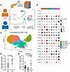
Effector CD8+ T cells are key drivers of type 1 diabetes (T1D) pathogenesis, yet questions remain regarding the molecular defects leading to altered cytotoxicity, peripheral tissue phenotype, and receptor specificity. We analyzed human pancreatic lymph nodes (pLNs) using mass cytometry and single-cell RNA-seq (scRNA-seq) with combined T cell receptor (TCR) profiling. Cytometric analysis revealed enrichment of T stem cell memory–like (TSCM-like) cells (CD8+CD45RA+CD27+CD28+CCR7+CXCR3+) in T1D pLNs. scRNA-seq indicated an elevated inflammatory cytokine gene signature (IFITM3, LTB) along with regulators of terminal differentiation (BCL6, BCL3), coupled with downregulation of exhaustion-associated genes (DUSP2, NR4A2, TSC22D3) in CD8+ T cells in T1D pLNs. Immune response enrichment analysis (IREA) indicated IL-15 signaling as a significant driver of these phenotypes. Integrated TCR and transcriptomics analysis revealed a cluster of diverse naive-like CD8+ T cell clones in T1D pLNs. Comparison of pLNs and pancreatic tissue slice isolates indicated sharing of effector CD8+ T cells, with enhanced terminal effector signatures within the pancreas relative to paired pLNs. Multiplex imaging revealed differential localization of T cell factor 1 (TCF1)- and thymocyte selection-associated high mobility group box protein (TOX)-expressing T cells in the pancreas, with islet-proximal TCF1+TOX+ cells displaying a mixture of activation and exhaustion-associated phenotypes. Thus, we provide multimodal cellular profiles enriched in T1D tissues for consideration in therapeutic targeting.
Authors
Leeana D. Peters, Howard R. Seay, Justin A. Smith, Amanda L. Posgai, Reed L. Berkowitz, Clive H. Wasserfall, Mark A. Atkinson, Rhonda Bacher, Maigan A. Brusko, Todd M. Brusko
Abstract
Membranous nephropathy (MN) is an autoimmune kidney disease and a major cause of nephrotic syndrome in adults. Although autoantibodies against phospholipase A2 receptor 1 (PLA2R) and complement activation are central to disease pathogenesis, the mechanisms by which anti-PLA2R antibodies activate complement at the podocyte surface remain incompletely defined. Here, we cloned 14 patient-derived anti-PLA2R monoclonal antibodies (mAbs) and found that they predominantly recognized the N-terminal cysteine-rich (CysR) and C-type lectin domain 1 (CTLD1) regions of PLA2R. Individual anti-PLA2R mAbs induced little or no complement-dependent cytotoxicity (CDC) of PLA2R-expressing podocytes in vitro. In contrast, paired mAbs targeting distinct epitopes, particularly CysR and CTLD1, markedly enhanced CDC. This effect was strongest for IgG1 and IgG3 antibodies, whereas IgG4 alone did not activate complement but modulated CDC in combination with IgG1. Purified IgG from patients with PLA2R-associated MN similarly induced CDC, which was augmented by addition of anti-PLA2R IgG1 and reduced by anti-PLA2R IgG4 or Fab fragments targeting CysR or CTLD1. In human PLA2R-expressing mice, paired anti-PLA2R antibodies increased glomerular complement deposition and induced albuminuria. These findings identify epitope pairing as a key determinant of complement activation in PLA2R-associated MN and support epitope-specific targeting strategies as a promising avenue for therapeutic intervention.
Authors
Tsai-Yi Wu, Kun-Hua Tu, Larissa Seifert, Kung-Wei Lin, Han-Po Shih, Yu-Fang Lo, Jhan-Jie Peng, Gunther Zahner, Oliver Kretz, You-Ning Lin, Chen-Xuan Kang, Jing-Ya Ding, Yi-Ran Tu, Li-Yi Ma, Ya-Ting Chuang, Chia-Chi Lo, Yu-Huan Tsai, Chih-Wei Yang, Nicola M. Tomas, Cheng-Lung Ku
Abstract
Multiple sclerosis (MS) is a complex inflammatory disease of the CNS resulting from an intricate interplay between genetic predisposition and environmental factors. Vitamin D (VD) deficiency is one of the established risk factors for MS. CD46 costimulation of CD4+ T cells induces a switch from Th1 to type I regulatory cells (Tr1), characterized by increased IL-10 production. This switch is impaired in MS T cells but can be restored by VD, which also strongly promotes expression of CD226 on CD46-activated T cells. The rs763361 polymorphism in the CD226 gene, resulting in a non-synonymous Gly307Ser variant, is associated with increased risk for MS. Herein, we show that expression of this CD226 risk allele disrupts the ability of CD46-activated T cells to operate the IFNγ/IL-10 switch upon VD exposure. Mechanistically, the risk variant impairs activation of the integrin LFA-1, which promotes the Tr1 phenotype. LFA-1-mediated Tr1 differentiation is also impaired in MS T cells expressing the CD226 risk allele upon CD46 and VD stimulation. Our study unveils how, in the context of MS susceptibility, a genetic polymorphism and an environmental factor act in concert to control the differentiation of Tr1 cells.
Authors
Saniya Kari, Aymeline Debonlier, Thibault Angles, Beatriz Chaves, Charles Grosjean, Florence Bucciarelli, Valérie Duplan-Eche, Lucie Nozeran, Jessica Lavery, Elena Morandi, Beatrice Pignolet, Max Mimpen, Joost Smolders, Roland Liblau, Abdelhadi Saoudi, Jan Damoiseaux, Frederick Masson, Loïc Dupré, Anne L. Astier
Abstract
Regulatory T (Treg) cells hold great promise as next-generation therapeutics for autoimmune diseases. However, maintaining their functional persistence within inflamed tissues remains a major translational challenge. Using an in vitro system that recapitulates the inflammatory CNS milieu of multiple sclerosis (MS), together with a pooled shRNA screen, we identify necroptotic signaling as a key driver of Treg cell death, thereby compromising Treg functional persistence under inflammatory conditions. We further demonstrate that Treg cells in both a mouse model of MS and patients with MS exhibit a preferential susceptibility to RIPK1 kinase-dependent necroptosis. Mechanistically, a FOXP3-driven low-glucose metabolic program renders Treg cells intrinsically susceptible to necroptosis by limiting O-GlcNAc modification on RIPK1. This vulnerability is not shared by conventional T cells under comparable inflammatory conditions. Finally, in combined with adoptive Treg cell transfer, we show that selective inhibition of necroptosis in Treg cells enhances their survival and suppressive function at sites of active inflammation, thereby reducing autoimmune pathology in mouse models of MS and systemic lupus erythematosus. Together, these findings identify necroptotic cell death as a barrier to Treg persistence within inflamed tissues and highlight the therapeutic potential of necroptosis-resistant Treg cells for the treatment of autoimmune diseases.
Authors
Qiaoyan Wu, Na Cui, Li Gao, Zhihui Lu, Xiang Ao, Rui Pang, Xingyan Li, Heling Pan, Daichao Xu, Peiying Li, Junying Yuan, Chengyu Zou
Abstract
Background Autoimmune diseases (ADs) often co-occur within individuals and families, indicating shared genetic risk factors. However, the composition of genetic overlap across autoimmunity is largely unknown. Methods This nationwide study included 6,336,615 individuals born in Sweden between 1932 and 1983, comprising 3,839,400 full-sibling pairs. Based on national heath-registers, 22 ADs were identified from 1969-2013. Aggregation and co-aggregation of ADs among siblings was used to estimate pairwise genetic correlations of ADs under a liability-threshold model. Network analysis and principal component analysis was used to characterize the structure of shared genetic risk across ADs. Results A total of 707,995 individuals (11.2%) were diagnosed with at least one AD. The studied ADs formed a network of significant genetic correlations (mean rg = 0.24, range 0.08-0.84) with clusters of more closely related ADs (rg ≥ 0.3). We found no evidence of a significant universal factor predisposing to autoimmunity. Conclusions This study demonstrates that ADs share substantial cluster-specific genetic overlap that largely aligns with affected tissue types, leading to distinct groupings of connective tissue diseases, gastrointestinal disorders, and endocrinopathies, whereas diseases of the nervous system show limited genetic cohesion. This suggests that shared biological mechanisms may drive coaggregation within disease groups. Clinically, these insights highlight the importance of monitoring patients and their relatives for related autoimmune disorders. Funding The Swedish Society of Medicine, Region Värmland's County Research Council, The Swedish Research Council, the Knut and Alice Wallenberg Foundation, The Regional Agreement on medical training and Clinical research (ALF) between Stockholm County Council and Karolinska Institutet
Authors
Daniel Eriksson, Ralf Kuja-Halkola, Marie E. Holmqvist, Henrik Larsson, Agnieszka Butwicka, Soffia Gudbjörnsdottir, Olle Kämpe, Sophie Bensing, Jakob Skov
Abstract
Systemic lupus erythematosus (SLE) is an autoimmune disease characterized by autoantibody production. Extrafollicular (EF) B cell responses contribute to SLE pathogenesis, with age-associated B cells (ABCs) giving rise to autoantibody-secreting plasmablasts (PBs). However, the migratory cues governing this EF trajectory remain unclear. Here, we identify a distinct ABC state with PB precursor characteristics (pre-PB ABCs) and reveal a migration-dependent program underlying their generation. Single-cell analysis of SLE patients and model mice showed that pre-PB ABCs were enriched in autoreactive clones and poised for PB differentiation. Their frequency correlated with autoantibody titers and disease activity, underscoring their pathogenic relevance. We further demonstrated that the oxysterol receptor EBI2 directed ABCs to EF niches within splenic bridging channels, promoting pre-PB ABC formation and autoreactive PB output. This process depended on the COMMD3/8 complex, a positive regulator of chemoattractant receptor signaling. Beyond EBI2-mediated ABC migration to EF niches, the COMMD3/8 complex was also required for trafficking of autoantibody-secreting cells to the bone marrow and infiltration of ABCs into the kidney. Accordingly, COMMD3/8 complex inhibition ameliorated disease in murine SLE models. These findings define a migration-dependent mechanism driving the EF differentiation of ABCs into autoreactive PBs and shaping the tissue distribution of pathogenic B cells, highlighting this program as a potential therapeutic target in SLE.
Authors
Taiichiro Shirai, Kentaro Kuzuya, Mizuki Kishi, Shinya Ichikawa, Shuhei Sakakibara, Akiko Nakai, Sarah Leach, Yu-Chen Liu, Daisuke Motooka, Daisuke Okuzaki, Masashi Narazaki, Atsushi Kumanogoh, Tomohiro Kurosaki, Jun Saegusa, Kazuhiro Suzuki
Abstract
CD20+ T cells are increasingly recognized as drivers of autoimmune and inflammatory diseases. However, their origin, development, and specific role in autoimmune skin diseases remain poorly understood. In this study, we observed an expansion of CD20+ T cells in the peripheral blood and skin lesions of patients with bullous pemphigoid (BP), which correlated with the levels of pathogenic autoantibodies and disease severity. Compared with CD20– T cells, CD20+ T cells exhibited enhanced metabolic and proinflammatory activities. In particular, antigen-specific BP180-NC16A-reactive T cells were enriched within the CD4+CD20+ subset. In both patients with BP and BP180-immunized mice, CD4+CD20+ T cells exhibited an antigen-specific follicular helper T (Tfh)-like phenotype, facilitating antibody production and B cell differentiation, whereas CD8+CD20+ T cells displayed cytotoxic and proinflammatory features. Mechanistically, we found that expression of the CD20-encoding gene MS4A1 in T cells was regulated by PAX5 in a DNA methylation-dependent manner. Therefore, our study elucidates the regulatory mechanisms governing CD20+ T cells and highlights their important role in the pathogenesis of BP.
Authors
Hui Fang, Shengxian Shen, Kang Li, Tianyu Cao, Bing Wang, Haijun Miao, Ke Xue, Yaxing Bai, Liang Li, Xia Li, Pei Qiao, Jieyu Zhang, Huanhuan Qu, Chen Zhang, Chunying Xiao, Bingyu Pang, Meng Fu, Hongjiang Qiao, Shuai Shao, Erle Dang, Gang Wang
Abstract
Background: Rheumatoid factor (RF) autoantibodies are highly prevalent, yet the molecular determinants of RF development and its progression to rheumatoid arthritis (RA) remain poorly understood. Here, we define the genetic, phenotypic, and molecular architecture of RF and its progression to RA. Methods: 469,036 UK Biobank participants with RF testing and 76 ALTRA cohort individuals were studied. Phenome-wide (PheWAS), genome-wide (GWAS), and proteome-wide association studies compared RF-positive individuals without autoimmune disease to RF-negative controls. Single-cell RNA sequencing enabled pseudobulk differential expression and cytokine signature enrichment analyses. Results: RF seroprevalence was 9.3% and longitudinally stable in 94.5% of individuals. PheWAS identified 48 significant associations, led by chronic viral hepatitis (OR 4.8), hypersensitivity pneumonitis (OR 3.6), bronchiectasis (OR 1.9), and COPD (OR 1.4). GWAS of 24,216 RF-positive individuals revealed 29 independent loci; the strongest signal was in the extended HLA region (OR 1.45, P-value=5.4×10-221). Non-HLA loci converged on B cell homeostasis genes (ETS1, BACH2, PAX5, TNFRSF13B, FCGR2A). RF-positive individuals did not carry elevated RA polygenic risk. Proteomic profiling identified 153 differentially abundant proteins enriched for humoral immunity and interferon-induced chemokines, with 79% showing dose-response relationships across titers. Progression to RA involved a shift toward activating tissue-damaging inflammatory pathways rather than amplification of the RF signature. Single-cell transcriptomics of RF-positive individuals without RA localized dysregulation to memory B cells, with downregulation of inhibitory genes (FCGR2B, BACH2, FOXP1) and upregulation of activation markers. Conclusion: RF production is governed by HLA class II and B cell regulatory loci, associated with mucosal inflammation, and is genetically and molecularly distinct from RA.
Authors
Mehmet Hocaoglu, Amr H. Sawalha

Abstract
Vitiligo is an autoimmune skin disease characterized by depigmentation, mainly due to CD8+ T cell–mediated destruction of melanocytes. Hyperglycemia exacerbates autoimmune responses and is associated with vitiligo; however, the underlying immunometabolic mechanisms are poorly understood. Here, we demonstrated the correlation between hyperglycemia and vitiligo in a case-control study and demonstrated that hyperglycemia aggravated vitiligo based on a mouse model. Targeted metabolomics identified succinate as the potential metabolite mediating hyperglycemia-aggravated vitiligo. Mechanistically, succinate promotes the activation of CD8+ T cells through succinate receptor 1 (SUCNR1) and promotes keratinocytes to secrete CXCL9 and CXCL10 by enhancing the stability and nuclear translocation of hypoxia-inducible factor-1α, facilitating the skin-homing of CD8+ T cells. Thus, hyperglycemia aggravates vitiligo through succinate/SUCNR1 axis–regulated CD8+ T cell hyperactivation. Our study provides insights into the long-observed yet previously unclear mechanism by which hyperglycemia accelerates vitiligo progression and highlights SUCNR1 as a potential therapeutic target.
Authors
Pan Kang, Yuqian Chang, Tingting Wang, Xiuli Yi, Yinghan Wang, Pengran Du, Jiaxi Chen, Baizhang Li, Shuli Li, Zhongjun Shao, Jianru Chen, Chunying Li
Abstract
BACKGROUND. Anti-nephrin autoantibodies have emerged as a putative pathogenic driver in a subset of patients with podocytopathies, including those with post-transplant disease recurrence. METHODS. We measured anti-nephrin autoantibodies in a cohort of 65 patients with podocytopathy associated with steroid-sensitive nephrotic syndrome (n = 39) and steroid-resistant nephrotic syndrome (n = 26), and in 34 patients with post-transplant podocytopathy recurrence. Fourteen patients with membranous nephropathy and 20 healthy volunteers served as controls. ELISA and immunoprecipitation assays were performed to detect anti-nephrin IgG using two different recombinant human nephrin proteins. Immunofluorescence analysis was performed to assess the deposition of IgG and their colocalization with nephrin in renal biopsies. RESULTS. When using murine antigen-based ELISA, the highest positivity was found in healthy volunteers (55%), correlating with levels of circulating natural anti-α-galactose-α-1,3-galactose antibodies. This cross-reactivity was abrogated with recombinant human nephrin expressed in human cells. In this setting, very low prevalence (<5%) of anti-nephrin antibody-positive patients was found in steroid-sensitive and steroid-resistant nephrotic syndrome cohorts and in patients with post-transplant disease recurrence. These frequencies were comparable to healthy volunteers. Using confocal and super-resolution microscopy, only trace amounts of IgM, but no IgG, were found in the glomeruli of analyzed biopsies, which did not colocalize with nephrin. CONCLUSIONS. With the methodology presented here, anti-nephrin reactivity was extremely rare and occurred at comparably low frequencies in healthy controls, native-kidney podocytopathies, and post-transplant disease recurrence. This suggests that these autoantibodies are not inherently disease-specific and may not serve as a broad biomarker across podocytopathies. TRIAL REGISTRATION. ClinicalTrials.gov NCT06334692. FUNDING. Private donation.
Authors
Francesco Pecoraro, Luca Perico, Federica Casiraghi, Paola Rizzo, Matias Trillini, Andrea Angeletti, Manuel Alfredo Podestà, Xhuliana Kajana, Agnese Spennacchio, Marta Todeschini, Marilena Mister, Giuseppe Castellano, Ariela Benigni, Giuseppe Remuzzi
No posts were found with this tag.


Copyright © 2026 American Society for Clinical Investigation
ISSN: 0021-9738 (print), 1558-8238 (online)