
Advertisement
Commentary
Open Access |  10.1172/JCI210364
c-KIT expands the tool kit for treating arteriovenous malformation
Ruilin Wu1,2 and Jason E. Fish1,2
1Peter Munk Cardiac Centre, UHN Research Institutes, University Health Network, Toronto, Ontario, Canada.
2Department of Laboratory Medicine and Pathobiology, University of Toronto, Toronto, Ontario, Canada.
Address correspondence to: Jason E. Fish, Peter Munk Cardiac Centre, UHN Research Institutes, University Health Network, Princess Margaret Cancer Research Tower, 3-309, 101 College Street, Toronto, Ontario, M5G 1L7, Canada. Phone: 416.522.8191; Email: jason.fish@utoronto.ca.
Find articles by Wu, R. in: PubMed | Google Scholar
1Peter Munk Cardiac Centre, UHN Research Institutes, University Health Network, Toronto, Ontario, Canada.
2Department of Laboratory Medicine and Pathobiology, University of Toronto, Toronto, Ontario, Canada.
Address correspondence to: Jason E. Fish, Peter Munk Cardiac Centre, UHN Research Institutes, University Health Network, Princess Margaret Cancer Research Tower, 3-309, 101 College Street, Toronto, Ontario, M5G 1L7, Canada. Phone: 416.522.8191; Email: jason.fish@utoronto.ca.
Find articles by
Fish, J.
in:
PubMed
|
Google Scholar
|

Published October 1, 2026 - More info
J Clin Invest. 2026;136(19):e210364. https://doi.org/10.1172/JCI210364.
© 2026 Wu, et al. This work is licensed under the Creative Commons Attribution 4.0 International License. To view a copy of this license, visit http://creativecommons.org/licenses/by/4.0/.

Related articles:
Abstract
Hereditary hemorrhagic telangiectasia type 2 (HHT2), caused by mutations in ACVRL1 (also known as ALK1), is characterized by brain arteriovenous malformations (bAVMs), abnormal artery–vein connections for which treatment options remain limited. Despite evidence of endothelial cell (EC) heterogeneity, its role in bAVM pathogenesis remains poorly defined. Using EC-specific inducible Alk1-knockout mice (Alk1iECKO) and regionally resolved single-cell RNA sequencing, we showed that loss of ALK1 signaling induces bAVMs predominantly in the perineural vascular plexus. This process is driven by the emergence of a KIT+ angiogenic EC population with human AVM-like transcriptional features, including tip cell markers and activation of PI3K and KRAS signaling pathways. Cross-species analyses and validation in human samples demonstrated that KIT expression is conserved in ECs from both sporadic and HHT2 bAVMs. Drug-repurposing analysis identified KIT as a top actionable target, and we showed that Kit is directly repressed by BMP9/ALK1/SMAD4 signaling. Pharmacological inhibition of KIT reduced angiogenic reprogramming and vascular malformations in vivo without affecting normal vasculature. These findings identify a pathogenic angiogenic EC state and position KIT signaling as a therapeutically actionable pathway in bAVMs.
Authors
Elise Drapé, Lauranne Carrier, Gael Cagnone, Atik Rohmana Maftuhatul Fuad, Mathilde Bizou, Damian Sanchez, Typhaine Anquetil, Jack Wang, Halima Drissi Touzani Walali, Adnan Gopinadhan, Patrick Piet van Vliet, Joel P. Howard, Mysha Ibnat, Gregor Andelfinger, Ethan Winkler, Bruno Larrivée, Alexandre Dubrac
Abstract
Vascular endothelial cells (ECs) encode a homeostatic fluid shear stress (FSS) set point that is essential for vascular stability. Deviations above or below this threshold trigger adaptive remodeling to restore physiological shear levels. Disruption of this control mechanism leads to enlarged arteriovenous malformations (AVMs) in hereditary hemorrhagic telangiectasia, a vascular disorder caused by heterozygous loss-of-function (LOF) mutation in ALK1, ENG, or SMAD4. Mechanistically, Smad4-deficient ECs are reset to a lower FSS set point value, resulting in AVMs that show characteristics of high-FSS remodeling with elevated KLF4 and high activation of the downstream Akt. Here, we investigated the KLF4/Akt upstream mechanisms by which SMAD4 sets the physiological FSS set point. We identified the receptor tyrosine kinase c-KIT as a component and regulator of the junctional mechanosensory receptor complex, which is highly upregulated in murine and human AVMs. SMAD4 restrains flow signaling by limiting c-KIT–dependent ERK5 activation and KLF4 induction. Thus, SMAD4 LOF leads to sustained c-KIT engagement in the sensory junctional apparatus, driving enhanced and prolonged activation of the ERK5/KLF4/Akt signaling axis. These results show that Smad4-LOF mutations induce malformations by disabling a key homeostatic mechanism and identify c-KIT as a potentially previously unrecognized therapeutic target.
Authors
Johannes Gahn, Fan Wu, Qing Zhang, Yuxi Di, Tanmaya Behera, Yonggang Ren, Zohrah Hashemi, Kuheli Banerjee, Julio Cordero, Claudia Gherman, Kornelia Andorfer, Caroline T. Seebauer, Fatemeh Mirzapour-Shafiyi, Gergana Dobreva, Martin A. Schwartz, Roxana Ola
-
An unexpected role for c-KIT in flow sensing
The junctional mechanosensing signaling complex, which includes VE-Cadherin, VEGFR2, and PECAM1, plays a key role in EC responses to the physical forces of blood flow (4). Although expression of c-KIT (encoded by the KIT gene) is negligible in normal ECs, this receptor tyrosine kinase is upregulated in cells with reduced SMAD4 (5) or ALK1 (6) and is induced in ECs of mouse and human AVMs (5, 6). The activity of c-KIT in ECs has previously been implicated in regulating angiogenesis, including through modulation of glycolysis (7, 8). In a study published in this issue of the JCI, Gahn et al. unexpectedly discovered that although c-KIT has no impact on normal flow responses and is downregulated by laminar flow or BMP9 stimulation, it is stably incorporated into the mechanosensing complex in cells lacking SMAD4, where it plays a key role in lowering the FSS set point and augmenting flow signaling (5). Through mechanistic analyses, they found that c-KIT signaling acted in conjunction with the mechanosensing complex to sustain the MEKK3/ERK5/KLF4/AKT axis (Figure 1). Blocking c-KIT signaling restored cell shape and alignment with flow and reduced proliferation in SMAD4-knockdown ECs. This effect appeared to be independent of c-KIT’s main ligand, stem cell factor (SCF).
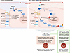 Figure 1
c-KIT as a mechanosensory component driving sustained outward remodeling and angiogenesis in HHT. Under physiological conditions, c-KIT expression in the endothelium is low, and c-KIT signaling is dispensable for blood flow responses and vascular development. However, related studies by Drapé et al. (6) and Gahn et al. (5) report that in the setting of HHT, loss of ALK1 or SMAD4 expression led to c-KIT upregulation and incorporation into the junctional mechanosensory complex. This resulted in excessive junctional signaling, leading to a lowered fluid shear stress set point in HHT ECs and sustained endothelial activation through the MEKK3/MEK5/ERK5/KLF4/AKT signaling axis. HHT ECs with high levels of c-KIT activity showed altered EC morphology and alignment, increased proliferation, and unique angiogenic signatures, driving continuous pathological vascular remodeling. Inhibition of c-KIT signaling in pathological ECs may restore these altered endothelial flow responses, offering a potential therapeutic direction for the management of AVMs in HHT.
Several questions remain regarding the role of c-KIT in flow sensing. How does c-KIT signaling affect the activity of the other components of this complex? Since blocking c-KIT appears to suppress activation of Src family kinases, it may have a proximal role in flow responses that are upstream of the other components, but this remains to be established. Furthermore, while immunoprecipitation experiments demonstrate interaction between c-KIT and other mechanosensory proteins, the precise localization and regulation of the subcellular distribution of c-KIT requires further analysis. Why does c-KIT not have a role in flow responses under normal conditions? Is this simply due to reduced expression levels, or are there other aspects of signaling that are only activated in HHT ECs? Indeed, it is not clear what activates c-KIT signaling in the context of SMAD4 deficiency, since it appears to be SCF independent.
Article tools
- Download citation information
- Send a comment
- Terms of use
- Standard abbreviations
- Need help? Email the journal
Metrics
Go to
- Top
- Abstract
- Defective flow-mediated responses in HHT
- An unexpected role for c-KIT in flow sensing
- Heterogeneity of HHT biology
- Can c-KIT inhibition reverse established AVMs?
- What is the specificity of c-KIT inhibition?
- Value of targeting c-KIT in other vascular anomalies
- Conflict of interest
- Funding support
- Footnotes
- References
- Version history


Copyright © 2026 American Society for Clinical Investigation
ISSN: 0021-9738 (print), 1558-8238 (online)