
Advertisement
Research ArticleBone biologyInflammationMetabolism
Open Access |  10.1172/JCI205829
Dietary omega-6 lipids promote postinjury aberrant bone formation in obesity
Stefanie L. Moye,1,2 Monisha Mittal,1 Tarun Srinivasan,2,3 Sneha Korlakunta,1 Chase A. Pagani,1,2 Ayelet Dar,1 Oromo Geshow,1 Dylan Feist,1 Lauren G. Zacharias,4 Zhao Li,5 Aaron W. James,5 Gerta Hoxhaj,4 Andrew M. Smith,6 Katherine A. Gallagher,7,8 Thomas P. Mathews,4 Robert J. Tower,1 and Benjamin Levi1
1Department of Surgery,
2Medical Scientist Training Program,
3Department of Immunology, and
4Children’s Medical Center Research Institute, University of Texas Southwestern Medical Center, Dallas, Texas, USA.
5Department of Pathology, Johns Hopkins School of Medicine, Baltimore, Maryland, USA.
6Department of Bioengineering, University of Illinois Urbana-Champaign, Urbana, Illinois, USA.
7Department of Surgery and
8Department of Microbiology, University of Michigan School of Medicine, Ann Arbor, Michigan, USA.
Address correspondence to: Benjamin Levi, Center for Organogenesis Regeneration and Trauma, University of Texas Southwestern Medical Center, 5323 Harry Hines Blvd., Dallas, Texas 75390, USA. Phone: 214.648.7230; Email: benjamin.levi@utsouthwestern.edu.
Find articles by
Moye, S.
in:
PubMed
|
Google Scholar
|

1Department of Surgery,
2Medical Scientist Training Program,
3Department of Immunology, and
4Children’s Medical Center Research Institute, University of Texas Southwestern Medical Center, Dallas, Texas, USA.
5Department of Pathology, Johns Hopkins School of Medicine, Baltimore, Maryland, USA.
6Department of Bioengineering, University of Illinois Urbana-Champaign, Urbana, Illinois, USA.
7Department of Surgery and
8Department of Microbiology, University of Michigan School of Medicine, Ann Arbor, Michigan, USA.
Address correspondence to: Benjamin Levi, Center for Organogenesis Regeneration and Trauma, University of Texas Southwestern Medical Center, 5323 Harry Hines Blvd., Dallas, Texas 75390, USA. Phone: 214.648.7230; Email: benjamin.levi@utsouthwestern.edu.
Find articles by Mittal, M. in: PubMed | Google Scholar
1Department of Surgery,
2Medical Scientist Training Program,
3Department of Immunology, and
4Children’s Medical Center Research Institute, University of Texas Southwestern Medical Center, Dallas, Texas, USA.
5Department of Pathology, Johns Hopkins School of Medicine, Baltimore, Maryland, USA.
6Department of Bioengineering, University of Illinois Urbana-Champaign, Urbana, Illinois, USA.
7Department of Surgery and
8Department of Microbiology, University of Michigan School of Medicine, Ann Arbor, Michigan, USA.
Address correspondence to: Benjamin Levi, Center for Organogenesis Regeneration and Trauma, University of Texas Southwestern Medical Center, 5323 Harry Hines Blvd., Dallas, Texas 75390, USA. Phone: 214.648.7230; Email: benjamin.levi@utsouthwestern.edu.
Find articles by Srinivasan, T. in: PubMed | Google Scholar
1Department of Surgery,
2Medical Scientist Training Program,
3Department of Immunology, and
4Children’s Medical Center Research Institute, University of Texas Southwestern Medical Center, Dallas, Texas, USA.
5Department of Pathology, Johns Hopkins School of Medicine, Baltimore, Maryland, USA.
6Department of Bioengineering, University of Illinois Urbana-Champaign, Urbana, Illinois, USA.
7Department of Surgery and
8Department of Microbiology, University of Michigan School of Medicine, Ann Arbor, Michigan, USA.
Address correspondence to: Benjamin Levi, Center for Organogenesis Regeneration and Trauma, University of Texas Southwestern Medical Center, 5323 Harry Hines Blvd., Dallas, Texas 75390, USA. Phone: 214.648.7230; Email: benjamin.levi@utsouthwestern.edu.
Find articles by Korlakunta, S. in: PubMed | Google Scholar
1Department of Surgery,
2Medical Scientist Training Program,
3Department of Immunology, and
4Children’s Medical Center Research Institute, University of Texas Southwestern Medical Center, Dallas, Texas, USA.
5Department of Pathology, Johns Hopkins School of Medicine, Baltimore, Maryland, USA.
6Department of Bioengineering, University of Illinois Urbana-Champaign, Urbana, Illinois, USA.
7Department of Surgery and
8Department of Microbiology, University of Michigan School of Medicine, Ann Arbor, Michigan, USA.
Address correspondence to: Benjamin Levi, Center for Organogenesis Regeneration and Trauma, University of Texas Southwestern Medical Center, 5323 Harry Hines Blvd., Dallas, Texas 75390, USA. Phone: 214.648.7230; Email: benjamin.levi@utsouthwestern.edu.
Find articles by
Pagani, C.
in:
PubMed
|
Google Scholar
|
1Department of Surgery,
2Medical Scientist Training Program,
3Department of Immunology, and
4Children’s Medical Center Research Institute, University of Texas Southwestern Medical Center, Dallas, Texas, USA.
5Department of Pathology, Johns Hopkins School of Medicine, Baltimore, Maryland, USA.
6Department of Bioengineering, University of Illinois Urbana-Champaign, Urbana, Illinois, USA.
7Department of Surgery and
8Department of Microbiology, University of Michigan School of Medicine, Ann Arbor, Michigan, USA.
Address correspondence to: Benjamin Levi, Center for Organogenesis Regeneration and Trauma, University of Texas Southwestern Medical Center, 5323 Harry Hines Blvd., Dallas, Texas 75390, USA. Phone: 214.648.7230; Email: benjamin.levi@utsouthwestern.edu.
Find articles by Dar, A. in: PubMed | Google Scholar
1Department of Surgery,
2Medical Scientist Training Program,
3Department of Immunology, and
4Children’s Medical Center Research Institute, University of Texas Southwestern Medical Center, Dallas, Texas, USA.
5Department of Pathology, Johns Hopkins School of Medicine, Baltimore, Maryland, USA.
6Department of Bioengineering, University of Illinois Urbana-Champaign, Urbana, Illinois, USA.
7Department of Surgery and
8Department of Microbiology, University of Michigan School of Medicine, Ann Arbor, Michigan, USA.
Address correspondence to: Benjamin Levi, Center for Organogenesis Regeneration and Trauma, University of Texas Southwestern Medical Center, 5323 Harry Hines Blvd., Dallas, Texas 75390, USA. Phone: 214.648.7230; Email: benjamin.levi@utsouthwestern.edu.
Find articles by Geshow, O. in: PubMed | Google Scholar
1Department of Surgery,
2Medical Scientist Training Program,
3Department of Immunology, and
4Children’s Medical Center Research Institute, University of Texas Southwestern Medical Center, Dallas, Texas, USA.
5Department of Pathology, Johns Hopkins School of Medicine, Baltimore, Maryland, USA.
6Department of Bioengineering, University of Illinois Urbana-Champaign, Urbana, Illinois, USA.
7Department of Surgery and
8Department of Microbiology, University of Michigan School of Medicine, Ann Arbor, Michigan, USA.
Address correspondence to: Benjamin Levi, Center for Organogenesis Regeneration and Trauma, University of Texas Southwestern Medical Center, 5323 Harry Hines Blvd., Dallas, Texas 75390, USA. Phone: 214.648.7230; Email: benjamin.levi@utsouthwestern.edu.
Find articles by Feist, D. in: PubMed | Google Scholar
1Department of Surgery,
2Medical Scientist Training Program,
3Department of Immunology, and
4Children’s Medical Center Research Institute, University of Texas Southwestern Medical Center, Dallas, Texas, USA.
5Department of Pathology, Johns Hopkins School of Medicine, Baltimore, Maryland, USA.
6Department of Bioengineering, University of Illinois Urbana-Champaign, Urbana, Illinois, USA.
7Department of Surgery and
8Department of Microbiology, University of Michigan School of Medicine, Ann Arbor, Michigan, USA.
Address correspondence to: Benjamin Levi, Center for Organogenesis Regeneration and Trauma, University of Texas Southwestern Medical Center, 5323 Harry Hines Blvd., Dallas, Texas 75390, USA. Phone: 214.648.7230; Email: benjamin.levi@utsouthwestern.edu.
Find articles by Zacharias, L. in: PubMed | Google Scholar
1Department of Surgery,
2Medical Scientist Training Program,
3Department of Immunology, and
4Children’s Medical Center Research Institute, University of Texas Southwestern Medical Center, Dallas, Texas, USA.
5Department of Pathology, Johns Hopkins School of Medicine, Baltimore, Maryland, USA.
6Department of Bioengineering, University of Illinois Urbana-Champaign, Urbana, Illinois, USA.
7Department of Surgery and
8Department of Microbiology, University of Michigan School of Medicine, Ann Arbor, Michigan, USA.
Address correspondence to: Benjamin Levi, Center for Organogenesis Regeneration and Trauma, University of Texas Southwestern Medical Center, 5323 Harry Hines Blvd., Dallas, Texas 75390, USA. Phone: 214.648.7230; Email: benjamin.levi@utsouthwestern.edu.
Find articles by Li, Z. in: PubMed | Google Scholar
1Department of Surgery,
2Medical Scientist Training Program,
3Department of Immunology, and
4Children’s Medical Center Research Institute, University of Texas Southwestern Medical Center, Dallas, Texas, USA.
5Department of Pathology, Johns Hopkins School of Medicine, Baltimore, Maryland, USA.
6Department of Bioengineering, University of Illinois Urbana-Champaign, Urbana, Illinois, USA.
7Department of Surgery and
8Department of Microbiology, University of Michigan School of Medicine, Ann Arbor, Michigan, USA.
Address correspondence to: Benjamin Levi, Center for Organogenesis Regeneration and Trauma, University of Texas Southwestern Medical Center, 5323 Harry Hines Blvd., Dallas, Texas 75390, USA. Phone: 214.648.7230; Email: benjamin.levi@utsouthwestern.edu.
Find articles by
James, A.
in:
PubMed
|
Google Scholar
|
1Department of Surgery,
2Medical Scientist Training Program,
3Department of Immunology, and
4Children’s Medical Center Research Institute, University of Texas Southwestern Medical Center, Dallas, Texas, USA.
5Department of Pathology, Johns Hopkins School of Medicine, Baltimore, Maryland, USA.
6Department of Bioengineering, University of Illinois Urbana-Champaign, Urbana, Illinois, USA.
7Department of Surgery and
8Department of Microbiology, University of Michigan School of Medicine, Ann Arbor, Michigan, USA.
Address correspondence to: Benjamin Levi, Center for Organogenesis Regeneration and Trauma, University of Texas Southwestern Medical Center, 5323 Harry Hines Blvd., Dallas, Texas 75390, USA. Phone: 214.648.7230; Email: benjamin.levi@utsouthwestern.edu.
Find articles by
Hoxhaj, G.
in:
PubMed
|
Google Scholar
|
1Department of Surgery,
2Medical Scientist Training Program,
3Department of Immunology, and
4Children’s Medical Center Research Institute, University of Texas Southwestern Medical Center, Dallas, Texas, USA.
5Department of Pathology, Johns Hopkins School of Medicine, Baltimore, Maryland, USA.
6Department of Bioengineering, University of Illinois Urbana-Champaign, Urbana, Illinois, USA.
7Department of Surgery and
8Department of Microbiology, University of Michigan School of Medicine, Ann Arbor, Michigan, USA.
Address correspondence to: Benjamin Levi, Center for Organogenesis Regeneration and Trauma, University of Texas Southwestern Medical Center, 5323 Harry Hines Blvd., Dallas, Texas 75390, USA. Phone: 214.648.7230; Email: benjamin.levi@utsouthwestern.edu.
Find articles by
Smith, A.
in:
PubMed
|
Google Scholar
|
1Department of Surgery,
2Medical Scientist Training Program,
3Department of Immunology, and
4Children’s Medical Center Research Institute, University of Texas Southwestern Medical Center, Dallas, Texas, USA.
5Department of Pathology, Johns Hopkins School of Medicine, Baltimore, Maryland, USA.
6Department of Bioengineering, University of Illinois Urbana-Champaign, Urbana, Illinois, USA.
7Department of Surgery and
8Department of Microbiology, University of Michigan School of Medicine, Ann Arbor, Michigan, USA.
Address correspondence to: Benjamin Levi, Center for Organogenesis Regeneration and Trauma, University of Texas Southwestern Medical Center, 5323 Harry Hines Blvd., Dallas, Texas 75390, USA. Phone: 214.648.7230; Email: benjamin.levi@utsouthwestern.edu.
Find articles by
Gallagher, K.
in:
PubMed
|
Google Scholar
|
1Department of Surgery,
2Medical Scientist Training Program,
3Department of Immunology, and
4Children’s Medical Center Research Institute, University of Texas Southwestern Medical Center, Dallas, Texas, USA.
5Department of Pathology, Johns Hopkins School of Medicine, Baltimore, Maryland, USA.
6Department of Bioengineering, University of Illinois Urbana-Champaign, Urbana, Illinois, USA.
7Department of Surgery and
8Department of Microbiology, University of Michigan School of Medicine, Ann Arbor, Michigan, USA.
Address correspondence to: Benjamin Levi, Center for Organogenesis Regeneration and Trauma, University of Texas Southwestern Medical Center, 5323 Harry Hines Blvd., Dallas, Texas 75390, USA. Phone: 214.648.7230; Email: benjamin.levi@utsouthwestern.edu.
Find articles by Mathews, T. in: PubMed | Google Scholar
1Department of Surgery,
2Medical Scientist Training Program,
3Department of Immunology, and
4Children’s Medical Center Research Institute, University of Texas Southwestern Medical Center, Dallas, Texas, USA.
5Department of Pathology, Johns Hopkins School of Medicine, Baltimore, Maryland, USA.
6Department of Bioengineering, University of Illinois Urbana-Champaign, Urbana, Illinois, USA.
7Department of Surgery and
8Department of Microbiology, University of Michigan School of Medicine, Ann Arbor, Michigan, USA.
Address correspondence to: Benjamin Levi, Center for Organogenesis Regeneration and Trauma, University of Texas Southwestern Medical Center, 5323 Harry Hines Blvd., Dallas, Texas 75390, USA. Phone: 214.648.7230; Email: benjamin.levi@utsouthwestern.edu.
Find articles by Tower, R. in: PubMed | Google Scholar
1Department of Surgery,
2Medical Scientist Training Program,
3Department of Immunology, and
4Children’s Medical Center Research Institute, University of Texas Southwestern Medical Center, Dallas, Texas, USA.
5Department of Pathology, Johns Hopkins School of Medicine, Baltimore, Maryland, USA.
6Department of Bioengineering, University of Illinois Urbana-Champaign, Urbana, Illinois, USA.
7Department of Surgery and
8Department of Microbiology, University of Michigan School of Medicine, Ann Arbor, Michigan, USA.
Address correspondence to: Benjamin Levi, Center for Organogenesis Regeneration and Trauma, University of Texas Southwestern Medical Center, 5323 Harry Hines Blvd., Dallas, Texas 75390, USA. Phone: 214.648.7230; Email: benjamin.levi@utsouthwestern.edu.
Find articles by Levi, B. in: PubMed | Google Scholar
Published June 11, 2026 - More info
J Clin Invest. 2026;136(17):e205829. https://doi.org/10.1172/JCI205829.
© 2026 Moye et al. This work is licensed under the Creative Commons Attribution 4.0 International License. To view a copy of this license, visit http://creativecommons.org/licenses/by/4.0/.
Received: February 24, 2026; Accepted: June 4, 2026

Related article:
Abstract
Bone formation in soft tissues, known as heterotopic ossification (HO), can occur as a complication of trauma or burn injury and can cause pain and functional limitations in the affected site. HO remains an unmet clinical challenge due to a general lack of specific medical therapies. In this issue of the JCI, a study by Moye et al. identified obesity as a risk factor for HO and further found that the association with obesity was driven not by caloric surplus, but instead by dietary omega-6 lipids, which are characteristically elevated in the Western diet. These omega-6 lipids accumulated directly at the incipient HO site, where they served as substrates for prostaglandin E2 (PGE2), fueling aberrant osteoblast differentiation. Overall, this work provides compelling support for dietary intervention or pharmacologic therapy directed at downstream PGE2 signaling as approaches to reduce HO in at-risk patients.
Authors
Peyton L. Carpen, Matthew B. Greenblatt

-
Results
Obesity enhances HO formation and impairs functional recovery after injury. To examine the association between obesity and HO, we analyzed a large retrospective cohort of patients who underwent total joint arthroplasty using the TriNetX database (>110 million US patients). After 1:1 propensity score matching for age, sex, and relevant comorbidities, we compared 622,009 patients with obesity (BMI > 35) and 622,009 nonobese controls (BMI = 18.5–24.9). We found that obesity was associated with a 28% increased risk of postoperative HO (0.997% vs. 0.774%; P < 0.0001, risk ratio = 1.289) (Figure 1A). These data extend the conclusions of prior case studies to a large, national patient cohort (13–15).
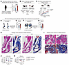 Figure 1
Obesity enhances HO formation and impairs functional recovery after injury. (A) TriNetX analysis of HO incidence after total joint arthroplasty in patients considered obese (BMI > 35) versus those with a normal weight (BMI 18.5–24.9) (n = 622,009 patients/cohort). (B) Experimental design: mice fed an HFD (60% fat) or control diet (10% fat) for 8 weeks prior to B/T or HA, followed for 9–12 weeks before μCT. (C) Body weight after diet initiation (n = 10–15 mice/group). Surgery performed at week 0. (D and E) Representative μCT images and quantification of ankle HO in HFD and control mice (9 weeks after B/T; n = 6–7 mice/group). (F and G) Representative μCT images and quantification of hip HO (12 weeks after HA; n = 7–9 mice/group). (H) Histological images of injured hind limbs (H&E, Masson’s trichrome) at 9 weeks, with outlined regions demonstrating newly formed HO in the calcaneus and Achilles tendon. Scale bars: 2.5 mm (left), 1 mm (right). (I) Histological images of injured hips at 12 weeks, with outlined regions demonstrating newly formed HO at the acetabulo-femoral joint. Scale bars: 1 mm. (J) Ankle range of motion over time in injured and uninjured limbs (3, 6, and 9 weeks; n = 5–12 mice/group; statistical analyses performed at 9 weeks). (K) Hip range of motion over time in injured and uninjured hips (3, 6, 9, and 12 weeks; n = 5–6 mice/group; statistical analyses performed at 12 weeks). Data represent at least 2 independent experiments. Means ± SEM are plotted. Statistics: χ2 (A), 2-tailed Student’s t test (C), or Mann-Whitney test (E, G, J, and K). *P < 0.05; ****P < 0.0001. See also Supplemental Figures 1 and 2.
To test whether diet-induced obesity causally enhances HO, we employed 2 well-established mouse models of traumatic and surgery-associated HO: burn/tenotomy (B/T) and hip arthroplasty (HA) (25–27). Mice were fed an HFD or control diet for 8 weeks prior to injury to induce obesity (Figure 1B and Supplemental Table 1; supplemental material available online with this article; https://doi.org/10.1172/JCI205829DS1). HFD-fed mice exhibited increased body weight, elevated adiposity, and hepatic steatosis without the loss of lean mass (Figure 1C and Supplemental Figure 1, A and C). Consistent with dysregulated metabolism, HFD mice had markedly higher serum leptin and resistin (Supplemental Figure 1B) (28).
After B/T, HFD mice developed significantly greater ectopic bone volume in the injured ankle compared with control-fed mice, as assessed by micro–computed tomography (μCT) at 9 weeks after injury (Figure 1, D and E). In the B/T model, the majority of HO forms around the calcaneus with additional bone formation adjacent to the Achilles tendon (25). Similar increases in HO were observed at the injured hip joint in the HA model after 12 weeks (Figure 1, F and G). In HA, ectopic bone forms in the periacetabular region, including around the subchondral bone (27). Histological analysis (3 weeks after injury) demonstrated increased Safranin O and trichrome staining in HFD mice, reflecting greater cartilage and collagen deposition at the injury site, consistent with increased endochondral ossification (Supplemental Figure 1E) (7). HFD mice also had increased ectopic bone formation at later time points (9 weeks B/T; 12 weeks HA) (Figure 1, H and I; HO regions outlined in black). Additionally, wound healing was markedly delayed with HFD (Supplemental Figure 1D).
Functionally, injured HFD mice exhibited impaired range of motion in the ankle and hip compared with injured controls (Figure 1, J and K). Injured HFD mice also had reduced locomotor activity and rearing, while uninjured mice performed similarly on functional assessments across diets (Supplemental Figure 1, F–H). These deficits were not explained by increased mechanical loading from excess body weight, as independent gait and black box pressure analyses demonstrated similar reductions in loading between HFD and control mice after injury (Supplemental Figure 1, I–K, and Supplemental Videos 1–4) (29, 30). Together, these findings demonstrate that HFD mice experience greater HO and worse functional recovery after injury and that these impairments occur independent of body weight–associated loading.
To test whether the increase in HO is a general feature of diet-induced obesity rather than unique to a single high-fat formulation, we evaluated mice fed a WD, which combines high fat with high sugar to induce obesity (Supplemental Table 1 and Supplemental Figure 2A) (31). WD mice also had increased HO volume compared with controls (B/T; Supplemental Figure 2, B and C). These findings show that distinct obesogenic diets similarly enhance HO, supporting a role for diet-driven metabolic state in predisposing injured tissues to aberrant bone formation.
HFD-induced obesity amplifies the injury-induced cytokine response at the site of HO formation. Having established that HFD enhanced HO and worsened functional recovery, we next asked whether obesity alters the early inflammatory response at the HO site. We profiled the cytokine content in lysates isolated from the HO site (injured Achilles tendon and surrounding soft tissue where ectopic bone forms) at baseline (day 0) and day 3 after injury (Figure 2A). Cytokines were nearly undetectable at baseline, whereas by day 3, HFD HO sites displayed substantially higher levels of CCL2, CCL7, CXCL1, CXCL2, IL-6, TNF-α, TGF-β1, and GM-CSF compared with injured controls (Figure 2, B and C). These factors coordinate leukocyte recruitment, monocyte/macrophage activation, and early matrix remodeling (32, 33), indicating that HFD intensifies the local inflammatory response to injury. In contrast, systemic changes were minimal, with only IL-6 elevated in HFD serum after injury (Supplemental Figure 3A).
 Figure 2
HFD-induced obesity amplifies the injury-induced cytokine response at the site of HO formation. (A) Schematic of HO-site cytokine profiling of HFD and control mice (day 0 and day 3 after B/T). (B) Heatmap of cytokines/chemokines in the HO site (z scores; n = 9–10 mice/group). (C) Quantification of key cytokines (CCL2, CCL7, CXCL1/2, IL-6, TNF-α, TGF-β1, GM-CSF) normalized to protein (n = 9–10 mice/group). (D) Schematic of HO-site immune cell profiling (day 3 after injury). (E–H) Representative flow cytometry plot (left) and quantification (right) of number of live CD45+ immune cells, macrophages (F4/80+ myeloid cells), monocytes (Ly6c+ myeloid cells), and neutrophils (Ly6g+ myeloid cells) (n = 5 mice/group). Data represent at least 2 independent experiments. Means ± SEM are plotted. Statistics: Mann-Whitney test (C and E–H). *P < 0.05; **P < 0.01; ***P < 0.001; ****P < 0.0001. See also Supplemental Figure 3.
To further define the composition of the HO microenvironment, we performed flow cytometry at 3 days after injury (Figure 2D). HFD HO sites contained increased numbers of immune cells, including macrophages, monocytes, and neutrophils (Figure 2, E–H, and Supplemental Figure 3B). These results define a heightened inflammatory microenvironment in HFD mice, characterized by amplified cytokine induction and increased recruitment of proinflammatory myeloid cells.
HFD-induced obesity enriches ω-6 fatty acids at the HO site. Given the established links between inflammation and lipid metabolism in obesity (34), we next profiled the metabolites in the HO site 7 days after injury (Figure 3A). Untargeted metabolomics revealed distinct clustering of HFD and control samples by principal component analysis (PCA; Figure 3B). Pathway analysis identified LA and AA metabolism among the most significantly upregulated pathways in HFD mice (Figure 3, C–F, and Supplemental Table 2). LA and AA are ω-6 PUFAs, with LA serving as the precursor to AA, a key substrate for inflammatory lipid mediators, including prostaglandins (35). Serum metabolomic analysis showed that HFD increased systemic LA but did not alter AA (Figure 3, G and H). In contrast, only the HFD injury site displayed increased AA (Figure 3F), despite comparable preinjury AA levels across diets (Supplemental Figure 4, A–C). This pattern suggests that HFD elevates LA globally, but AA is selectively enriched at the HO site, suggesting that local conversion of LA to AA primarily occurs at the site of aberrant tissue repair.
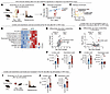 Figure 3
HFD-induced obesity enriches ω-6 fatty acids at the HO site. (A) Schematic of metabolomics workflow for the B/T HO site in HFD and control mice (day 7 after injury). (B) PCA of HO-site metabolite profiles (PC1, 38%; PC2, 24.2%; n = 4–5 mice/group). (C) Pathway analysis. (D) Heatmap of differential metabolites in HFD versus control (z scores; n = 4–5 mice/group). (E) Volcano plot. (F) Quantification of LA and AA abundance at the HO site (n = 4–5 mice/group). (G) Volcano plot of serum metabolites at day 7 after B/T (LA and AA highlighted; n = 5 mice/group). (H) Quantification of LA and AA abundance in serum (n = 5 mice/group). (I) Schematic of metabolomics workflow for hip HO site (day 7 after injury). (J and K) Quantification of LA and AA abundance at the hip HO site (n = 5 mice/group). Means ± SEM are plotted. Statistics: Mann-Whitney test (F, H, J, and K). *P < 0.05; **P < 0.01. See also Supplemental Figure 4.
These trends were conserved in the HA model, where LA and AA were significantly elevated at the hip HO site (periarticular soft tissue surrounding the injured hip joint) in HFD mice compared with injured controls (Figure 3, I–K). Additionally, we compared the metabolite levels in the injured and contralateral uninjured hips within the same animals. In obese mice, injury increased the abundance of AA at the HO site, without altering LA, while control mice showed no injury-associated changes (Supplemental Figure 4, D and E).
Lipidomics of the HO site 7 days after injury revealed broad diet-dependent lipid remodeling, with HFD HO sites showing increased lysophospholipids across lysophosphatidylethanolamine, lysophosphatidylcholine, and lysophosphatidylinositol subclasses (Supplemental Figure 4, F–J). Several of the most enriched species were lysophospholipids containing AA (20:4) acyl chains (Supplemental Figure 4I). AA-bearing lipids are generated by cytosolic phospholipase A2α–independent (cPLA2α, Pla2g4a) cleavage of membrane phospholipids, a process that increases during tissue injury and inflammation (36). Notably, these lipids were elevated only in the HFD HO site, indicating that AA release is specific to obese states. Together, these findings indicate that obesity not only increases ω-6 lipid abundance, but also enhances membrane lipid remodeling, allowing for injury-specific AA release and accumulation within the HO site.
Myeloid COX-2 promotes local PGE2 production and HO in HFD-induced obesity. Elevated inflammatory signals and increased AA in HFD HO sites suggested enhanced substrate availability for COX activity (Figure 4A) (35). COX enzymes convert AA into prostaglandins, bioactive lipids that regulate inflammation and tissue remodeling (35) with well-established roles in bone formation and fracture repair (37). We next assessed prostaglandin production to determine whether this pathway promotes HO formation in HFD mice. COX-2, the inducible COX isoform activated during injury and inflammatory stress (35), was strongly upregulated at the HFD HO site, accompanied by increased local PGE2 levels, while serum PGE2 remained unchanged (Figure 4, B–D). These data indicate that HFD-driven enhancement of COX-2 activity was restricted to the HO site.
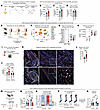 Figure 4
Myeloid COX-2 promotes local PGE2 production and HO in obesity. (A) Overview: dietary LA from an HFD serves as a precursor for AA, which is metabolized by COX enzymes into prostaglandins, including PGE2. (B–D) COX-2 and PGE2 levels at the HO site and serum PGE2 levels in HFD and control mice (day 3 after B/T; n = 5 mice/group). (E) Schematic of scRNA-seq workflow for HO-site tissue before and after B/T injury. (F) UMAP of HO-site cell populations annotated by canonical marker expression. (G and H) Ptgs1 and Ptgs2 expression by cell type and within myeloid cells split by diet and injury status. (I) Schematic of CD11b+ myeloid cell enrichment. (J) Ptgs2 expression in CD11b+ myeloid and CD11b– nonmyeloid cells (n = 4–5 mice/group). (K and L) Confocal imaging and quantification of COX-2 colocalization with CD11b+ myeloid cells in injured hind limbs at 1 week after B/T (n = 3–4 mice/group). Each point represents the proportion of CD11b+ cells expressing COX-2 within individual fields of view. White arrows denote Cox2+ CD11b+ myeloid cells. Scale bars: 1,000 μm (×10), 100 μm (×20), 50 μm (×63). (M) Schematic for 1-week celecoxib or vehicle treatment. (N) HO-site PGE2 levels in control, control + celecoxib, HFD, and HFD + celecoxib mice (day 3 after B/T; n = 5 mice/group). (O) Heatmap of chemokine and cytokine expression at day 3 after B/T. Control and HFD data from Figure 2B were reanalyzed with celecoxib-treated groups for comparison (n = 8–10 mice/group). (P and Q) μCT images and HO quantification (9 weeks after B/T; n = 6–8 mice/group). Statistics: Mann-Whitney test (B–D and L) or 1-way ANOVA (J, N, and Q). Data represent at least 2 independent experiments. Means ± SEM are plotted. *P < 0.05; **P < 0.01; ***P < 0.001. See also Supplemental Figure 5.
To more clearly define the cellular and molecular landscape of the HO site in HFD-induced obesity, we performed scRNA-seq of uninjured tendon and HO-site tissue from HFD- and control-fed mice (Figure 4E). Unsupervised clustering and UMAP visualization identified distinct cell populations, with annotations made using canonical marker genes (Figure 4F and Supplemental Figure 5A). Following injury, we found that myeloid cells expressed the highest levels of Ptgs2 (COX-2) and that Ptgs2 increased selectively in HFD myeloid cells, whereas Ptgs1 (COX-1) decreased (Figure 4, G and H). These findings indicate that HFD drives early myeloid-specific activation of COX-2 at the HO site after injury. qPCR of isolated HO-site myeloid cells and immunofluorescence of injured mouse hind limbs supported our findings, demonstrating selective enrichment of COX-2 in CD11b+ myeloid cells at the HFD HO site (Figure 4, I–L).
Expression of other key enzymes involved in AA production (delta-5 desaturase Fads1 and delta-6 desaturase Fads2) and prostaglandin synthesis (Ptges, microsomal PGE synthase) (38, 39) was increased in HFD myeloid cells at the HO site (Supplemental Figure 5B). Pla2g4a (cPLA2α), which releases AA from membrane phospholipids (36), was also significantly elevated (Supplemental Figure 5B), consistent with our lipidomic data showing enrichment of AA-containing lysophospholipids (Supplemental Figure 4I). Importantly, preinjury tendon and serum PGE2 levels did not differ between control and HFD mice (Supplemental Figure 5C). These results demonstrate that HFD-induced obesity specifically enhances COX-2 expression in myeloid cells after injury, resulting in increased PGE2 production at the HO site.
To test whether COX-2 activity was responsible for the increased HO in HFD mice, we treated HFD mice with celecoxib, an FDA-approved selective COX-2 inhibitor, for 1 week after injury (Figure 4M). We chose this time course to model HO prophylaxis after surgery and minimize off-target effects (40, 41). Celecoxib reduced HO-site and serum PGE2 levels (Figure 4N and Supplemental Figure 5D) and selectively lowered local inflammatory cytokines (e.g., CXCL1, IL-6, CCL2, CXCL2, TNF-α) without altering systemic levels (Figure 4O and Supplemental Figure 5E). The antiinflammatory effects of celecoxib were specific to the HFD HO site, consistent with the selective elevation in COX-2 activity observed in HFD mice. Importantly, celecoxib significantly reduced HO in HFD mice but had no effect in control mice (Figure 4, P and Q). These data imply that COX-2 signaling is required for the increased HO in HFD mice and that transient inhibition during the inflammatory phase is sufficient to prevent ectopic bone formation in obesity.
Given the known off-target effects of celecoxib (42), we next tested a cell-specific COX-2 inhibition strategy. Given our scRNA-seq finding that COX-2 is enriched in myeloid cells, we used an FDA-approved polysaccharide nanocarrier composed of dextran, which is selective for these cells (43–45). Local delivery of COX-2 inhibitor-loaded nanoparticles after injury led to reduced HO volume and HO-site PGE2 (Supplemental Figure 5, F–H) in HFD mice, suggesting that myeloid targeting of the COX-2/PGE2 pathway represents a novel cell-specific therapeutic strategy to attenuate HO.
Consistent with our preclinical findings, TriNetX analysis showed that obese patients who underwent total joint arthroplasty and were prescribed celecoxib postoperatively had reduced HO incidence compared with obese patients not receiving COX-2 inhibition (1.12% vs. 1.46%; P = 0.0027; risk ratio = 0.791), with no significant effect in nonobese patients (1.21% vs. 1.46%; P = 0.103; risk ratio = 0.828) (Supplemental Figure 5I).
Together, these data show that obesity selectively amplifies myeloid COX-2–mediated conversion of injury-site AA to PGE2, promoting inflammation and HO formation. These findings highlight an immunometabolic therapeutic opportunity for obese patients at risk of HO.
HFD-induced obesity induces an osteochondral transcriptional program in MPCs. We next evaluated how the prostaglandin-rich microenvironment at the HFD HO site alters the transcriptomic profile of MPCs, which give rise to ectopic bone (46). scRNA-seq analysis revealed that MPCs from injured HFD HO sites adopted a distinct transcriptional program primed for ectopic bone formation (Figure 5, A and B). These HFD MPCs were enriched for ECM (Col12a1, Plod2, Tnn, Thbs1), chondrogenic (Sox6, Acan), and osteogenic genes (Runx2, Spp1, Ibsp), while adipogenic genes (Pparg, Cebpa) were suppressed (Figure 5, A and B) (46, 47). In contrast, MPCs from uninjured mice displayed similar gene expression regardless of diet (Figure 5B). GSEA (using normalized enrichment scores) supported these findings, showing that injured HFD MPCs upregulated pathways linked to ectopic bone formation, including ECM remodeling and collagen biosynthesis (Supplemental Figure 6A) (26).
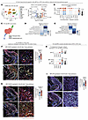 Figure 5
HFD-induced obesity induces an osteochondral transcriptional program in MPCs. (A) UMAP of HO-site cell populations (left). Volcano plot (right) showing the top differentially expressed genes in HFD versus control MPCs after injury (red: log2FC > 0.5, P < 0.001; blue: log2FC < –0.5, P < 0.001). (B) Expression of chondrogenic/ECM, osteogenic, and adipogenic genes in MPCs. (C) UMAP of MPC subclusters, including MPC-Tenos and MPC-OCs before and after injury. (D and E) Pathway analysis in MPC-OCs and MPC-Tenos after injury. (F) Confocal images showing colocalization of SOX9 (associated with chondrogenesis) with PDGFRα (MPCs) and quantification in injured HFD and control hind limbs (1 week after B/T; n = 3–4 mice/group). Each data point represents the proportion of SOX9+PDGFRα+ cells within individual fields of view at the HO site. Scale bars: 1,000 μm (×10), 50 μm (×63). White arrows denote SOX9+ PDGFRα+ MPCs. (G) Confocal images showing colocalization of RUNX2 (osteoblast precursors) with PDGFRα and quantification (1 week after B/T; n = 3–4 mice/group). Each data point represents the proportion of RUNX2+PDGFRα+ cells within individual fields of view at the HO site. Scale bars: 1,000 μm (×10), 50 μm (×63). White arrows denote RUNX2+ PDGFRα+ MPCs. (H) PGE2 receptor (Ptger1-4) gene expression in MPCs and MPC subclusters. (I) Confocal images showing colocalization of EP4 (Ptger4) with PDGFRα and quantification in injured hind limbs (1 week after B/T; n = 3–4 mice/group). Each data point represents the proportion of EP4+PDGFRα+ cells within individual fields of view at the HO site. Scale bars: 1,000 μm (×10), 50 μm (×63). EP4, PGE2 receptor 4. Statistics: Mann-Whitney test (F, G, and I). Data represent at least 2 independent experiments. Means ± SEM are plotted. *P < 0.05; **P < 0.01. See also Supplemental Figure 6.
Subclustering of MPCs into MPC-tenocytes (MPC-Tenos), MPC–osteochondral cells (MPC-OCs), and undifferentiated MPCs enabled more detailed analysis of distinct cellular programs after injury, corresponding to tendon repair (MPC-Teno: Scx, Tnmd) or aberrant osteogenesis (MPC-OC: Acan, Sox6, Runx2) (Figure 5C and Supplemental Figure 6, B–D) (46). HFD MPC-OCs were enriched for ECM organization, cartilage development, endochondral ossification, and growth factor signaling, pathways characteristic of cells primed for osteochondral differentiation (Figure 5D) (26). Even MPC-Teno clusters from HFD mice exhibited increased ECM and collagen-associated pathways (Figure 5E), consistent with a broader shift toward a pro-ossification transcriptional program. Immunofluorescence analysis supported our scRNA-seq findings, revealing an increased proportion of SOX9+ (associated with chondrogenesis) and RUNX2+ (osteoblast precursor marker) MPCs at the HFD HO site (Figure 5, F and G), consistent with increased osteochondral differentiation. Together, these findings suggest that obesity promotes pro-osteogenic signaling in HO-site MPCs, providing a cellular basis for the increased HO phenotype.
Next, we assessed whether PGE2 signaling mediates the transcriptomic changes observed in MPCs. PGE2 signals through 4 receptors: EP1, EP2, EP3, and EP4 (Ptger1-4) (48). Among Ptger1-4, Ptger4, implicated by prior studies in bone formation and collagen–ECM interactions (49–51), was the dominant receptor in MPCs and was selectively enriched in MPC-OCs (Figure 5H). We also noted an increased percentage of EP4+ MPCs at the HFD HO site (Figure 5I), suggesting enhanced activation of PGE2/EP4 signaling.
To determine whether inflammatory COX-2/PGE2 activity spatially localizes with osteochondral signaling at the HO site, we performed HD spatial transcriptomics of the injury site from HFD and control mice 7 days after B/T. Spatial analyses revealed higher expression of AA/COX-2 and prostaglandin synthesis pathway genes within the HO-developing site of HFD mice (distal end of transected Achilles tendon) compared with controls (Supplemental Figure 6E). These regions of COX-2hi cells were located near cells enriched for osteochondral differentiation (Supplemental Figure 6E), supporting a distinct immunometabolic microenvironment. Corresponding scRNA-seq analysis showed enrichment of the same gene modules in injured HFD cells (Supplemental Figure 6F). Consistent with our earlier scRNA-seq analyses, demonstrating enhanced Ptgs2 expression in myeloid cells (Figure 4, H and J) and enrichment of osteochondral genes in HFD MPCs (Figure 5B), these data support a spatially defined COX-2/PGE2/EP4 signaling pathway linking myeloid-derived prostaglandins to aberrant MPC differentiation in obesity.
To directly test whether PGE2 was sufficient to drive transcriptional changes in MPCs, we treated MPCs isolated from the HO site with PGE2 and assessed downstream gene expression (Supplemental Figure 6G). PGE2 enhanced the expression of key collagen and ECM-remodeling genes (Col3a1, Itga5, Plod2) and osteochondral genes (Sox9, Runx2) (Supplemental Figure 6H), similar to the HFD MPC transcriptional program observed in vivo. This effect was significantly reduced by cotreatment with an EP4 inhibitor (L-161,982) (Supplemental Figure 6H) (52). Additionally, Ptger4 expression was induced with PGE2 and suppressed by EP4 inhibition, suggesting that PGE2 potentiates the transcription of its receptor on MPCs. These data indicate that PGE2/EP4 signaling directly regulates MPC gene expression to promote HO.
Dietary LA depletion mitigates obesity-associated HO. Given that LA metabolism and downstream AA/PGE2 pathways are increased in HFD-associated HO, we next tested whether dietary LA depletion attenuates HO formation. We generated an isocaloric low-LA HFD (Supplemental Table 1) by replacing soybean oil with a mixture of fats (tallow, palm oil, and olive oil) which are lower in ω-6 PUFAs, while maintaining the same amount of total fat. The amount of dietary LA and the ω-6/ω-3 ratio of the low-LA HFD were similar to the control diet despite the increased total fat content (Supplemental Table 3).
Mice were maintained on the low-LA HFD for 8 weeks before injury and gained body weight and fat mass at a similar distribution and rate to the HFD mice (Figure 6, A and B). Metabolomic analysis of the HO site revealed that LA levels were markedly reduced in low-LA HFD–fed mice and were comparable with controls (Figure 6C). Consistent with reduced ω-6 substrate availability, HO-site PGE2 production was also suppressed in low-LA HFD–fed mice (Figure 6D). Accordingly, low-LA HFD–fed mice exhibited significantly reduced total HO volume after injury, comparable with control mice (Figure 6, E and F). Together, these data uncouple diet-induced obesity from ω-6 lipid availability and demonstrate that dietary LA is required to activate COX-2/PGE2 signaling and promote aberrant ectopic bone formation.
 Figure 6
Dietary LA depletion mitigates obesity-associated HO. (A) Body weight after diet initiation (n = 8 mice/group). Surgery performed at week 0. (B) Preinjury fat mass and lean mass (NMR; n = 5 mice/group). (C) Quantification of LA abundance at the HO site (7 days after B/T; n = 4–5 mice/group). (D) HO-site PGE2 levels normalized to protein (3 days after B/T; n = 4–5 mice/group). (E and F) Representative μCT images and quantification of HO (9 weeks after B/T; n = 7–8 mice/group). Data represent at least 2 independent experiments. Means ± SEM are plotted. Statistics: 2-tailed Student’s t test (A) or 1-way ANOVA (B–D and F). *P < 0.05; **P < 0.01; ****P < 0.0001.


Copyright © 2026 American Society for Clinical Investigation
ISSN: 0021-9738 (print), 1558-8238 (online)