
Advertisement
Research ArticleCell biologyClinical ResearchHematology
Open Access |  10.1172/JCI204701
XY0206 targets FLT3-dependent resistance states in acute myeloid leukemia
Long Shen,1,2 Yang Yang,1,2 Chenghua Xu,3 Bo Jiang,1,2 Xiaoxiao Duan,1,2 Xianfeng Shao,1,2 Simeng Li,1,2 Siyi Liu,1,2 Chao Huang,4 Lin Song,1,2 Mingyuan Sun,1,2 Jinting Fan,1,2 Ning Wang,1,2 Dong Zhang,1,2 Youyang Fang,1,2 Lichun Kang,5 Yajun Jiang,4 Mingming Niu,1,2 Junyuan Qi,1,2 Tao Cheng,1,2 and Hong Wang1,2
1State Key Laboratory of Experimental Hematology, National Clinical Research Center for Blood Diseases, Haihe Laboratory of Cell Ecosystem, Institute of Hematology & Blood Diseases Hospital, Chinese Academy of Medical Sciences & Peking Union Medical College, Tianjin, China.
2Tianjin Institutes of Health Science, Tianjin, China.
3Haihe Laboratory of Cell Ecosystem, Tianjin Medical University, Tianjin, China.
4State Key Laboratory of Pharmaceutical Biotechnology, School of Life Sciences, Chemistry and Biomedicine Innovation Center (ChemBIC), Nanjing University, Nanjing, China.
5Department of Clinical Laboratory, Tianjin First Central Hospital, School of Medicine, Nankai University, Tianjin, China.
Address correspondence to: Hong Wang, Tao Cheng, Junyuan Qi, or Mingming Niu, State Key Laboratory of Experimental Hematology, Institute of Hematology & Blood Diseases Hospital, 288 Nanjing Road, Tianjin 300020, China. Phone: 86.22.23909114; Email: wanghong@ihcams.ac.cn (HW); chengtao@ihcams.ac.cn (TC); qijy@ihcams.ac.cn (JQ); niumingming@ihcams.ac.cn (MN).
Authorship note: LS, YY, CX, and BJ contributed equally to this study.
Find articles by Shen, L. in: PubMed | Google Scholar
1State Key Laboratory of Experimental Hematology, National Clinical Research Center for Blood Diseases, Haihe Laboratory of Cell Ecosystem, Institute of Hematology & Blood Diseases Hospital, Chinese Academy of Medical Sciences & Peking Union Medical College, Tianjin, China.
2Tianjin Institutes of Health Science, Tianjin, China.
3Haihe Laboratory of Cell Ecosystem, Tianjin Medical University, Tianjin, China.
4State Key Laboratory of Pharmaceutical Biotechnology, School of Life Sciences, Chemistry and Biomedicine Innovation Center (ChemBIC), Nanjing University, Nanjing, China.
5Department of Clinical Laboratory, Tianjin First Central Hospital, School of Medicine, Nankai University, Tianjin, China.
Address correspondence to: Hong Wang, Tao Cheng, Junyuan Qi, or Mingming Niu, State Key Laboratory of Experimental Hematology, Institute of Hematology & Blood Diseases Hospital, 288 Nanjing Road, Tianjin 300020, China. Phone: 86.22.23909114; Email: wanghong@ihcams.ac.cn (HW); chengtao@ihcams.ac.cn (TC); qijy@ihcams.ac.cn (JQ); niumingming@ihcams.ac.cn (MN).
Authorship note: LS, YY, CX, and BJ contributed equally to this study.
Find articles by Yang, Y. in: PubMed | Google Scholar
1State Key Laboratory of Experimental Hematology, National Clinical Research Center for Blood Diseases, Haihe Laboratory of Cell Ecosystem, Institute of Hematology & Blood Diseases Hospital, Chinese Academy of Medical Sciences & Peking Union Medical College, Tianjin, China.
2Tianjin Institutes of Health Science, Tianjin, China.
3Haihe Laboratory of Cell Ecosystem, Tianjin Medical University, Tianjin, China.
4State Key Laboratory of Pharmaceutical Biotechnology, School of Life Sciences, Chemistry and Biomedicine Innovation Center (ChemBIC), Nanjing University, Nanjing, China.
5Department of Clinical Laboratory, Tianjin First Central Hospital, School of Medicine, Nankai University, Tianjin, China.
Address correspondence to: Hong Wang, Tao Cheng, Junyuan Qi, or Mingming Niu, State Key Laboratory of Experimental Hematology, Institute of Hematology & Blood Diseases Hospital, 288 Nanjing Road, Tianjin 300020, China. Phone: 86.22.23909114; Email: wanghong@ihcams.ac.cn (HW); chengtao@ihcams.ac.cn (TC); qijy@ihcams.ac.cn (JQ); niumingming@ihcams.ac.cn (MN).
Authorship note: LS, YY, CX, and BJ contributed equally to this study.
Find articles by Xu, C. in: PubMed | Google Scholar
1State Key Laboratory of Experimental Hematology, National Clinical Research Center for Blood Diseases, Haihe Laboratory of Cell Ecosystem, Institute of Hematology & Blood Diseases Hospital, Chinese Academy of Medical Sciences & Peking Union Medical College, Tianjin, China.
2Tianjin Institutes of Health Science, Tianjin, China.
3Haihe Laboratory of Cell Ecosystem, Tianjin Medical University, Tianjin, China.
4State Key Laboratory of Pharmaceutical Biotechnology, School of Life Sciences, Chemistry and Biomedicine Innovation Center (ChemBIC), Nanjing University, Nanjing, China.
5Department of Clinical Laboratory, Tianjin First Central Hospital, School of Medicine, Nankai University, Tianjin, China.
Address correspondence to: Hong Wang, Tao Cheng, Junyuan Qi, or Mingming Niu, State Key Laboratory of Experimental Hematology, Institute of Hematology & Blood Diseases Hospital, 288 Nanjing Road, Tianjin 300020, China. Phone: 86.22.23909114; Email: wanghong@ihcams.ac.cn (HW); chengtao@ihcams.ac.cn (TC); qijy@ihcams.ac.cn (JQ); niumingming@ihcams.ac.cn (MN).
Authorship note: LS, YY, CX, and BJ contributed equally to this study.
Find articles by Jiang, B. in: PubMed | Google Scholar
1State Key Laboratory of Experimental Hematology, National Clinical Research Center for Blood Diseases, Haihe Laboratory of Cell Ecosystem, Institute of Hematology & Blood Diseases Hospital, Chinese Academy of Medical Sciences & Peking Union Medical College, Tianjin, China.
2Tianjin Institutes of Health Science, Tianjin, China.
3Haihe Laboratory of Cell Ecosystem, Tianjin Medical University, Tianjin, China.
4State Key Laboratory of Pharmaceutical Biotechnology, School of Life Sciences, Chemistry and Biomedicine Innovation Center (ChemBIC), Nanjing University, Nanjing, China.
5Department of Clinical Laboratory, Tianjin First Central Hospital, School of Medicine, Nankai University, Tianjin, China.
Address correspondence to: Hong Wang, Tao Cheng, Junyuan Qi, or Mingming Niu, State Key Laboratory of Experimental Hematology, Institute of Hematology & Blood Diseases Hospital, 288 Nanjing Road, Tianjin 300020, China. Phone: 86.22.23909114; Email: wanghong@ihcams.ac.cn (HW); chengtao@ihcams.ac.cn (TC); qijy@ihcams.ac.cn (JQ); niumingming@ihcams.ac.cn (MN).
Authorship note: LS, YY, CX, and BJ contributed equally to this study.
Find articles by Duan, X. in: PubMed | Google Scholar
1State Key Laboratory of Experimental Hematology, National Clinical Research Center for Blood Diseases, Haihe Laboratory of Cell Ecosystem, Institute of Hematology & Blood Diseases Hospital, Chinese Academy of Medical Sciences & Peking Union Medical College, Tianjin, China.
2Tianjin Institutes of Health Science, Tianjin, China.
3Haihe Laboratory of Cell Ecosystem, Tianjin Medical University, Tianjin, China.
4State Key Laboratory of Pharmaceutical Biotechnology, School of Life Sciences, Chemistry and Biomedicine Innovation Center (ChemBIC), Nanjing University, Nanjing, China.
5Department of Clinical Laboratory, Tianjin First Central Hospital, School of Medicine, Nankai University, Tianjin, China.
Address correspondence to: Hong Wang, Tao Cheng, Junyuan Qi, or Mingming Niu, State Key Laboratory of Experimental Hematology, Institute of Hematology & Blood Diseases Hospital, 288 Nanjing Road, Tianjin 300020, China. Phone: 86.22.23909114; Email: wanghong@ihcams.ac.cn (HW); chengtao@ihcams.ac.cn (TC); qijy@ihcams.ac.cn (JQ); niumingming@ihcams.ac.cn (MN).
Authorship note: LS, YY, CX, and BJ contributed equally to this study.
Find articles by Shao, X. in: PubMed | Google Scholar
1State Key Laboratory of Experimental Hematology, National Clinical Research Center for Blood Diseases, Haihe Laboratory of Cell Ecosystem, Institute of Hematology & Blood Diseases Hospital, Chinese Academy of Medical Sciences & Peking Union Medical College, Tianjin, China.
2Tianjin Institutes of Health Science, Tianjin, China.
3Haihe Laboratory of Cell Ecosystem, Tianjin Medical University, Tianjin, China.
4State Key Laboratory of Pharmaceutical Biotechnology, School of Life Sciences, Chemistry and Biomedicine Innovation Center (ChemBIC), Nanjing University, Nanjing, China.
5Department of Clinical Laboratory, Tianjin First Central Hospital, School of Medicine, Nankai University, Tianjin, China.
Address correspondence to: Hong Wang, Tao Cheng, Junyuan Qi, or Mingming Niu, State Key Laboratory of Experimental Hematology, Institute of Hematology & Blood Diseases Hospital, 288 Nanjing Road, Tianjin 300020, China. Phone: 86.22.23909114; Email: wanghong@ihcams.ac.cn (HW); chengtao@ihcams.ac.cn (TC); qijy@ihcams.ac.cn (JQ); niumingming@ihcams.ac.cn (MN).
Authorship note: LS, YY, CX, and BJ contributed equally to this study.
Find articles by Li, S. in: PubMed | Google Scholar
1State Key Laboratory of Experimental Hematology, National Clinical Research Center for Blood Diseases, Haihe Laboratory of Cell Ecosystem, Institute of Hematology & Blood Diseases Hospital, Chinese Academy of Medical Sciences & Peking Union Medical College, Tianjin, China.
2Tianjin Institutes of Health Science, Tianjin, China.
3Haihe Laboratory of Cell Ecosystem, Tianjin Medical University, Tianjin, China.
4State Key Laboratory of Pharmaceutical Biotechnology, School of Life Sciences, Chemistry and Biomedicine Innovation Center (ChemBIC), Nanjing University, Nanjing, China.
5Department of Clinical Laboratory, Tianjin First Central Hospital, School of Medicine, Nankai University, Tianjin, China.
Address correspondence to: Hong Wang, Tao Cheng, Junyuan Qi, or Mingming Niu, State Key Laboratory of Experimental Hematology, Institute of Hematology & Blood Diseases Hospital, 288 Nanjing Road, Tianjin 300020, China. Phone: 86.22.23909114; Email: wanghong@ihcams.ac.cn (HW); chengtao@ihcams.ac.cn (TC); qijy@ihcams.ac.cn (JQ); niumingming@ihcams.ac.cn (MN).
Authorship note: LS, YY, CX, and BJ contributed equally to this study.
Find articles by Liu, S. in: PubMed | Google Scholar
1State Key Laboratory of Experimental Hematology, National Clinical Research Center for Blood Diseases, Haihe Laboratory of Cell Ecosystem, Institute of Hematology & Blood Diseases Hospital, Chinese Academy of Medical Sciences & Peking Union Medical College, Tianjin, China.
2Tianjin Institutes of Health Science, Tianjin, China.
3Haihe Laboratory of Cell Ecosystem, Tianjin Medical University, Tianjin, China.
4State Key Laboratory of Pharmaceutical Biotechnology, School of Life Sciences, Chemistry and Biomedicine Innovation Center (ChemBIC), Nanjing University, Nanjing, China.
5Department of Clinical Laboratory, Tianjin First Central Hospital, School of Medicine, Nankai University, Tianjin, China.
Address correspondence to: Hong Wang, Tao Cheng, Junyuan Qi, or Mingming Niu, State Key Laboratory of Experimental Hematology, Institute of Hematology & Blood Diseases Hospital, 288 Nanjing Road, Tianjin 300020, China. Phone: 86.22.23909114; Email: wanghong@ihcams.ac.cn (HW); chengtao@ihcams.ac.cn (TC); qijy@ihcams.ac.cn (JQ); niumingming@ihcams.ac.cn (MN).
Authorship note: LS, YY, CX, and BJ contributed equally to this study.
Find articles by Huang, C. in: PubMed | Google Scholar
1State Key Laboratory of Experimental Hematology, National Clinical Research Center for Blood Diseases, Haihe Laboratory of Cell Ecosystem, Institute of Hematology & Blood Diseases Hospital, Chinese Academy of Medical Sciences & Peking Union Medical College, Tianjin, China.
2Tianjin Institutes of Health Science, Tianjin, China.
3Haihe Laboratory of Cell Ecosystem, Tianjin Medical University, Tianjin, China.
4State Key Laboratory of Pharmaceutical Biotechnology, School of Life Sciences, Chemistry and Biomedicine Innovation Center (ChemBIC), Nanjing University, Nanjing, China.
5Department of Clinical Laboratory, Tianjin First Central Hospital, School of Medicine, Nankai University, Tianjin, China.
Address correspondence to: Hong Wang, Tao Cheng, Junyuan Qi, or Mingming Niu, State Key Laboratory of Experimental Hematology, Institute of Hematology & Blood Diseases Hospital, 288 Nanjing Road, Tianjin 300020, China. Phone: 86.22.23909114; Email: wanghong@ihcams.ac.cn (HW); chengtao@ihcams.ac.cn (TC); qijy@ihcams.ac.cn (JQ); niumingming@ihcams.ac.cn (MN).
Authorship note: LS, YY, CX, and BJ contributed equally to this study.
Find articles by Song, L. in: PubMed | Google Scholar
1State Key Laboratory of Experimental Hematology, National Clinical Research Center for Blood Diseases, Haihe Laboratory of Cell Ecosystem, Institute of Hematology & Blood Diseases Hospital, Chinese Academy of Medical Sciences & Peking Union Medical College, Tianjin, China.
2Tianjin Institutes of Health Science, Tianjin, China.
3Haihe Laboratory of Cell Ecosystem, Tianjin Medical University, Tianjin, China.
4State Key Laboratory of Pharmaceutical Biotechnology, School of Life Sciences, Chemistry and Biomedicine Innovation Center (ChemBIC), Nanjing University, Nanjing, China.
5Department of Clinical Laboratory, Tianjin First Central Hospital, School of Medicine, Nankai University, Tianjin, China.
Address correspondence to: Hong Wang, Tao Cheng, Junyuan Qi, or Mingming Niu, State Key Laboratory of Experimental Hematology, Institute of Hematology & Blood Diseases Hospital, 288 Nanjing Road, Tianjin 300020, China. Phone: 86.22.23909114; Email: wanghong@ihcams.ac.cn (HW); chengtao@ihcams.ac.cn (TC); qijy@ihcams.ac.cn (JQ); niumingming@ihcams.ac.cn (MN).
Authorship note: LS, YY, CX, and BJ contributed equally to this study.
Find articles by Sun, M. in: PubMed | Google Scholar
1State Key Laboratory of Experimental Hematology, National Clinical Research Center for Blood Diseases, Haihe Laboratory of Cell Ecosystem, Institute of Hematology & Blood Diseases Hospital, Chinese Academy of Medical Sciences & Peking Union Medical College, Tianjin, China.
2Tianjin Institutes of Health Science, Tianjin, China.
3Haihe Laboratory of Cell Ecosystem, Tianjin Medical University, Tianjin, China.
4State Key Laboratory of Pharmaceutical Biotechnology, School of Life Sciences, Chemistry and Biomedicine Innovation Center (ChemBIC), Nanjing University, Nanjing, China.
5Department of Clinical Laboratory, Tianjin First Central Hospital, School of Medicine, Nankai University, Tianjin, China.
Address correspondence to: Hong Wang, Tao Cheng, Junyuan Qi, or Mingming Niu, State Key Laboratory of Experimental Hematology, Institute of Hematology & Blood Diseases Hospital, 288 Nanjing Road, Tianjin 300020, China. Phone: 86.22.23909114; Email: wanghong@ihcams.ac.cn (HW); chengtao@ihcams.ac.cn (TC); qijy@ihcams.ac.cn (JQ); niumingming@ihcams.ac.cn (MN).
Authorship note: LS, YY, CX, and BJ contributed equally to this study.
Find articles by Fan, J. in: PubMed | Google Scholar
1State Key Laboratory of Experimental Hematology, National Clinical Research Center for Blood Diseases, Haihe Laboratory of Cell Ecosystem, Institute of Hematology & Blood Diseases Hospital, Chinese Academy of Medical Sciences & Peking Union Medical College, Tianjin, China.
2Tianjin Institutes of Health Science, Tianjin, China.
3Haihe Laboratory of Cell Ecosystem, Tianjin Medical University, Tianjin, China.
4State Key Laboratory of Pharmaceutical Biotechnology, School of Life Sciences, Chemistry and Biomedicine Innovation Center (ChemBIC), Nanjing University, Nanjing, China.
5Department of Clinical Laboratory, Tianjin First Central Hospital, School of Medicine, Nankai University, Tianjin, China.
Address correspondence to: Hong Wang, Tao Cheng, Junyuan Qi, or Mingming Niu, State Key Laboratory of Experimental Hematology, Institute of Hematology & Blood Diseases Hospital, 288 Nanjing Road, Tianjin 300020, China. Phone: 86.22.23909114; Email: wanghong@ihcams.ac.cn (HW); chengtao@ihcams.ac.cn (TC); qijy@ihcams.ac.cn (JQ); niumingming@ihcams.ac.cn (MN).
Authorship note: LS, YY, CX, and BJ contributed equally to this study.
Find articles by Wang, N. in: PubMed | Google Scholar
1State Key Laboratory of Experimental Hematology, National Clinical Research Center for Blood Diseases, Haihe Laboratory of Cell Ecosystem, Institute of Hematology & Blood Diseases Hospital, Chinese Academy of Medical Sciences & Peking Union Medical College, Tianjin, China.
2Tianjin Institutes of Health Science, Tianjin, China.
3Haihe Laboratory of Cell Ecosystem, Tianjin Medical University, Tianjin, China.
4State Key Laboratory of Pharmaceutical Biotechnology, School of Life Sciences, Chemistry and Biomedicine Innovation Center (ChemBIC), Nanjing University, Nanjing, China.
5Department of Clinical Laboratory, Tianjin First Central Hospital, School of Medicine, Nankai University, Tianjin, China.
Address correspondence to: Hong Wang, Tao Cheng, Junyuan Qi, or Mingming Niu, State Key Laboratory of Experimental Hematology, Institute of Hematology & Blood Diseases Hospital, 288 Nanjing Road, Tianjin 300020, China. Phone: 86.22.23909114; Email: wanghong@ihcams.ac.cn (HW); chengtao@ihcams.ac.cn (TC); qijy@ihcams.ac.cn (JQ); niumingming@ihcams.ac.cn (MN).
Authorship note: LS, YY, CX, and BJ contributed equally to this study.
Find articles by Zhang, D. in: PubMed | Google Scholar
1State Key Laboratory of Experimental Hematology, National Clinical Research Center for Blood Diseases, Haihe Laboratory of Cell Ecosystem, Institute of Hematology & Blood Diseases Hospital, Chinese Academy of Medical Sciences & Peking Union Medical College, Tianjin, China.
2Tianjin Institutes of Health Science, Tianjin, China.
3Haihe Laboratory of Cell Ecosystem, Tianjin Medical University, Tianjin, China.
4State Key Laboratory of Pharmaceutical Biotechnology, School of Life Sciences, Chemistry and Biomedicine Innovation Center (ChemBIC), Nanjing University, Nanjing, China.
5Department of Clinical Laboratory, Tianjin First Central Hospital, School of Medicine, Nankai University, Tianjin, China.
Address correspondence to: Hong Wang, Tao Cheng, Junyuan Qi, or Mingming Niu, State Key Laboratory of Experimental Hematology, Institute of Hematology & Blood Diseases Hospital, 288 Nanjing Road, Tianjin 300020, China. Phone: 86.22.23909114; Email: wanghong@ihcams.ac.cn (HW); chengtao@ihcams.ac.cn (TC); qijy@ihcams.ac.cn (JQ); niumingming@ihcams.ac.cn (MN).
Authorship note: LS, YY, CX, and BJ contributed equally to this study.
Find articles by Fang, Y. in: PubMed | Google Scholar
1State Key Laboratory of Experimental Hematology, National Clinical Research Center for Blood Diseases, Haihe Laboratory of Cell Ecosystem, Institute of Hematology & Blood Diseases Hospital, Chinese Academy of Medical Sciences & Peking Union Medical College, Tianjin, China.
2Tianjin Institutes of Health Science, Tianjin, China.
3Haihe Laboratory of Cell Ecosystem, Tianjin Medical University, Tianjin, China.
4State Key Laboratory of Pharmaceutical Biotechnology, School of Life Sciences, Chemistry and Biomedicine Innovation Center (ChemBIC), Nanjing University, Nanjing, China.
5Department of Clinical Laboratory, Tianjin First Central Hospital, School of Medicine, Nankai University, Tianjin, China.
Address correspondence to: Hong Wang, Tao Cheng, Junyuan Qi, or Mingming Niu, State Key Laboratory of Experimental Hematology, Institute of Hematology & Blood Diseases Hospital, 288 Nanjing Road, Tianjin 300020, China. Phone: 86.22.23909114; Email: wanghong@ihcams.ac.cn (HW); chengtao@ihcams.ac.cn (TC); qijy@ihcams.ac.cn (JQ); niumingming@ihcams.ac.cn (MN).
Authorship note: LS, YY, CX, and BJ contributed equally to this study.
Find articles by Kang, L. in: PubMed | Google Scholar
1State Key Laboratory of Experimental Hematology, National Clinical Research Center for Blood Diseases, Haihe Laboratory of Cell Ecosystem, Institute of Hematology & Blood Diseases Hospital, Chinese Academy of Medical Sciences & Peking Union Medical College, Tianjin, China.
2Tianjin Institutes of Health Science, Tianjin, China.
3Haihe Laboratory of Cell Ecosystem, Tianjin Medical University, Tianjin, China.
4State Key Laboratory of Pharmaceutical Biotechnology, School of Life Sciences, Chemistry and Biomedicine Innovation Center (ChemBIC), Nanjing University, Nanjing, China.
5Department of Clinical Laboratory, Tianjin First Central Hospital, School of Medicine, Nankai University, Tianjin, China.
Address correspondence to: Hong Wang, Tao Cheng, Junyuan Qi, or Mingming Niu, State Key Laboratory of Experimental Hematology, Institute of Hematology & Blood Diseases Hospital, 288 Nanjing Road, Tianjin 300020, China. Phone: 86.22.23909114; Email: wanghong@ihcams.ac.cn (HW); chengtao@ihcams.ac.cn (TC); qijy@ihcams.ac.cn (JQ); niumingming@ihcams.ac.cn (MN).
Authorship note: LS, YY, CX, and BJ contributed equally to this study.
Find articles by Jiang, Y. in: PubMed | Google Scholar
1State Key Laboratory of Experimental Hematology, National Clinical Research Center for Blood Diseases, Haihe Laboratory of Cell Ecosystem, Institute of Hematology & Blood Diseases Hospital, Chinese Academy of Medical Sciences & Peking Union Medical College, Tianjin, China.
2Tianjin Institutes of Health Science, Tianjin, China.
3Haihe Laboratory of Cell Ecosystem, Tianjin Medical University, Tianjin, China.
4State Key Laboratory of Pharmaceutical Biotechnology, School of Life Sciences, Chemistry and Biomedicine Innovation Center (ChemBIC), Nanjing University, Nanjing, China.
5Department of Clinical Laboratory, Tianjin First Central Hospital, School of Medicine, Nankai University, Tianjin, China.
Address correspondence to: Hong Wang, Tao Cheng, Junyuan Qi, or Mingming Niu, State Key Laboratory of Experimental Hematology, Institute of Hematology & Blood Diseases Hospital, 288 Nanjing Road, Tianjin 300020, China. Phone: 86.22.23909114; Email: wanghong@ihcams.ac.cn (HW); chengtao@ihcams.ac.cn (TC); qijy@ihcams.ac.cn (JQ); niumingming@ihcams.ac.cn (MN).
Authorship note: LS, YY, CX, and BJ contributed equally to this study.
Find articles by Niu, M. in: PubMed | Google Scholar
1State Key Laboratory of Experimental Hematology, National Clinical Research Center for Blood Diseases, Haihe Laboratory of Cell Ecosystem, Institute of Hematology & Blood Diseases Hospital, Chinese Academy of Medical Sciences & Peking Union Medical College, Tianjin, China.
2Tianjin Institutes of Health Science, Tianjin, China.
3Haihe Laboratory of Cell Ecosystem, Tianjin Medical University, Tianjin, China.
4State Key Laboratory of Pharmaceutical Biotechnology, School of Life Sciences, Chemistry and Biomedicine Innovation Center (ChemBIC), Nanjing University, Nanjing, China.
5Department of Clinical Laboratory, Tianjin First Central Hospital, School of Medicine, Nankai University, Tianjin, China.
Address correspondence to: Hong Wang, Tao Cheng, Junyuan Qi, or Mingming Niu, State Key Laboratory of Experimental Hematology, Institute of Hematology & Blood Diseases Hospital, 288 Nanjing Road, Tianjin 300020, China. Phone: 86.22.23909114; Email: wanghong@ihcams.ac.cn (HW); chengtao@ihcams.ac.cn (TC); qijy@ihcams.ac.cn (JQ); niumingming@ihcams.ac.cn (MN).
Authorship note: LS, YY, CX, and BJ contributed equally to this study.
Find articles by Qi, J. in: PubMed | Google Scholar
1State Key Laboratory of Experimental Hematology, National Clinical Research Center for Blood Diseases, Haihe Laboratory of Cell Ecosystem, Institute of Hematology & Blood Diseases Hospital, Chinese Academy of Medical Sciences & Peking Union Medical College, Tianjin, China.
2Tianjin Institutes of Health Science, Tianjin, China.
3Haihe Laboratory of Cell Ecosystem, Tianjin Medical University, Tianjin, China.
4State Key Laboratory of Pharmaceutical Biotechnology, School of Life Sciences, Chemistry and Biomedicine Innovation Center (ChemBIC), Nanjing University, Nanjing, China.
5Department of Clinical Laboratory, Tianjin First Central Hospital, School of Medicine, Nankai University, Tianjin, China.
Address correspondence to: Hong Wang, Tao Cheng, Junyuan Qi, or Mingming Niu, State Key Laboratory of Experimental Hematology, Institute of Hematology & Blood Diseases Hospital, 288 Nanjing Road, Tianjin 300020, China. Phone: 86.22.23909114; Email: wanghong@ihcams.ac.cn (HW); chengtao@ihcams.ac.cn (TC); qijy@ihcams.ac.cn (JQ); niumingming@ihcams.ac.cn (MN).
Authorship note: LS, YY, CX, and BJ contributed equally to this study.
Find articles by
Cheng, T.
in:
PubMed
|
Google Scholar
|

1State Key Laboratory of Experimental Hematology, National Clinical Research Center for Blood Diseases, Haihe Laboratory of Cell Ecosystem, Institute of Hematology & Blood Diseases Hospital, Chinese Academy of Medical Sciences & Peking Union Medical College, Tianjin, China.
2Tianjin Institutes of Health Science, Tianjin, China.
3Haihe Laboratory of Cell Ecosystem, Tianjin Medical University, Tianjin, China.
4State Key Laboratory of Pharmaceutical Biotechnology, School of Life Sciences, Chemistry and Biomedicine Innovation Center (ChemBIC), Nanjing University, Nanjing, China.
5Department of Clinical Laboratory, Tianjin First Central Hospital, School of Medicine, Nankai University, Tianjin, China.
Address correspondence to: Hong Wang, Tao Cheng, Junyuan Qi, or Mingming Niu, State Key Laboratory of Experimental Hematology, Institute of Hematology & Blood Diseases Hospital, 288 Nanjing Road, Tianjin 300020, China. Phone: 86.22.23909114; Email: wanghong@ihcams.ac.cn (HW); chengtao@ihcams.ac.cn (TC); qijy@ihcams.ac.cn (JQ); niumingming@ihcams.ac.cn (MN).
Authorship note: LS, YY, CX, and BJ contributed equally to this study.
Find articles by Wang, H. in: PubMed | Google Scholar
Published September 1, 2026 - More info
J Clin Invest. 2026;136(20):e204701. https://doi.org/10.1172/JCI204701.
© 2026 Shen et al. This work is licensed under the Creative Commons Attribution 4.0 International License. To view a copy of this license, visit http://creativecommons.org/licenses/by/4.0/.
Received: January 21, 2026; Accepted: August 26, 2026

Related article:
Abstract
Authors
Yiping Yang
-
Results
Biochemical and multi-omics profiling define an FLT3-centered inhibitory profile of XY0206. XY0206, originally reported as compound 10d, was developed through structure-guided optimization of the sunitinib scaffold to improve FLT3-directed potency and tolerability (Figure 1A). This optimization focused on the solvent-interface region of sunitinib, where modification of the N-diethylaminoethyl carboxamide tail was intended to improve the potency and tolerability of the sunitinib scaffold while preserving activity against FLT3-dependent AML. Prior work demonstrated potent FLT3/FLT3-ITD inhibition, preferential activity in FLT3-ITD AML models, favorable oral pharmacokinetics, and improved tolerability compared with sunitinib (26).
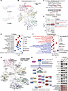 Figure 1
Biochemical and multi-omics profiling define an FLT3-centered inhibitory profile of XY0206. (A) Chemical structures of sunitinib and XY0206, with structural differences highlighted in blue and red. (B) Kinase inhibition profiling of XY0206 at 100 nM across 217 kinases. (C) Workflow for proteomic and phosphoproteomic profiling of FLT3-ITD AML cells treated with sunitinib or XY0206. (D) Scatterplots of differentially expressed proteins after sunitinib or XY0206 treatment. Red and blue dots indicate upregulated and downregulated proteins, respectively; selected proteins are labeled. (E) Pathway enrichment analysis of proteins regulated by XY0206 or sunitinib. NES, normalized enrichment score. (F) GSEA of phosphosite alterations after XY0206 or sunitinib treatment. Canonical pathways were obtained from MSigDB. (G) Kinase activity inference based on tyrosine-enriched phosphoproteomics using KSEA and RoKAI. Kinases shown were identified by both methods, with P < 0.05 and at least 2 substrate sites. Node shapes indicate treatment specificity: triangles, sunitinib-specific; pentagons, XY0206-specific; circles, shared. (H) Integrated FLT3 pathway map showing MS-derived protein and phosphosite-level changes after treatment. Heatmap values indicate log2 fold changes (FCs) from the corresponding proteomic or phosphoproteomic datasets; they should not be interpreted as phosphorylation (Phospho) occupancy, absolute phosphoprotein abundance, or mathematically combinable values across datasets. (I) Immunoblot analysis of total and phosphorylated FLT3, STAT5, AKT, ERK1/2, and c-MYC in MOLM13 cells treated with XY0206 or sunitinib (10 nM) for 3 hours. Numbers below blots indicate densitometric ratios normalized to total protein or GAPDH and scaled to DMSO. Immunoblots were repeated independently at least twice with similar results.
To further define the biochemical activity spectrum of XY0206, we performed single-concentration kinase profiling at 100 nM against a panel of 217 kinases. This assay was used to broadly assess kinase coverage at a concentration substantially above the reported FLT3/FLT3-ITD IC50 values, allowing identification of additional potential kinase targets. Under this condition, XY0206 strongly inhibited FLT3, along with other RTKs, including RET and PDGFRβ, with near-complete inhibition observed (Figure 1B and Supplemental Table 1; supplemental material available online with this article; https://doi.org/10.1172/JCI204701DS1), whereas the majority of kinases in the panel showed lower levels of inhibition. These data support an FLT3-centered kinase inhibition profile for XY0206, with additional activity against selected kinases under this screening condition.
We then integrated previously reported and newly generated enzymatic IC50 data for representative RTKs, selected based on the kinase profiling results and the known sunitinib target spectrum (26). XY0206 most potently inhibited FLT3-ITD (IC50 = 0.70 nM), followed by FLT3 and RET (IC50 = 5.27 nM and 6.92 nM, respectively). Activity against PDGFRβ was lower (IC50 = 26.00 nM), whereas inhibition of PDGFRα, KDR, KIT, and ALK was substantially weaker (IC50 = 158–675 nM) (Table 1). Compared with FLT3-ITD, XY0206 was ~37-fold less potent against PDGFRβ and >200-fold less potent against PDGFRα, KDR, KIT, and ALK, highlighting its preferential biochemical inhibition of FLT3-ITD while retaining modest activity against RET and PDGFRβ. Because RET has been implicated in supporting FLT3-ITD stability and PDGFRβ can activate downstream AKT and ERK signaling pathways that overlap with FLT3 signaling (27, 28), these data support preferential targeting of an FLT3-centered RTK signaling network.
To define the cellular consequences of XY0206-mediated FLT3-centered kinase inhibition, we performed integrated proteomic and phosphoproteomic analyses in FLT3-ITD AML cells treated with matched concentrations of XY0206 or sunitinib (Figure 1C). Proteomic profiling identified distinct drug-regulated protein expression programs following XY0206 or sunitinib treatment (Figure 1D and Supplemental Table 2). Pathway-level analysis of the proteomic data indicated suppression of AML- and proliferation-associated signaling programs, including cell-cycle, FLT3, MAPK, and PI3K/AKT/mTOR pathways (Figure 1E), consistent with attenuation of FLT3-dependent proliferative and survival signaling. Under matched treatment conditions, sunitinib showed comparatively stronger modulation of broader RTK- and NOTCH-associated pathways, consistent with its multi-kinase activity profile.
Global phosphoproteomic profiling was then used to define kinase-substrate-level signaling changes. XY0206 treatment was associated with reduced phosphorylation of multiple FLT3 downstream intermediates, including MAP2K2, PTPN11, SOS1, and STAT5A (Supplemental Figure 1, A and B, and Supplemental Table 3). These site-specific phosphorylation changes were further integrated at the pathway level, revealing coordinated modulation of apoptosis, cell death, cell differentiation, cell division, and cell proliferation programs, together with canonical FLT3 downstream signaling (Figure 1F), indicating coordinated modulation of antileukemic biological programs. Under matched treatment conditions, sunitinib had less pronounced effects on these FLT3-associated phosphoproteomic programs.
Kinase activity was inferred from phosphosite dynamics using the KSEA and RoKAI apps. To more directly interrogate FLT3/RTK-associated signaling, we additionally performed phosphotyrosine-enriched phosphoproteomic profiling, which increased the number of detected tyrosine-phosphorylated sites from 1,783 in the global phosphoproteomic dataset to 7,515 (Supplemental Figure 1C and Supplemental Table 4). The resulting phosphotyrosine-enriched kinase analysis showed that XY0206-modulated kinases were primarily concentrated in FLT3/RTK-associated nodes and downstream branches, whereas sunitinib affected a broader kinase spectrum, consistent with its multi-kinase profile (Figure 1G and Supplemental Figure 1D). Integration of proteomic and phosphoproteomic datasets onto a canonical FLT3 pathway map associated XY0206 treatment with reduced MYC-associated signaling, a key downstream program controlling leukemic cell proliferation, differentiation, and survival (Figure 1H). Immunoblot assays confirmed that XY0206 more effectively suppressed phosphorylation of canonical FLT3 downstream effectors, including STAT5, AKT, and ERK1/2, compared with sunitinib at equivalent concentrations (Figure 1I). Collectively, these data support a model in which XY0206 preferentially suppresses FLT3-centered signaling and downstream antileukemic programs in AML cells.
XY0206 exhibits preferential antileukemic activity in FLT3-ITD–mutated AML cells. We next systematically evaluated the antileukemic activity of XY0206 across a panel of AML and FLT3-driven cell models. Based on DepMap transcriptomic data and validation by immunoblotting (Supplemental Figure 2, A and B), we selected human AML cell lines with relatively high FLT3 expression (OCI-AML2), very low FLT3 expression (HEL), BCR-ABL–driven K562 cells as an FLT3-independent kinase-driven control, and FLT3-ITD mutations (MV4-11 and MOLM13), as well as murine Ba/F3 cells lacking endogenous FLT3 expression and Ba/F3 cells engineered to express FLT3-ITD. Dose response analyses were performed to determine IC50 values, measured using cell viability and proliferation assays, for XY0206, its parental compound sunitinib, and clinically used or approved FLT3is, including gilteritinib, midostaurin, and quizartinib/AC220.
Across the tested models, XY0206 exhibited growth-inhibitory activity comparable to that of approved FLT3is in FLT3-ITD-mutated cells. XY0206 suppressed the growth of FLT3-ITD-mutated cell lines, including MV4-11, MOLM13, and Ba/F3-FLT3-ITD, with IC50 values of 5.88 nM, 14.97 nM, and 4.35 nM, respectively. In contrast, higher IC50 values were observed in FLT3–wild-type (FLT3–WT) or FLT3-low– or negative-expressing cell lines, including OCI-AML2, HEL, parental Ba/F3 cells, and K562 cells (Table 2 and Supplemental Figure 2C). Together, these findings are consistent with preferential growth-inhibitory activity of XY0206 in FLT3-ITD–driven leukemia models, and underscore the influence of FLT3 expression status in non–FLT3-dependent cellular contexts.
We next assessed the functional consequences of XY0206 treatment in FLT3-ITD–mutated cells. Exposure to XY0206 for 24 hours resulted in cell-cycle perturbation, characterized by an increased proportion of cells in the G0/G1 phase (Figure 2A and Supplemental Figure 2, D–F). Apoptosis assays further demonstrated a dose-dependent increase in annexin V–positive cells (Figure 2B and Supplemental Figure 2, G and H), consistent with induction of apoptotic cell death. In line with these phenotypic changes, XY0206 treatment was associated with concentration-dependent suppression of FLT3 and downstream signaling, as evidenced by reduced phosphorylation of FLT3, STAT5, AKT, and ERK1/2. The inhibitory effects of XY0206 on these phosphorylation events were comparable to those observed with gilteritinib and were more pronounced than those induced by sunitinib at matched concentrations. Notably, the reduction in mTOR phosphorylation was less prominent than the inhibition of phosphorylated FLT3, STAT5, AKT, and ERK1/2, arguing against broad suppression of downstream kinase pathways as the primary mechanism of XY0206 activity (Figure 2C). Furthermore, in FLT3-independent, BCR-ABL–driven K562 cells, XY0206 at the same concentrations did not suppress phosphorylation of FLT3, STAT5, AKT, ERK1/2, or mTOR (Supplemental Figure 2I). These results support the interpretation that XY0206 suppresses STAT5, AKT, and ERK1/2 signaling primarily through inhibition of mutant FLT3 rather than through direct off-target inhibition of these downstream signaling nodes.
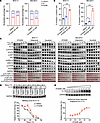 Figure 2
XY0206 exhibits preferential antileukemic activity in FLT3-ITD—mutated AML cells. (A) Cell cycle distribution of MV4-11 and MOLM13 cells treated with DMSO or XY0206 for 24 hours, assessed by PI staining and flow cytometry. (B) Apoptosis of MV4-11 and MOLM13 cells treated with DMSO, 10 nM XY0206, or 40 nM XY0206 for 48 hours, assessed by annexin V/PI staining and flow cytometry. (C) Immunoblot analysis of total and phosphorylated FLT3, STAT5, AKT, ERK1/2, and mTOR in FLT3-ITD–mutated MV4-11 and MOLM13 cells treated with XY0206, gilteritinib, or sunitinib at the indicated concentrations for 3 hours. Numbers below blots indicate densitometric ratios of phosphorylated to total protein normalized to vehicle-treated controls. (D) CETSA analysis of FLT3 thermal stabilization in MOLM13 cells treated with XY0206 (100 nM) or DMSO for 1 hour across the indicated temperature gradient. (E) Isothermal dose-response CETSA analysis of FLT3 thermal stabilization in MOLM13 cells treated with increasing concentrations of XY0206 for 1 hour, followed by heating at 50.4°C for 3 minutes. Data are shown as mean ± SD. For A, n = 3 biological replicates; for B, n = 3 independent biological experiments. Statistical analyses were performed using 2-tailed unpaired Student’s t test for A and 1-way ANOVA followed by Dunnett’s test for B. *P < 0.05, **P < 0.01, ***P < 0.001. All immunoblots were repeated independently at least twice with similar results, and representative blots are shown.
To assess target engagement in intact cells, we performed a cellular thermal shift assay (CETSA) (29). XY0206 treatment increased the thermal stability of FLT3 relative to DMSO control, resulting in a rightward shift of the FLT3 protein melting curve (Figure 2D). This stabilization effect increased with escalating concentrations of XY0206 (0–1,000 nM) (Figure 2E), supporting direct engagement of FLT3 by XY0206 in a cellular context. Collectively, these results indicate that XY0206 directly engages FLT3 and preferentially suppresses FLT3-ITD–mutated AML cells through inhibition of FLT3-dependent signaling pathways.
XY0206 retains activity against FLT3-ITD AML cells harboring the FLT3-F691L gatekeeper mutation. Acquired resistance to FLT3is frequently arises from treatment-emergent mutations within the TKD of FLT3 (5, 15, 30, 31). To evaluate the activity of XY0206 against clinically relevant TKD variants, we engineered Ba/F3 cells, which lack endogenous FLT3 expression, to express FLT3-ITD alone or in combination with representative TKD mutations (Supplemental Figure 3A). Gilteritinib retained activity against FLT3-ITD-D835V/Y and FLT3-ITD-Y842C but showed markedly reduced activity in cells expressing FLT3-ITD-F691L. In contrast, XY0206 maintained robust growth-inhibitory activity against Ba/F3-FLT3-ITD-F691L cells, whereas the parental compound, sunitinib, exhibited intermediate inhibition. The measured IC50 values were as follows: XY0206, 16.69 nM; gilteritinib, 382.97 nM; and sunitinib, 30.17 nM (Figure 3A and Supplemental Figure 3B), highlighting the superior potency of XY0206 in this clinically challenging mutant context. Functional assays further demonstrated that XY0206 treatment induced cell-cycle perturbation and increased apoptosis in Ba/F3-FLT3-ITD-F691L cells, effects that were more pronounced than those observed with gilteritinib or sunitinib under matched conditions (Figure 3, B and C, and Supplemental Figure 3, C and D). Immunoblot analysis confirmed that XY0206 effectively reduced phosphorylation of FLT3 and downstream signaling effectors, including STAT5, AKT, and ERK1/2, whereas gilteritinib and sunitinib showed only limited inhibition of these pathways (Figure 3D).
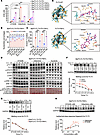 Figure 3
XY0206 retains activity against FLT3-ITD AML cells harboring the FLT3-F691L gatekeeper mutation. (A) IC50 values of XY0206, gilteritinib, and sunitinib in Ba/F3-FLT3-ITD cells with or without FLT3-TKD mutations after 72 hours of treatment. (B) Cell-cycle distribution of Ba/F3-FLT3-ITD-F691L cells treated with increasing concentrations of XY0206 (XY), gilteritinib (Gil), or sunitinib (Suni) for 24 hours, analyzed by PI staining and flow cytometry; comparisons were performed between XY0206 and gilteritinib at matched concentrations. (C) Apoptosis of Ba/F3-FLT3-ITD-F691L cells treated with XY0206, gilteritinib, or sunitinib for 48 hours, analyzed by annexin V/PI staining and flow cytometry. (D) Immunoblot analysis of total and phosphorylated FLT3, STAT5, AKT, and ERK1/2 in Ba/F3-FLT3-ITD-F691L cells treated with XY0206, gilteritinib, or sunitinib for 3 hours. Numbers below blots indicate densitometric ratios of phosphorylated to total protein normalized to vehicle-treated controls. (E) Molecular docking analysis of XY0206 bound to FLT3; dashed lines indicate hydrogen bonds. (F) CETSA analysis of FLT3 thermal stabilization in Ba/F3-FLT3-ITD-F691L cells treated with XY0206 (100 nM) or DMSO across the indicated temperature gradient. (G) CETSA analysis of FLT3 thermal stabilization in Ba/F3-FLT3-ITD and Ba/F3-FLT3-ITD-F691L cells treated with XY0206. (H) Isothermal dose-response CETSA analysis of FLT3 thermal stabilization in Ba/F3-FLT3-ITD-F691L cells treated with increasing concentrations of XY0206 or gilteritinib for 1 hour, followed by heating at 47.8°C for 3 minutes. Data are shown as mean ± SD from 3 independent biological experiments for A–C. Statistical analyses were performed using 2-tailed unpaired Student’s t test for A and 1-way ANOVA followed by Tukey’s multiple-comparison test for B and C. **P < 0.01, ***P < 0.001. All immunoblots were repeated independently at least twice with similar results, and representative blots are shown.
To elucidate the structural basis underlying this activity, we performed molecular docking and molecular dynamics simulations of XY0206 bound to both FLT3-WT and the F691L mutant (FLT3-F691L). Analyses of root mean square deviation, root mean square fluctuation, radius of gyration, and hydrogen-bonding dynamics indicated stable protein–ligand interactions over the simulation period (Supplemental Figure 3, E–H). Molecular mechanics/Poisson–Boltzmann surface area (MM-PBSA) calculations showed binding free energies of –52.894 kcal/mol for FLT3-WT and –60.144 kcal/mol for FLT3-F691L during the final 50 ns of equilibration, suggesting comparable binding affinity of XY0206 for both forms of FLT3. Molecular surface and close-up analyses revealed that XY0206 forms hydrogen bonds with key residues, including Leu616, Glu692, and Asp698, within the FLT3 active site and engages in hydrophobic interactions with Val624, Val675, Leu818, and Phe830. Although substitution of Phe691 with Leu alters 1 hydrophobic interaction, XY0206 maintains multiple stabilizing contacts, including hydrogen bonding with Asp698 and Arg834 and hydrophobic interactions with Val624, Ala642, Val675, Leu818, and Phe830 (Figure 3E).
To experimentally validate these predictions, CETSAs were performed. XY0206 treatment increased the thermal stability of FLT3-ITD-F691L compared with DMSO-treated controls, as reflected by a rightward shift in the protein melting curve (Figure 3F). No appreciable difference in FLT3 stabilization was observed between FLT3-ITD cells with or without the F691L mutation, indicating that this substitution does not substantially alter XY0206 binding stability in cells (Figure 3G). In contrast, gilteritinib induced only modest thermal stabilization, whereas XY0206 produced a concentration-dependent increase in FLT3 stability (Figure 3H).
XY0206 retains activity in FLT3-ITD AML with growth factor–associated and acquired resistance. Elevated levels of growth factors and cytokines have been implicated in attenuating the response of FLT3-ITD AML cells to FLT3is (13, 15). To examine drug activity under such conditions, MOLM13 and MV4-11 cells were treated with increasing concentrations of XY0206, gilteritinib, or sunitinib in the presence or absence of FGF2, which has been used as a simplified, growth factor–based model to assess cytokine-associated modulation of drug response. In cell viability assays, exposure to FGF2 reduced the sensitivity of both cell lines to gilteritinib and sunitinib, resulting in a modest rightward shift of the dose-response curve. In contrast, XY0206 displayed a largely overlapping dose-response profile under FGF2 stimulation, without an evident rightward shift (Figure 4A). Consistent results were observed in cell proliferation assays, in which treatment with XY0206 at its IC50 continued to suppress the proliferation of FGF2-stimulated FLT3-ITD AML cells, whereas gilteritinib and sunitinib exhibited reduced efficacy under the same conditions (Figure 4B). At the signaling level, immunoblot analysis indicated that FGF2 partially attenuated gilteritinib- and sunitinib-mediated inhibition of phosphorylated FLT3 (p-FLT3) and p-ERK1/2, whereas suppression of these signaling components by XY0206 was preserved (Figure 4C). These findings suggest that XY0206 retains antileukemic activity under growth factor–enriched conditions that diminish the response to gilteritinib and sunitinib.
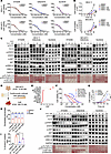 Figure 4
XY0206 retains activity in FLT3-ITD AML with growth factor—associated and acquired resistance. (A) CCK-8 viability assays of MV4-11 and MOLM13 cells treated with XY0206, gilteritinib, or sunitinib for 3 days with or without FGF2 (10 ng/mL). (B) Growth kinetics of MV4-11 and MOLM13 cells cultured with or without FGF2 (20 ng/mL) and treated at respective IC50 concentrations. Cells were counted every 2 or 3 days for up to 10 days (MV4-11) or 8 days (MOLM13). (C) Immunoblot analysis of phosphorylated and total FLT3, STAT5, AKT, and ERK1/2 in MV4-11 and MOLM13 cells treated with XY0206, gilteritinib, or sunitinib for 10 minutes with or without FGF2. Conc, concentration. (D) Schematic of GilR cell generation and gilteritinib IC50 determination. (E) WES analysis of GilR cells showing acquired genetic alterations relative to MOLM13. VAF, variant allele frequency. (F) CCK-8 dose-response curves of parental MOLM13 and GilR cells treated with increasing concentrations of XY0206, gilteritinib, or sunitinib for 72 hours. (G) Proliferation of GilR cells treated with XY0206, gilteritinib, or sunitinib (80 nM), measured every 2 days for 8 days. (H) Cell cycle distribution of GilR cells treated with XY0206 (XY), gilteritinib (Gil), or sunitinib (Suni) for 24 hours. (I) Apoptosis of GilR cells treated with XY0206, gilteritinib, or sunitinib for 72 hours. (J) Immunoblot analysis of total and phosphorylated FLT3, STAT5, AKT, and ERK1/2 in GilR cells treated for 4 hours. Numbers below blots in C and J indicate densitometric ratios normalized to vehicle controls. Data are mean ± SD from 3 independent biological experiments for A, B, and F–I. Statistical analyses were performed using 2-way ANOVA with Tukey’s test for B and G and 1-way ANOVA with Tukey’s test for H and I. *P < 0.05, ***P < 0.001. Immunoblots in C and J were repeated independently at least twice with similar results.
We next established a cellular model of acquired resistance induced by prolonged exposure to an FLT3i to evaluate the activity of XY0206. MOLM13 cells were continuously cultured in low-dose gilteritinib for approximately 10 months, leading to the establishment of a polyclonal gilteritinib-tolerant subline, designated GilR. Compared with parental MOLM13 cells, GilR cells had a 21-fold increase in gilteritinib IC50, from 14.58 nM to 308.70 nM (Figure 4D). Whole-exome sequencing (WES) revealed the accumulation of multiple genetic alterations in GilR cells relative to the parental line, including mutations in genes previously associated with AML progression and drug response, such as FLT3, CCND3, CHD7, CBL, ARID1A, and IRS2 (Figure 4E and Supplemental Table 5). Among these alterations, FLT3 mutations were prominently represented by the F691L gatekeeper substitution, consistent with continued FLT3 pathway engagement in this resistant model. Despite reduced sensitivity to gilteritinib, XY0206 remained active in GilR cells. Treatment with XY0206 resulted in sustained suppression of cell viability (Figure 4F) and was associated with greater inhibition of cell proliferation (Figure 4G), increased accumulation of cells in the G0/G1 phase, and higher levels of apoptosis compared with gilteritinib at matched concentrations. The effects of sunitinib were intermediate, showing stronger cytotoxicity than gilteritinib but weaker than XY0206 (Figure 4, H and I, and Supplemental Figure 4, A and B). Immunoblot analysis confirmed that XY0206 more completely suppressed phosphorylation of FLT3 and downstream effectors, including STAT5, AKT, and ERK1/2, whereas these pathways remained partially active after gilteritinib or sunitinib treatment (Figure 4J).
Consistent with emerging evidence, a recent conference abstract suggested that FLT3-F691L may sustain MAPK signaling through a GRB2-dependent, kinase-independent mechanism under FLT3i treatment (32). To explore whether GRB2-associated signaling might be relevant to XY0206 activity, we examined GRB2 expression in FLT3-F691L–resistant models. XY0206 treatment reduced total GRB2 protein levels in both GilR and Ba/F3-FLT3-ITD-F691L cells (Supplemental Figure 4, C and D). Moreover, GRB2 depletion further decreased the IC50 of XY0206 in GilR cells and enhanced XY0206-mediated killing of resistant cells (Supplemental Figure 4, E and F). These data indicate that GRB2-associated signaling may contribute to the maintenance of FLT3-F691L–driven resistant states and can modulate cellular responsiveness to FLT3 inhibition by XY0206. However, these findings do not establish GRB2 as a direct molecular target of XY0206, nor do they support direct disruption of FLT3-GRB2 interaction or kinase-independent FLT3 signaling. Therefore, GRB2 is interpreted here as a functional component of the FLT3-F691L–associated signaling network rather than a direct pharmacological target of XY0206.
To further examine whether resistance mechanisms downstream of RTK signaling could limit XY0206 activity, we generated a second long-term, low-dose, gilteritinib-resistant MOLM13 subline, designated GilR2. The IC50 for gilteritinib increased from 14.58 nM to 1,174.00 nM (Supplemental Figure 4G). WES analysis revealed, in addition to FLT3 mutations, a clinically relevant NRAS mutation associated with FLT3i resistance (Supplemental Figure 4H and Supplemental Table 5). Drug testing in GilR2 indicated that XY0206 also exhibited reduced activity (Supplemental Figure 4I), suggesting that NRAS-mutant subclones may bypass upstream FLT3/RTK-associated signaling and thereby diminish sensitivity to XY0206. These findings further support the presence of heterogeneous resistance mechanisms and indicate that XY0206 activity is most evident in FLT3-dependent resistance contexts, whereas resistance driven by downstream RAS pathway activation may require additional therapeutic strategies.
Overall, these data indicate that XY0206 retains activity in FLT3-dependent AML models exhibiting reduced responsiveness to gilteritinib, including both growth factor–associated and treatment-emergent resistant contexts, while revealing the impact of multiple resistance mechanisms and subclonal heterogeneity on drug sensitivity.
XY0206 demonstrates antileukemic activity with favorable tolerability in patient-derived AML blasts and preclinical mouse models. We next evaluated the activity of XY0206 in patient-derived AML blasts harboring FLT3-ITD mutations. Two independent patient samples were analyzed. AML01 was obtained from a patient with relapsed AML refractory to multiple prior therapies, including gilteritinib, whereas AML02 was derived from an unrelated patient with FLT3-ITD AML. Targeted sequencing was performed to provide molecular context for these samples. In addition to FLT3-ITD, AML01 harbored co-occurring mutations in DNMT3A, IDH2, and CEBPA, and AML02 carried multiple additional AML-associated alterations, including mutations in WT1, TET2, and SETDB1 (Supplemental Table 6), highlighting the genetic heterogeneity characteristic of FLT3-ITD AML. Across both samples, cell viability assays, direct cell counting, and apoptosis analyses consistently showed that XY0206 produced stronger cytotoxic effects than clinically used FLT3is, including sorafenib and gilteritinib (Figure 5, A–C, and Supplemental Figure 5). Consistent with these functional results, immunoblot analysis demonstrated suppression of p-FLT3 and downstream effectors STAT5, AKT, and ERK1/2, supporting effective inhibition of FLT3-associated signaling in primary AML blasts (Figure 5D).
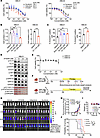 Figure 5
XY0206 demonstrates antileukemic activity with favorable tolerability in patient-derived AML blasts and preclinical mouse models. (A) Cell viability of primary FLT3-ITD AML blasts (AML01 and AML02) treated with sorafenib, gilteritinib, or XY0206 for 72 hours; IC50 values were calculated using GraphPad Prism. (B) Viable cell counts of primary AML blasts after 48-hour treatment with sorafenib (Sora), gilteritinib (Gil), or XY0206 (XY). (C) Apoptosis of primary AML blasts after 48-hour treatment with sorafenib, gilteritinib, or XY0206, assessed by annexin V/PI staining and flow cytometry. (D) Immunoblot analysis of phosphorylated and total FLT3, STAT5, AKT, and ERK1/2 in AML01 cells treated with XY0206 for 24 hours. Numbers below blots indicate densitometric ratios normalized to total protein and scaled to vehicle control. (E) Viability of CBMC01 and CBMC02 treated with increasing concentrations of XY0206 for 72 hours. (F) Schematic of the cell line-derived xenograft model and treatment schedule. D, day. (G and H) Representative longitudinal bioluminescence images and quantification of leukemia burden in mice treated with vehicle, XY0206, gilteritinib, or sunitinib. Imaging was performed every 3 or 4 days. (I) Body-weight monitoring during treatment. (J) Kaplan-Meier survival analysis from an independent cohort treated with the same dosing schedule. n = 6 mice per group. Data are mean ± SD from 3 independent experiments for A–C and E, and from 5 mice/group for H and I. For B and C, multigroup comparisons were analyzed by 1-way ANOVA with Tukey’s test and 2-group comparisons by 2-tailed unpaired Student’s t test. For data reported in H and I, 2-way ANOVA with Tukey’s test was used. For data reported in J, a log-rank test was used. *P < 0.05, **P < 0.01, ***P < 0.001. Immunoblots in D were repeated independently at least twice with similar results.
To assess potential effects on normal hematopoietic cells, cord blood mononuclear cells (CBMCs) from healthy donors were exposed to increasing concentrations of XY0206. CBMC viability remained largely preserved across the tested concentration range, indicating selectivity of XY0206 for leukemic blasts over normal hematopoietic cells (Figure 5E). Antileukemic activity was further examined in vivo using a disseminated leukemia model established by intravenous injection of luciferase-labeled GilR AML cells into NOD.Cg-Prkdcscid Il2rgtm1Wjl/SzJ (NSG) mice (Figure 5F). XY0206 treatment led to a marked reduction in leukemia burden compared with vehicle, gilteritinib, and sunitinib, as assessed by bioluminescence imaging (Figure 5, G and H). Throughout the treatment period, body weight remained stable and comparable between XY0206-treated and vehicle-treated mice (Figure 5I), suggesting a favorable tolerability profile. In a parallel survival analysis, XY0206 treatment also prolonged mouse survival compared with gilteritinib or sunitinib treatment (Figure 5J). Together, these data demonstrate that XY0206 exhibits potent antileukemic activity in primary FLT3-ITD AML blasts and improves leukemia control and survival in preclinical resistant-AML models.
Phase I/II clinical evaluation of XY0206 monotherapy in patients with R/R AML. Building on the preclinical activity observed in cellular and mouse models, a phase I/II clinical study was conducted to evaluate the safety, tolerability, pharmacokinetics, and preliminary antileukemic activity of XY0206 in adult patients with R/R AML. The study included an initial dose-escalation stage followed by dose expansion. Patients received continuous oral XY0206 monotherapy until unacceptable toxicity, disease progression, relapse, death, voluntary withdrawal, or transition to subsequent therapy. A total of 35 eligible patients with available genomic profiling were included in the analysis (Figure 6A and Supplemental Figure 6). The median age was 49 years (range, 20–68). This was a heavily pretreated cohort: 33 patients (94.3%) had received anthracycline-containing induction or salvage regimens before enrollment, and 10 patients (28.6%) had prior exposure to FLT3i-based therapy, predominantly sorafenib-based treatment. One patient had received both sorafenib and gilteritinib, whereas 25 patients (71.4%) were FLT3i naive. Detailed patient-level pretreatment histories are provided in Supplemental Table 7. Genomic profiling revealed that 27 patients (77.1%) harbored FLT3 mutations, including 23 patients (65.7%) with FLT3-ITD alone and 2 patients (5.7%) with FLT3-TKD alone; 2 patients (5.7%) carried both FLT3-ITD and FLT3-TKD mutations, and 8 patients (22.9%) carried FLT3-WT (Table 3).
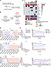 Figure 6
Phase I/II clinical evaluation of XY0206 monotherapy in patients with R/R AML. (A) Consolidated Standards of Reporting Trials (CONSORT) diagram of the single-center phase I/II trial. (B) Overview of treatment efficacy in patients with FLT3i-exposed R/R AML who received XY0206 monotherapy, with a median treatment duration of 34 days. (C) Baseline clinical and molecular characteristics of patients with FLT3i-exposed R/R AML enrolled in the trial, including age, pathogenic gene mutations, and bone marrow blast percentage. (D) Representative case illustrating bone marrow blast percentage before and after XY0206 treatment. Prior regimens included azacitidine (AZA), homoharringtonine plus cytarabine (HA), daunorubicin plus cytarabine (DA), sorafenib (Sora), venetoclax (Ven), homoharringtonine plus cytarabine plus daunorubicin (HAD), intermediate-dose cytarabine (IDAC), and decitabine (DAC). Patients 13 and 14 proceeded to HSCT. (E) Dynamic changes in bone marrow (BM) and peripheral blood (PB) blast percentages and hematologic parameters, including hemoglobin (HGB), platelet count (PLT), WBC count, and RBC count, during XY0206 treatment in 3 representative patients. Measurements were collected at baseline and multiple time points after treatment initiation.
During dose escalation, XY0206 was initially administered at planned once-daily dose levels of 12.5, 25, 50, and 62.5 mg. The 50 mg once-daily dose was associated with reduced tolerability, and 1 patient in this cohort experienced a protocol-defined dose-limiting toxicity of colitis. An alternative 25 mg twice-daily schedule was also explored; however, this schedule did not meaningfully improve the tolerability profile. Therefore, an intermediate 37.5 mg once-daily dose level was evaluated to optimize the balance among tolerability, pharmacokinetic exposure, and preliminary antileukemic activity. Pharmacokinetic analyses demonstrated that XY0206 achieved approximately dose-proportional systemic exposure across the evaluated dose range of 12.5 to 62.5 mg. After a single oral dose, the median time to maximum serum concentration (Tmax) ranged from 2.00 to 6.00 hours, with a modest prolongation to 2.00 to 8.00 hours after repeated dosing (Table 4). At steady state, systemic exposure, as assessed by AUC0–t, increased by approximately 1.51- to 3.55-fold relative to single-dose administration, and steady-state peak concentrations increased by 1.55- to 3.17-fold, indicating moderate accumulation with repeated dosing (Table 5).
Within this exposure range, XY0206 demonstrated clinically relevant antileukemic activity. According to the 2022 European LeukemiaNet (ELN) response criteria, 2 patients (5.7%) achieved complete remission, 3 patients (8.6%) achieved CRi, and 11 patients (31.4%) achieved an MLFS, resulting in an overall CRc rate, defined as the sum of CR, CRi, and MLFS, of 45.7% (n = 16 of 35). Among patients with FLT3-ITD mutations, the CRc rate was 60.0% (n = 15 of 25). The median duration of response for patients achieving CRc was 45.5 days (range, 10–448 days). The median duration of XY0206 therapy across the full cohort was 57 days (range, 16–506 days). Patient-level treatment exposure, response duration, reason for treatment discontinuation, subsequent HSCT, and grade ≥3 adverse events are summarized in Supplemental Table 7.
Treatment-emergent adverse events (TEAEs) of grade 3 or higher were predominantly hematologic and consistent with the underlying disease, including leukopenia, neutropenia, thrombocytopenia, and anemia, and prior treatment history. Nonhematologic grade ≥3 events were less frequent and included infectious pneumonia, hypokalemia, hypertension, hypertriglyceridemia, rash, bone pain, diarrhea, and colitis. Detailed safety data by dose group are provided in Table 6 and Supplemental Table 8. Reasons for treatment discontinuation included lack of response, relapse after initial response, TEAEs, transition to HSCT, loss to follow-up, voluntary withdrawal, and poor compliance. Achievement of MLFS did not require protocol-mandated interruption or dose reduction of XY0206; treatment continued unless predefined discontinuation criteria were met or patients proceeded to subsequent therapy.
Consistent with these clinical responses, reductions in bone marrow blast percentages were observed in a subset of patients with R/R AML, independent of prior chemotherapy exposure (Figure 6, D and E, and Supplemental Figure 7). Because this phase I/II trial enrolled a broader R/R AML population rather than specifically selecting patients with prior FLT3i exposure, prior FLT3i use was present only in a subset of patients and reflected heterogeneous pre-enrollment treatment histories. Although 10 patients had prior exposure to FLT3i-based therapies, 1 had FLT3-TKD mutation without FLT3-ITD at enrollment and 1 discontinued XY0206 before completing the first treatment cycle. Thus, 8 patients with FLT3-ITD mutations who had received at least 1 prior cycle of FLT3i-based therapy were included in the post-FLT3i efficacy analysis. Among these 8 patients, 3 achieved CRc following XY0206 treatment, and 2 of these patients subsequently proceeded to HSCT (Figure 6, B–E). These observations suggest that XY0206 can achieve sustained systemic exposure and meaningful antileukemic activity in heavily pretreated AML, including the evaluable post-FLT3i FLT3-ITD subgroup.


Copyright © 2026 American Society for Clinical Investigation
ISSN: 0021-9738 (print), 1558-8238 (online)