
Advertisement
Research ArticleBone biologyClinical ResearchGenetics
Open Access |  10.1172/JCI199048
MEK inhibitor mirdametinib promotes fracture healing in osteofibrous dysplasia RASopathy
Aysha B. Khalid,1 Kristin Denton,1 Nandina Paria,1 Ila Oxendine,1 Meghan Wassell,1 Reuel Cornelia,1 Sasidhar Uppuganti,2 Jeffry S. Nyman,2,3,4 G. Jayashree Jagadeesh,5 Carlos R. Ferreira,5 Simon J. Conway,6 Robert E. Hammer,7 John Ritter,7 Mylinh Nguyen,7 David A. Podeszwa,8,9 Laura J. Klesse,10,11,12 Carol A. Wise,1,9,10,13 and Jonathan J. Rios1,9,10,11,13
1Center for Translational Research, Scottish Rite for Children, Dallas, Texas, USA.
2Department of Orthopaedic Surgery, Vanderbilt University Medical Center, Nashville, Tennessee, USA.
3Department of Biomedical Engineering, Vanderbilt University, Nashville, Tennessee, USA.
4United States Department of Veterans Affairs, Tennessee Valley Healthcare System, Nashville, Tennessee, USA.
5Unit on Skeletal Genomics, Eunice Kennedy Shriver National Institute of Child Health and Human Development, NIH, Bethesda, Maryland, USA.
6Herman B. Wells Center for Pediatric Research, Indiana University School of Medicine, Indianapolis, Indiana, USA.
7Department of Biochemistry, UT Southwestern Medical Center, Dallas, Texas, USA.
8Department of Orthopaedics, Scottish Rite for Children, Dallas, Texas, USA.
9Department of Orthopaedic Surgery,
10Department of Pediatrics, and
11Simmons Comprehensive Cancer Center, UT Southwestern Medical Center, Dallas, Texas, USA.
12Department of Pediatrics, Children’s Health, Dallas, Texas, USA.
13McDermott Center for Human Growth and Development, UT Southwestern Medical Center, Dallas, Texas, USA.
Address correspondence to: Jonathan J. Rios, Scottish Rite for Children, 2222 Welborn Street, Dallas, Texas 75219, USA. Phone: 214.559.7877; Email: Jonathan.Rios@tsrh.org.
Find articles by Khalid, A. in: PubMed | Google Scholar
1Center for Translational Research, Scottish Rite for Children, Dallas, Texas, USA.
2Department of Orthopaedic Surgery, Vanderbilt University Medical Center, Nashville, Tennessee, USA.
3Department of Biomedical Engineering, Vanderbilt University, Nashville, Tennessee, USA.
4United States Department of Veterans Affairs, Tennessee Valley Healthcare System, Nashville, Tennessee, USA.
5Unit on Skeletal Genomics, Eunice Kennedy Shriver National Institute of Child Health and Human Development, NIH, Bethesda, Maryland, USA.
6Herman B. Wells Center for Pediatric Research, Indiana University School of Medicine, Indianapolis, Indiana, USA.
7Department of Biochemistry, UT Southwestern Medical Center, Dallas, Texas, USA.
8Department of Orthopaedics, Scottish Rite for Children, Dallas, Texas, USA.
9Department of Orthopaedic Surgery,
10Department of Pediatrics, and
11Simmons Comprehensive Cancer Center, UT Southwestern Medical Center, Dallas, Texas, USA.
12Department of Pediatrics, Children’s Health, Dallas, Texas, USA.
13McDermott Center for Human Growth and Development, UT Southwestern Medical Center, Dallas, Texas, USA.
Address correspondence to: Jonathan J. Rios, Scottish Rite for Children, 2222 Welborn Street, Dallas, Texas 75219, USA. Phone: 214.559.7877; Email: Jonathan.Rios@tsrh.org.
Find articles by Denton, K. in: PubMed | Google Scholar
1Center for Translational Research, Scottish Rite for Children, Dallas, Texas, USA.
2Department of Orthopaedic Surgery, Vanderbilt University Medical Center, Nashville, Tennessee, USA.
3Department of Biomedical Engineering, Vanderbilt University, Nashville, Tennessee, USA.
4United States Department of Veterans Affairs, Tennessee Valley Healthcare System, Nashville, Tennessee, USA.
5Unit on Skeletal Genomics, Eunice Kennedy Shriver National Institute of Child Health and Human Development, NIH, Bethesda, Maryland, USA.
6Herman B. Wells Center for Pediatric Research, Indiana University School of Medicine, Indianapolis, Indiana, USA.
7Department of Biochemistry, UT Southwestern Medical Center, Dallas, Texas, USA.
8Department of Orthopaedics, Scottish Rite for Children, Dallas, Texas, USA.
9Department of Orthopaedic Surgery,
10Department of Pediatrics, and
11Simmons Comprehensive Cancer Center, UT Southwestern Medical Center, Dallas, Texas, USA.
12Department of Pediatrics, Children’s Health, Dallas, Texas, USA.
13McDermott Center for Human Growth and Development, UT Southwestern Medical Center, Dallas, Texas, USA.
Address correspondence to: Jonathan J. Rios, Scottish Rite for Children, 2222 Welborn Street, Dallas, Texas 75219, USA. Phone: 214.559.7877; Email: Jonathan.Rios@tsrh.org.
Find articles by Paria, N. in: PubMed | Google Scholar
1Center for Translational Research, Scottish Rite for Children, Dallas, Texas, USA.
2Department of Orthopaedic Surgery, Vanderbilt University Medical Center, Nashville, Tennessee, USA.
3Department of Biomedical Engineering, Vanderbilt University, Nashville, Tennessee, USA.
4United States Department of Veterans Affairs, Tennessee Valley Healthcare System, Nashville, Tennessee, USA.
5Unit on Skeletal Genomics, Eunice Kennedy Shriver National Institute of Child Health and Human Development, NIH, Bethesda, Maryland, USA.
6Herman B. Wells Center for Pediatric Research, Indiana University School of Medicine, Indianapolis, Indiana, USA.
7Department of Biochemistry, UT Southwestern Medical Center, Dallas, Texas, USA.
8Department of Orthopaedics, Scottish Rite for Children, Dallas, Texas, USA.
9Department of Orthopaedic Surgery,
10Department of Pediatrics, and
11Simmons Comprehensive Cancer Center, UT Southwestern Medical Center, Dallas, Texas, USA.
12Department of Pediatrics, Children’s Health, Dallas, Texas, USA.
13McDermott Center for Human Growth and Development, UT Southwestern Medical Center, Dallas, Texas, USA.
Address correspondence to: Jonathan J. Rios, Scottish Rite for Children, 2222 Welborn Street, Dallas, Texas 75219, USA. Phone: 214.559.7877; Email: Jonathan.Rios@tsrh.org.
Find articles by Oxendine, I. in: PubMed | Google Scholar
1Center for Translational Research, Scottish Rite for Children, Dallas, Texas, USA.
2Department of Orthopaedic Surgery, Vanderbilt University Medical Center, Nashville, Tennessee, USA.
3Department of Biomedical Engineering, Vanderbilt University, Nashville, Tennessee, USA.
4United States Department of Veterans Affairs, Tennessee Valley Healthcare System, Nashville, Tennessee, USA.
5Unit on Skeletal Genomics, Eunice Kennedy Shriver National Institute of Child Health and Human Development, NIH, Bethesda, Maryland, USA.
6Herman B. Wells Center for Pediatric Research, Indiana University School of Medicine, Indianapolis, Indiana, USA.
7Department of Biochemistry, UT Southwestern Medical Center, Dallas, Texas, USA.
8Department of Orthopaedics, Scottish Rite for Children, Dallas, Texas, USA.
9Department of Orthopaedic Surgery,
10Department of Pediatrics, and
11Simmons Comprehensive Cancer Center, UT Southwestern Medical Center, Dallas, Texas, USA.
12Department of Pediatrics, Children’s Health, Dallas, Texas, USA.
13McDermott Center for Human Growth and Development, UT Southwestern Medical Center, Dallas, Texas, USA.
Address correspondence to: Jonathan J. Rios, Scottish Rite for Children, 2222 Welborn Street, Dallas, Texas 75219, USA. Phone: 214.559.7877; Email: Jonathan.Rios@tsrh.org.
Find articles by Wassell, M. in: PubMed | Google Scholar
1Center for Translational Research, Scottish Rite for Children, Dallas, Texas, USA.
2Department of Orthopaedic Surgery, Vanderbilt University Medical Center, Nashville, Tennessee, USA.
3Department of Biomedical Engineering, Vanderbilt University, Nashville, Tennessee, USA.
4United States Department of Veterans Affairs, Tennessee Valley Healthcare System, Nashville, Tennessee, USA.
5Unit on Skeletal Genomics, Eunice Kennedy Shriver National Institute of Child Health and Human Development, NIH, Bethesda, Maryland, USA.
6Herman B. Wells Center for Pediatric Research, Indiana University School of Medicine, Indianapolis, Indiana, USA.
7Department of Biochemistry, UT Southwestern Medical Center, Dallas, Texas, USA.
8Department of Orthopaedics, Scottish Rite for Children, Dallas, Texas, USA.
9Department of Orthopaedic Surgery,
10Department of Pediatrics, and
11Simmons Comprehensive Cancer Center, UT Southwestern Medical Center, Dallas, Texas, USA.
12Department of Pediatrics, Children’s Health, Dallas, Texas, USA.
13McDermott Center for Human Growth and Development, UT Southwestern Medical Center, Dallas, Texas, USA.
Address correspondence to: Jonathan J. Rios, Scottish Rite for Children, 2222 Welborn Street, Dallas, Texas 75219, USA. Phone: 214.559.7877; Email: Jonathan.Rios@tsrh.org.
Find articles by Cornelia, R. in: PubMed | Google Scholar
1Center for Translational Research, Scottish Rite for Children, Dallas, Texas, USA.
2Department of Orthopaedic Surgery, Vanderbilt University Medical Center, Nashville, Tennessee, USA.
3Department of Biomedical Engineering, Vanderbilt University, Nashville, Tennessee, USA.
4United States Department of Veterans Affairs, Tennessee Valley Healthcare System, Nashville, Tennessee, USA.
5Unit on Skeletal Genomics, Eunice Kennedy Shriver National Institute of Child Health and Human Development, NIH, Bethesda, Maryland, USA.
6Herman B. Wells Center for Pediatric Research, Indiana University School of Medicine, Indianapolis, Indiana, USA.
7Department of Biochemistry, UT Southwestern Medical Center, Dallas, Texas, USA.
8Department of Orthopaedics, Scottish Rite for Children, Dallas, Texas, USA.
9Department of Orthopaedic Surgery,
10Department of Pediatrics, and
11Simmons Comprehensive Cancer Center, UT Southwestern Medical Center, Dallas, Texas, USA.
12Department of Pediatrics, Children’s Health, Dallas, Texas, USA.
13McDermott Center for Human Growth and Development, UT Southwestern Medical Center, Dallas, Texas, USA.
Address correspondence to: Jonathan J. Rios, Scottish Rite for Children, 2222 Welborn Street, Dallas, Texas 75219, USA. Phone: 214.559.7877; Email: Jonathan.Rios@tsrh.org.
Find articles by Uppuganti, S. in: PubMed | Google Scholar
1Center for Translational Research, Scottish Rite for Children, Dallas, Texas, USA.
2Department of Orthopaedic Surgery, Vanderbilt University Medical Center, Nashville, Tennessee, USA.
3Department of Biomedical Engineering, Vanderbilt University, Nashville, Tennessee, USA.
4United States Department of Veterans Affairs, Tennessee Valley Healthcare System, Nashville, Tennessee, USA.
5Unit on Skeletal Genomics, Eunice Kennedy Shriver National Institute of Child Health and Human Development, NIH, Bethesda, Maryland, USA.
6Herman B. Wells Center for Pediatric Research, Indiana University School of Medicine, Indianapolis, Indiana, USA.
7Department of Biochemistry, UT Southwestern Medical Center, Dallas, Texas, USA.
8Department of Orthopaedics, Scottish Rite for Children, Dallas, Texas, USA.
9Department of Orthopaedic Surgery,
10Department of Pediatrics, and
11Simmons Comprehensive Cancer Center, UT Southwestern Medical Center, Dallas, Texas, USA.
12Department of Pediatrics, Children’s Health, Dallas, Texas, USA.
13McDermott Center for Human Growth and Development, UT Southwestern Medical Center, Dallas, Texas, USA.
Address correspondence to: Jonathan J. Rios, Scottish Rite for Children, 2222 Welborn Street, Dallas, Texas 75219, USA. Phone: 214.559.7877; Email: Jonathan.Rios@tsrh.org.
Find articles by Nyman, J. in: PubMed | Google Scholar
1Center for Translational Research, Scottish Rite for Children, Dallas, Texas, USA.
2Department of Orthopaedic Surgery, Vanderbilt University Medical Center, Nashville, Tennessee, USA.
3Department of Biomedical Engineering, Vanderbilt University, Nashville, Tennessee, USA.
4United States Department of Veterans Affairs, Tennessee Valley Healthcare System, Nashville, Tennessee, USA.
5Unit on Skeletal Genomics, Eunice Kennedy Shriver National Institute of Child Health and Human Development, NIH, Bethesda, Maryland, USA.
6Herman B. Wells Center for Pediatric Research, Indiana University School of Medicine, Indianapolis, Indiana, USA.
7Department of Biochemistry, UT Southwestern Medical Center, Dallas, Texas, USA.
8Department of Orthopaedics, Scottish Rite for Children, Dallas, Texas, USA.
9Department of Orthopaedic Surgery,
10Department of Pediatrics, and
11Simmons Comprehensive Cancer Center, UT Southwestern Medical Center, Dallas, Texas, USA.
12Department of Pediatrics, Children’s Health, Dallas, Texas, USA.
13McDermott Center for Human Growth and Development, UT Southwestern Medical Center, Dallas, Texas, USA.
Address correspondence to: Jonathan J. Rios, Scottish Rite for Children, 2222 Welborn Street, Dallas, Texas 75219, USA. Phone: 214.559.7877; Email: Jonathan.Rios@tsrh.org.
Find articles by Jagadeesh, G. in: PubMed | Google Scholar
1Center for Translational Research, Scottish Rite for Children, Dallas, Texas, USA.
2Department of Orthopaedic Surgery, Vanderbilt University Medical Center, Nashville, Tennessee, USA.
3Department of Biomedical Engineering, Vanderbilt University, Nashville, Tennessee, USA.
4United States Department of Veterans Affairs, Tennessee Valley Healthcare System, Nashville, Tennessee, USA.
5Unit on Skeletal Genomics, Eunice Kennedy Shriver National Institute of Child Health and Human Development, NIH, Bethesda, Maryland, USA.
6Herman B. Wells Center for Pediatric Research, Indiana University School of Medicine, Indianapolis, Indiana, USA.
7Department of Biochemistry, UT Southwestern Medical Center, Dallas, Texas, USA.
8Department of Orthopaedics, Scottish Rite for Children, Dallas, Texas, USA.
9Department of Orthopaedic Surgery,
10Department of Pediatrics, and
11Simmons Comprehensive Cancer Center, UT Southwestern Medical Center, Dallas, Texas, USA.
12Department of Pediatrics, Children’s Health, Dallas, Texas, USA.
13McDermott Center for Human Growth and Development, UT Southwestern Medical Center, Dallas, Texas, USA.
Address correspondence to: Jonathan J. Rios, Scottish Rite for Children, 2222 Welborn Street, Dallas, Texas 75219, USA. Phone: 214.559.7877; Email: Jonathan.Rios@tsrh.org.
Find articles by Ferreira, C. in: PubMed | Google Scholar
1Center for Translational Research, Scottish Rite for Children, Dallas, Texas, USA.
2Department of Orthopaedic Surgery, Vanderbilt University Medical Center, Nashville, Tennessee, USA.
3Department of Biomedical Engineering, Vanderbilt University, Nashville, Tennessee, USA.
4United States Department of Veterans Affairs, Tennessee Valley Healthcare System, Nashville, Tennessee, USA.
5Unit on Skeletal Genomics, Eunice Kennedy Shriver National Institute of Child Health and Human Development, NIH, Bethesda, Maryland, USA.
6Herman B. Wells Center for Pediatric Research, Indiana University School of Medicine, Indianapolis, Indiana, USA.
7Department of Biochemistry, UT Southwestern Medical Center, Dallas, Texas, USA.
8Department of Orthopaedics, Scottish Rite for Children, Dallas, Texas, USA.
9Department of Orthopaedic Surgery,
10Department of Pediatrics, and
11Simmons Comprehensive Cancer Center, UT Southwestern Medical Center, Dallas, Texas, USA.
12Department of Pediatrics, Children’s Health, Dallas, Texas, USA.
13McDermott Center for Human Growth and Development, UT Southwestern Medical Center, Dallas, Texas, USA.
Address correspondence to: Jonathan J. Rios, Scottish Rite for Children, 2222 Welborn Street, Dallas, Texas 75219, USA. Phone: 214.559.7877; Email: Jonathan.Rios@tsrh.org.
Find articles by
Conway, S.
in:
PubMed
|
Google Scholar
|

1Center for Translational Research, Scottish Rite for Children, Dallas, Texas, USA.
2Department of Orthopaedic Surgery, Vanderbilt University Medical Center, Nashville, Tennessee, USA.
3Department of Biomedical Engineering, Vanderbilt University, Nashville, Tennessee, USA.
4United States Department of Veterans Affairs, Tennessee Valley Healthcare System, Nashville, Tennessee, USA.
5Unit on Skeletal Genomics, Eunice Kennedy Shriver National Institute of Child Health and Human Development, NIH, Bethesda, Maryland, USA.
6Herman B. Wells Center for Pediatric Research, Indiana University School of Medicine, Indianapolis, Indiana, USA.
7Department of Biochemistry, UT Southwestern Medical Center, Dallas, Texas, USA.
8Department of Orthopaedics, Scottish Rite for Children, Dallas, Texas, USA.
9Department of Orthopaedic Surgery,
10Department of Pediatrics, and
11Simmons Comprehensive Cancer Center, UT Southwestern Medical Center, Dallas, Texas, USA.
12Department of Pediatrics, Children’s Health, Dallas, Texas, USA.
13McDermott Center for Human Growth and Development, UT Southwestern Medical Center, Dallas, Texas, USA.
Address correspondence to: Jonathan J. Rios, Scottish Rite for Children, 2222 Welborn Street, Dallas, Texas 75219, USA. Phone: 214.559.7877; Email: Jonathan.Rios@tsrh.org.
Find articles by Hammer, R. in: PubMed | Google Scholar
1Center for Translational Research, Scottish Rite for Children, Dallas, Texas, USA.
2Department of Orthopaedic Surgery, Vanderbilt University Medical Center, Nashville, Tennessee, USA.
3Department of Biomedical Engineering, Vanderbilt University, Nashville, Tennessee, USA.
4United States Department of Veterans Affairs, Tennessee Valley Healthcare System, Nashville, Tennessee, USA.
5Unit on Skeletal Genomics, Eunice Kennedy Shriver National Institute of Child Health and Human Development, NIH, Bethesda, Maryland, USA.
6Herman B. Wells Center for Pediatric Research, Indiana University School of Medicine, Indianapolis, Indiana, USA.
7Department of Biochemistry, UT Southwestern Medical Center, Dallas, Texas, USA.
8Department of Orthopaedics, Scottish Rite for Children, Dallas, Texas, USA.
9Department of Orthopaedic Surgery,
10Department of Pediatrics, and
11Simmons Comprehensive Cancer Center, UT Southwestern Medical Center, Dallas, Texas, USA.
12Department of Pediatrics, Children’s Health, Dallas, Texas, USA.
13McDermott Center for Human Growth and Development, UT Southwestern Medical Center, Dallas, Texas, USA.
Address correspondence to: Jonathan J. Rios, Scottish Rite for Children, 2222 Welborn Street, Dallas, Texas 75219, USA. Phone: 214.559.7877; Email: Jonathan.Rios@tsrh.org.
Find articles by Ritter, J. in: PubMed | Google Scholar
1Center for Translational Research, Scottish Rite for Children, Dallas, Texas, USA.
2Department of Orthopaedic Surgery, Vanderbilt University Medical Center, Nashville, Tennessee, USA.
3Department of Biomedical Engineering, Vanderbilt University, Nashville, Tennessee, USA.
4United States Department of Veterans Affairs, Tennessee Valley Healthcare System, Nashville, Tennessee, USA.
5Unit on Skeletal Genomics, Eunice Kennedy Shriver National Institute of Child Health and Human Development, NIH, Bethesda, Maryland, USA.
6Herman B. Wells Center for Pediatric Research, Indiana University School of Medicine, Indianapolis, Indiana, USA.
7Department of Biochemistry, UT Southwestern Medical Center, Dallas, Texas, USA.
8Department of Orthopaedics, Scottish Rite for Children, Dallas, Texas, USA.
9Department of Orthopaedic Surgery,
10Department of Pediatrics, and
11Simmons Comprehensive Cancer Center, UT Southwestern Medical Center, Dallas, Texas, USA.
12Department of Pediatrics, Children’s Health, Dallas, Texas, USA.
13McDermott Center for Human Growth and Development, UT Southwestern Medical Center, Dallas, Texas, USA.
Address correspondence to: Jonathan J. Rios, Scottish Rite for Children, 2222 Welborn Street, Dallas, Texas 75219, USA. Phone: 214.559.7877; Email: Jonathan.Rios@tsrh.org.
Find articles by Nguyen, M. in: PubMed | Google Scholar
1Center for Translational Research, Scottish Rite for Children, Dallas, Texas, USA.
2Department of Orthopaedic Surgery, Vanderbilt University Medical Center, Nashville, Tennessee, USA.
3Department of Biomedical Engineering, Vanderbilt University, Nashville, Tennessee, USA.
4United States Department of Veterans Affairs, Tennessee Valley Healthcare System, Nashville, Tennessee, USA.
5Unit on Skeletal Genomics, Eunice Kennedy Shriver National Institute of Child Health and Human Development, NIH, Bethesda, Maryland, USA.
6Herman B. Wells Center for Pediatric Research, Indiana University School of Medicine, Indianapolis, Indiana, USA.
7Department of Biochemistry, UT Southwestern Medical Center, Dallas, Texas, USA.
8Department of Orthopaedics, Scottish Rite for Children, Dallas, Texas, USA.
9Department of Orthopaedic Surgery,
10Department of Pediatrics, and
11Simmons Comprehensive Cancer Center, UT Southwestern Medical Center, Dallas, Texas, USA.
12Department of Pediatrics, Children’s Health, Dallas, Texas, USA.
13McDermott Center for Human Growth and Development, UT Southwestern Medical Center, Dallas, Texas, USA.
Address correspondence to: Jonathan J. Rios, Scottish Rite for Children, 2222 Welborn Street, Dallas, Texas 75219, USA. Phone: 214.559.7877; Email: Jonathan.Rios@tsrh.org.
Find articles by Podeszwa, D. in: PubMed | Google Scholar
1Center for Translational Research, Scottish Rite for Children, Dallas, Texas, USA.
2Department of Orthopaedic Surgery, Vanderbilt University Medical Center, Nashville, Tennessee, USA.
3Department of Biomedical Engineering, Vanderbilt University, Nashville, Tennessee, USA.
4United States Department of Veterans Affairs, Tennessee Valley Healthcare System, Nashville, Tennessee, USA.
5Unit on Skeletal Genomics, Eunice Kennedy Shriver National Institute of Child Health and Human Development, NIH, Bethesda, Maryland, USA.
6Herman B. Wells Center for Pediatric Research, Indiana University School of Medicine, Indianapolis, Indiana, USA.
7Department of Biochemistry, UT Southwestern Medical Center, Dallas, Texas, USA.
8Department of Orthopaedics, Scottish Rite for Children, Dallas, Texas, USA.
9Department of Orthopaedic Surgery,
10Department of Pediatrics, and
11Simmons Comprehensive Cancer Center, UT Southwestern Medical Center, Dallas, Texas, USA.
12Department of Pediatrics, Children’s Health, Dallas, Texas, USA.
13McDermott Center for Human Growth and Development, UT Southwestern Medical Center, Dallas, Texas, USA.
Address correspondence to: Jonathan J. Rios, Scottish Rite for Children, 2222 Welborn Street, Dallas, Texas 75219, USA. Phone: 214.559.7877; Email: Jonathan.Rios@tsrh.org.
Find articles by Klesse, L. in: PubMed | Google Scholar
1Center for Translational Research, Scottish Rite for Children, Dallas, Texas, USA.
2Department of Orthopaedic Surgery, Vanderbilt University Medical Center, Nashville, Tennessee, USA.
3Department of Biomedical Engineering, Vanderbilt University, Nashville, Tennessee, USA.
4United States Department of Veterans Affairs, Tennessee Valley Healthcare System, Nashville, Tennessee, USA.
5Unit on Skeletal Genomics, Eunice Kennedy Shriver National Institute of Child Health and Human Development, NIH, Bethesda, Maryland, USA.
6Herman B. Wells Center for Pediatric Research, Indiana University School of Medicine, Indianapolis, Indiana, USA.
7Department of Biochemistry, UT Southwestern Medical Center, Dallas, Texas, USA.
8Department of Orthopaedics, Scottish Rite for Children, Dallas, Texas, USA.
9Department of Orthopaedic Surgery,
10Department of Pediatrics, and
11Simmons Comprehensive Cancer Center, UT Southwestern Medical Center, Dallas, Texas, USA.
12Department of Pediatrics, Children’s Health, Dallas, Texas, USA.
13McDermott Center for Human Growth and Development, UT Southwestern Medical Center, Dallas, Texas, USA.
Address correspondence to: Jonathan J. Rios, Scottish Rite for Children, 2222 Welborn Street, Dallas, Texas 75219, USA. Phone: 214.559.7877; Email: Jonathan.Rios@tsrh.org.
Find articles by
Wise, C.
in:
PubMed
|
Google Scholar
|
1Center for Translational Research, Scottish Rite for Children, Dallas, Texas, USA.
2Department of Orthopaedic Surgery, Vanderbilt University Medical Center, Nashville, Tennessee, USA.
3Department of Biomedical Engineering, Vanderbilt University, Nashville, Tennessee, USA.
4United States Department of Veterans Affairs, Tennessee Valley Healthcare System, Nashville, Tennessee, USA.
5Unit on Skeletal Genomics, Eunice Kennedy Shriver National Institute of Child Health and Human Development, NIH, Bethesda, Maryland, USA.
6Herman B. Wells Center for Pediatric Research, Indiana University School of Medicine, Indianapolis, Indiana, USA.
7Department of Biochemistry, UT Southwestern Medical Center, Dallas, Texas, USA.
8Department of Orthopaedics, Scottish Rite for Children, Dallas, Texas, USA.
9Department of Orthopaedic Surgery,
10Department of Pediatrics, and
11Simmons Comprehensive Cancer Center, UT Southwestern Medical Center, Dallas, Texas, USA.
12Department of Pediatrics, Children’s Health, Dallas, Texas, USA.
13McDermott Center for Human Growth and Development, UT Southwestern Medical Center, Dallas, Texas, USA.
Address correspondence to: Jonathan J. Rios, Scottish Rite for Children, 2222 Welborn Street, Dallas, Texas 75219, USA. Phone: 214.559.7877; Email: Jonathan.Rios@tsrh.org.
Find articles by Rios, J. in: PubMed | Google Scholar
Published February 26, 2026 - More info
J Clin Invest. 2026;136(9):e199048. https://doi.org/10.1172/JCI199048.
© 2026 Khalid et al. This work is licensed under the Creative Commons Attribution 4.0 International License. To view a copy of this license, visit http://creativecommons.org/licenses/by/4.0/.
Received: August 12, 2025; Accepted: February 19, 2026

-
Results
MetΔJMD expression promotes ERK pathway activation in skeletal progenitor cells. To investigate the effect of MetΔJMD expression on skeletal progenitor cell differentiation and skeletal development, we deleted the JMD-encoding Met exon 15 in mice using CRISPR/Cas9 (Supplemental Figure 1A; supplemental material available online with this article; https://doi.org/10.1172/JCI199048DS1). Heterozygous Met+/ΔJMD mice (herein referred to as MetΔJMD) were viable and fertile with no overt phenotype. Similar to mice harboring constitutive activating Met kinase domain variants (16), homozygous MetΔJMD/ΔJMD mice were embryonically lethal. Ligand-dependent gain of function due to loss of the MET JMD in lung cancer cell lines results in prolonged MAPK pathway activation following stimulation with HGF (17). Therefore, we isolated bone marrow stromal cells (BMSCs) from adult control and MetΔJMD mice, treated the cells with HGF, and quantified relative ERK activation and normalization. In both control and MetΔJMD BMSCs, the ERK pathway was quickly activated in response to HGF, although normalization following stimulation was significantly delayed in MetΔJMD BMSCs compared with the control (Figure 1, A and B). Thus, MetΔJMD mice harbor a heterozygous germline deletion of the endogenous Met JMD-encoding exon leading to ligand-dependent gain of function in MAPK signaling.
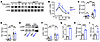 Figure 1
Characterization of MetΔJMD osteoprogenitor cells. (A and B) Representative Western blot time course (A) and quantification (B) of ERK activation following HGF stimulation of BMSCs from control (black; n = 3) and MetΔJMD (blue; n = 5) mice. Statistical analysis was conducted by 2-way ANOVA with Dunnett’s multiple-test correction including only 5- and 15-minute time points. (C and D) Relative (Rel.) expression (exp.) of Met (C) and Hgf (D) in BMSCs and PECs from control (n = 8–9) and MetΔJMD (n = 7–9) mice. Statistically significant differences were determined by 2-way ANOVA with Šidák’s multiple-test correction. (E and F) Representative Western blot (E) and quantification (F) of ERK pathway activation of serum-starved BMSCs and PECs from control (n = 5) and MetΔJMD (n = 5) mice. Statistically significant differences were determined by 2-way ANOVA with Šidák’s multiple-test correction. (G) Cell count quantification of PECs from control (n = 4) and MetΔJMD (n = 4) mice. Statistically significant differences were determined by t test. (H and I) Relative expression of the mesenchymal adhesion genes Ncad (H) and Vim (I) in PECs from control (n = 6) and MetΔJMD (n = 6) mice. (G–I) Statistically significant differences were determined by 2-tailed t test. RT-qPCR values from cultured cells and other quantification data are presented as the mean ± SEM. *P < 0.05, ***P < 0.001, and ****P < 0.0001.
OFD lesions occur at the periosteal surface of bone, and Met expression was previously detected at the periosteal surface of mouse embryonic long bones (4). Therefore, we cultured leptin receptor–expressing (LepR-expressing) BMSCs and periostin-expressing (Postn-expressing) periosteal explant cells (PECs) from control and MetΔJMD mice (Supplemental Figure 1, B and C) and tested expression of Met and Hgf in both cell types. Met expression was significantly higher in PECs compared with BMSCs (P < 0.0001). Compared with control cells, we found that Met expression was higher in MetΔJMD BMSCs and PECs (Figure 1C). This difference was predominantly due to the relative overexpression of the MetΔJMD allele (Supplemental Figure 1, D and E), implicating, for the first time to our knowledge, an allele-specific feedback mechanism associated with the MetΔJMD allele. While Hgf expression was modestly elevated in MetΔJMD BMSCs compared with the control, Hgf expression was significantly higher in PECs from both control and MetΔJMD mice (Figure 1D). Consistent with the higher Met and Hgf expression in PECs, ERK activation was significantly higher in serum-starved control and MetΔJMD PECs compared with BMSCs (Figure 1, E and F). We observed no differences in AKT/mTOR pathway activation, as assayed by phosphorylated S6 (p-S6) (Supplemental Figure 1F). These results implicate a molecular basis for the periosteal localization of OFD lesions observed in patients.
Last, MET activation enhances tumor growth and invasion, in part, through promotion of epithelial-mesenchymal transition (18–21). Therefore, we tested cell adhesion and the expression of mesenchymal cell adhesion–associated genes in control and MetΔJMD PECs. Compared with control cells, MetΔJMD PECs demonstrated significantly greater adherence in culture concomitant with significantly higher expression of the cell adhesion genes N-cadherin (Ncad) and vimentin (Vim) (Figure 1, G–I), although no difference in cell proliferation was observed (Supplemental Figure 1, G and H). Taken together, these results demonstrated molecular and cellular defects in skeletal progenitor cells from MetΔJMD mice associated with ERK pathway activation.
Impaired progenitor cell differentiation and skeletal development in MetΔJMD mice. In osteoprogenitor cells, MAPK homeostasis is essential for proper osteogenic differentiation and mineralization. Preclinical cellular and rodent models of skeletal RASopathies, such as in neurofibromatosis type 1 (NF1) (22, 23) and others (24), have implicated impaired osteogenic differentiation and/or mineralization in the pathogenesis of disease. Therefore, we tested osteogenic differentiation of MetΔJMD PECs by measuring the relative expression of early and late osteogenic genes following differentiation, including integrin-binding sialoprotein (Ibsp), RUNX family transcription factor 2 (Runx2), osterix (Osx), osteocalcin (Ocn), and dentin matrix acidic phosphoprotein 1 (Dmp1). While expression of all osteogenic genes was significantly increased in control PECs following differentiation, expression of these genes was mostly unchanged in MetΔJMD PECs, suggesting impaired osteogenic differentiation in MetΔJMD PECs (Figure 2, A and B, and Supplemental Figure 2, A–C). Consistent with this, mineralization measured by alizarin red staining was significantly reduced following differentiation of MetΔJMD PECs compared with the control (Figure 2, C and D). These results demonstrate that expression of the MetΔJMD allele impaired osteogenic differentiation and mineralization of periosteal osteoprogenitor cells, potentially due to MAPK activation.
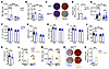 Figure 2
Impaired osteogenic differentiation of PECs from MetΔJMD mice. (A and B) Relative expression of the osteogenic genes Ibsp (A) and Dmp1 (B) in control (n = 4–9) and MetΔJMD (n = 4–9) PECs prior to (undifferentiated [Undiff.], black) and following osteogenic differentiation (Osteo., blue). Statistically significant differences were determined by 2-way ANOVA with Šidák’s multiple-test correction. (C and D) Representative alizarin red and crystal violet (CV) staining (C) and quantification (D) following osteogenic differentiation of control (Met+/+, n = 6) and MetΔJMD (n = 6) PECs. Statistically significant differences were determined by t test. (E–J) Quantification of cortical porosity (E) (Ct. Por.), cortical thickness (F) (Ct. Th.), pMOI (G), periosteal perimeter (H) (Ps. Pm), endosteal perimeter (I) (Es. Pm.), and stiffness (J) of femurs from 4-month-old male and female control (black; n = 15–49) and MetΔJMD (blue; n = 13–47) male and female mice. Statistically significant differences were determined by 2-way ANOVA with Šidák’s multiple-test correction. (K) Quantification of PYT of femurs from 4-month-old male control (n = 17) and MetΔJMD (n = 11) mice. Statistically significant differences were determined by t test. (L and M) Relative expression of the osteogenic genes Ibsp (L) and Dmp1 (M) prior to differentiation (Undiff., black) or following standard osteogenic differentiation (Osteo., blue) or osteogenic differentiation with BMP2 (Osteo.+BMP2, orange) in control (n = 5) and MetΔJMD (n = 5–6) PECs. Statistically significant differences were determined by 2-way ANOVA with Tukey’s multiple-test correction. (N and O) Representative alizarin red staining (N) and quantification (O) following osteogenic differentiation of MetΔJMD PECs. (D, K, and N) Statistically significant differences were determined by 2-tailed t test (n = 4 per group). RT-qPCR from cultured cells and other quantification data are presented as the mean ± SEM. *P < 0.05, **P < 0.01, ***P < 0.001, and ****P < 0.0001.
To evaluate whether in vitro defects in osteoblast differentiation and mineralization correlate with skeletal deficiencies in vivo, we analyzed the microstructure and biomechanical properties of the long bones from adult control and MetΔJMD mice. Trabecular bone microarchitecture was unchanged in MetΔJMD mice compared with the control (Supplemental Figure 2, D–G), and we observed no differences in surface osteoblasts or osteoclasts between control and MetΔJMD mice (Supplemental Figure 2, H and I). In contrast, cortical porosity, but not cortical thickness, was significantly increased at the femur mid-diaphysis in MetΔJMD mice, with greater differences observed in male versus female mice (Figure 2, E and F). The polar moment of inertia (pMOI) of the femur mid-diaphyseal cortex, a structural parameter sensitive to the periosteal circumference, was lower in 4-month-old male MetΔJMD mice (Figure 2G). Likewise, both the periosteal and endosteal circumferences were reduced in male MetΔJMD mice (Figure 2, H and I). Consistent with increased cortical porosity, biomechanical testing further demonstrated reduced stiffness and post-yield toughness (PYT) in the femurs of male MetΔJMD mice compared with controls (Figure 2, J and K). Taken together, MetΔJMD expression inhibited osteogenic differentiation of periosteal progenitor cells, resulting in impaired cortical bone development in mice, leading to decreased resistance to an applied load.
OFD lesions may progress to fracture, which is often treated with recombinant bone morphogenetic protein 2 (BMP2) applied to the outer periosteal bone surface. Therefore, we tested whether exogenous BMP2 can rescue osteogenic differentiation defects inherent in MetΔJMD PECs. Osteogenic gene expression was significantly increased following osteogenic differentiation with BMP2 of both control and MetΔJMD PECs (Figure 2, L and M, and Supplemental Figure 2, J–L). Alizarin red staining confirmed the rescue of osteoblast mineralization following differentiation of MetΔJMD PECs with BMP2 (Figure 2, N and O). These results demonstrate that MetΔJMD-expressing skeletal progenitor cells remained responsive to the anabolic effects of BMP2 despite activated MAPK signaling. The intact responsiveness to BMP2 is consistent with the lack of overt skeletal deformity in MetΔJMD mice.
Reduced Met expression enhances osteogenic differentiation of PECs. MetΔJMD expression significantly inhibited osteogenic differentiation of PECs; therefore, we next tested whether loss of Met promotes PEC osteogenesis. To test this, we cultured PECs following conditional deletion of Met in the bone periosteum of Postn-Cre Metfl/fl mice (herein referred to as MetPostn). Expression of Met, and to a lesser extent Hgf, was significantly reduced in MetPostn PECs compared with the control (Figure 3, A and B). To test osteogenic differentiation, we measured the expression of the osteogenic genes Dmp1 and Alpl in control and MetPostn PECs. Following differentiation, the expression of osteogenic genes was significantly higher in MetPostn PECs than in differentiated control PECs (Figure 3, C–F). Likewise, there was suggestive evidence (P = 0.08) for increased mineralization following differentiation of MetPostn PECs compared with control (Figure 3, G and H). Taken together with results from MetΔJMD PECs, these results suggest that Met regulates osteogenic differentiation of cultured PECs and that inhibition of Met, or downstream MAPK signaling, may rescue osteogenic differentiation of MetΔJMD PECs.
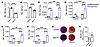 Figure 3
Osteogenic differentiation of PECs from MetPostn mice. (A and B) Relative expression of Met (A) and Hgf (B) in PECs from control (n = 6) and MetPostn (n = 7) mice. Statistically significant differences were determined by t test. (C–F) Relative expression of the osteogenic marker genes Dmp1 (C), Alpl (D), Runx2 (E), and Ocn (F) prior to (Undiff., black) and following osteogenic differentiation (Osteogenesis, blue) of PECs from control (n = 3) and MetPostn (n = 4–6) mice. Statistically significant differences were determined by 2-way ANOVA with Šidák’s multiple-test correction. (G and H) Representative crystal violet and alizarin red staining (G) and quantification (H) of PECs from control and MetPostn mice following osteogenic differentiation (n = 6 per group). A–C, H, and K) Statistically significant differences were determined by t test. RT-qPCR from cultured cells and other quantification data are presented as the mean ± SEM. **P < 0.01, ***P < 0.001, and ****P < 0.0001.
MEK inhibition rescues differentiation and mineralization of MetΔJMD PECs. The elevated MAPK activation associated with MetΔJMD expression implicates the use of targeted therapies, such as the MET inhibitor (METi) capmatinib or the MEK inhibitors (MEKi) mirdametinib and selumetinib to treat OFD (Figure 4A). Each compound was tested for its ability to rescue the cell adhesion and osteogenic differentiation defects inherent in MetΔJMD PECs. Capmatinib is approved by the FDA to treat lung cancers expressing the METΔJMD allele or those with a high MET copy number (copy number >10), both of which convey the MET oncogene addiction (14, 25). In contrast, MET inhibition was ineffective in treating lung cancer with low-level MET amplification (copy number <10) (14). These results suggest that the efficacy of METi is context dependent, and the effectiveness of MET inhibition in OFD remains untested. As expected, cell adhesion of vehicle-treated MetΔJMD PECs was significantly higher compared with vehicle-treated control cells, while capmatinib treatment significantly reduced cell adhesion of MetΔJMD PECs to control levels (Supplemental Figure 3A). Likewise, expression of the cell adhesion genes Ncad and Vim was significantly increased in vehicle-treated MetΔJMD PECs but normalized to control levels with capmatinib treatment (Supplemental Figure 3, B and C). To test mineralization, control and MetΔJMD PECs were treated with capmatinib throughout in vitro osteogenic differentiation. Following osteogenic differentiation, capmatinib failed to rescue mineralization of MetΔJMD PECs (Supplemental Figure 3, D and E). These results suggest that, unlike in MET-addicted lung cancer, the expression of MetΔJMD in PECs is not sufficient to permit therapeutic rescue with a METi. In support of this, the level of Met expression in MetΔJMD PECs was 5 times lower than in METi-sensitive sarcomas expressing the MetΔJMD allele (our unpublished observations).
 Figure 4
MEK inhibition rescues osteogenic differentiation of MetΔJMD PECs. (A) Schematic of the MET receptor (blue) bound by HGF (yellow) to activate the downstream MEK/ERK signaling cascade, resulting in the regulation of osteogenic differentiation of skeletal progenitor cells. Pharmacologic inhibitors of MET (capmatinib) and MEK (mirdametinib, selumetinib) are shown in red. (B and C) Representative Western blot (B) and quantification (C) of ERK pathway activation in MetΔJMD PECs treated with vehicle (n = 8) or mirdametinib (mirda.) (n = 8). Statistically significant differences were determined by t test. (D) Cell counts of PECs from control (n = 14) and MetΔJMD (n = 14) mice. Statistically significant differences were determined by 1-way ANOVA with Tukey’s multiple-test correction. (E and F) Relative expression of the cell adhesion genes Ncad (E) and Vim (F) in control (Met+/+; n = 9–10) and MetΔJMD (n = 7–10) PECs treated with vehicle (Veh.) or mirdametinib. Statistically significant differences were determined by 1-way ANOVA with Tukey’s multiple-test correction. (G–I) Relative expression of the osteogenic genes Alpl (G), Ibsp (H), and Dmp1 (I) in MetΔJMD PECs prior to (Undiff., black) or following osteogenic differentiation in the presence of vehicle (Osteo.+vehicle, blue) or mirdametinib (Osteo.+mirda., orange) (n = 3–8 per group). Statistically significant differences were determined by 1-way ANOVA with Tukey’s multiple-test correction. (J and K) Representative alizarin red staining (J) and quantification (K) of MetΔJMD PECs following osteogenic differentiation in the presence of vehicle (blue, n = 6) or mirdametinib (orange, n = 6). Statistically significant differences were determined by t test. RT-qPCR from cultured cells and other quantification data are presented as the mean ± SEM. **P < 0.01, ***P < 0.001, and ****P < 0.0001.
We next tested whether directly targeting MAPK pathway activation with the MEKi mirdametinib rescues osteogenic differentiation and mineralization of MetΔJMD PECs. Mirdametinib (PD0325901) is a non-ATP-competitive and selective small-molecule inhibitor of both MEK1 and MEK2 phosphorylation (26). In adults and children with NF1, mirdametinib reduced plexiform neurofibroma (pNF) tumor volume, leading to its recent approval by the FDA and the European Medicines Agency (27). To confirm the molecular response of MetΔJMD PECs to mirdametinib treatment, we compared MAPK pathway activation of treated cells with vehicle-treated MetΔJMD PECs. Compared with vehicle treatment, mirdametinib significantly reduced ERK pathway activation in MetΔJMD PECs (Figure 4, B and C). Mirdametinib rescued cell adhesion of treated MetΔJMD PECs, which was further confirmed with the significant reduction in expression of Ncad and Vim (Figure 4, D–F). We next tested the rescue of osteogenic differentiation and mineralization with mirdametinib treatment. Compared with vehicle, mirdametinib treatment significantly rescued osteogenic differentiation of MetΔJMD PECs, as evidenced by increases in the expression of osteogenic genes (Figure 4, G–I, and Supplemental Figure 4, A–C). Consistent with the rescue in differentiation, mirdametinib also significantly rescued mineralization in differentiated MetΔJMD PECs (Figure 4, J and K).
To ensure that the rescue of cell adhesion, osteogenic differentiation, and mineralization of MetΔJMD PECs was indeed associated with MAPK pathway inhibition, we independently repeated all experiments with selumetinib, another MEKi approved by the FDA to treat pNF in children with NF1 (28). As expected, we found that selumetinib significantly reduced ERK pathway activation in MetΔJMD PECs (Supplemental Figure 5, A and B) and rescued the expression of the cell adhesion genes Ncad and Vim (Supplemental Figure 5, C and D). Compared with vehicle-treated cells, selumetinib treatment throughout differentiation resulted in significantly increased the expression of osteogenic genes, consistent with rescue of osteogenesis (Supplemental Figure 5, E–J). Finally, alizarin red staining demonstrated significant rescue of mineralization of selumetinib-treated MetΔJMD PECs (Supplemental Figure 5, K and L). These results show that inhibition of MEK pathway activation rescued cellular and osteogenic defects inherent in MetΔJMD PECs and further suggest the therapeutic potential of MEKi in the treatment of children with OFD.
MEKi enhance the expression of skeletal gene signatures in OFD patient–derived primary cells. We previously identified a somatic p.(Tyr1003Ser) variant in primary cells cultured from a patient’s OFD fracture lesion, implicating somatic MET mutations in the pathogenesis of sporadic OFD–associated pseudarthrosis (4). To further evaluate somatic MET mutations as a cause of OFD fracture pseudarthrosis, we tested METΔJMD expression by reverse transcription PCR (RT-PCR) and sequenced the JMD-encoding exon for Tyr1003 mutations in primary cells cultured from pseudarthroses of 4 patients with OFD. RT-PCR detected an alternatively spliced MET transcript in lesion-derived primary cells from 1 patient (Supplemental Figure 6A). No alternative splicing or MET JMD or kinase domain mutations were detected in the remaining 3 patient samples, possibly due to somatic mosaicism below the level of detection or to mutations present in other genes not previously associated with OFD. The alternatively spliced variant was cloned and confirmed by Sanger sequencing as the METΔJMD allele (Supplemental Figure 6B). Sequencing of DNA from lesion-derived primary cells identified a somatic c.3028+2T>C splice donor variant that was not detected in DNA from a blood sample from the patient (Supplemental Figure 6C). The same allele was previously reported as a somatic mutation in lung adenocarcinomas (29, 30). We performed droplet digital PCR (ddPCR) to confirm and quantify the mutation using DNA from primary lesion cells and blood (control). The mutation burden (variant allele fraction) was 19% in the lesion-derived cells but was undetected in the blood sample (Supplemental Figure 6D), further confirming the somatic occurrence of this variant resulting in expression of the METΔJMD allele. These results independently confirm that somatic MET mutations of the JMD domain are a cause of sporadic OFD and raise the possibility that mutations in other genes may also contribute to OFD.
To begin investigating the therapeutic potential of METi or MEKi for the treatment of OFD, we tested the effect of METi or MEKi monotherapy on OFD lesion–derived primary cells, including those from patients with and without detectable MET mutations. We first tested the molecular effect of the METi capmatinib on patient lesion–derived primary cells. We found that apmatinib treatment failed to significantly inhibit ERK pathway activation, and RNA-seq analysis demonstrated a minimal molecular response to METi, with few genes being differentially expressed compared with vehicle treatment (Supplemental Figure 7, A–C, and Supplemental Table 1). As with the MetΔJMD PECs, these results do not support a therapeutic potential for METi in OFD.
We next tested the response of OFD patient lesion–derived primary cells to MEKi, including cells from patients with and without detectable MET gene variants. Mirdametinib significantly, and in a dose-dependent manner, inhibited ERK pathway activation (Figure 5, A and B). RNA-seq profiling distinguished vehicle-treated from mirdametinib-treated samples, identifying 333 transcripts with reduced expression and 152 transcripts with increased expression following mirdametinib treatment (Figure 5, C–E, and Supplemental Table 2). We performed gene ontology (GO) analysis to summarize the biologic response of mirdametinib in OFD lesion–derived primary cells. GO analysis results demonstrated that genes with increased expression following mirdametinib treatment were significantly associated with biologic processes required for skeletal development and fracture healing (Figure 5F and Supplemental Table 3). Genes with reduced expression following treatment were significantly enriched in MAPK pathway processes as well as biologic processes expected to be affected by MAPK pathway inhibition, such as cell-cycle regulation and cell proliferation (Figure 5G and Supplemental Table 4). These results are similar to those of a recent study of NF1 patient pseudarthrosis–derived primary cells treated with the MEKi selumetinib, which demonstrated that the molecular response to treatment was specific to MAPK hyperactive primary lesion cells, whereas patient-matched iliac crest control primary cells showed a minimal response to treatment (31).
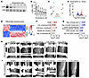 Figure 5
Mirdametinib improves pseudarthrosis healing in a patient with OFD. (A and B) Representative Western blot (A) and quantification (B) demonstrating a dose-dependent reduction in ERK pathway activation in OFD patient pseudarthrosis–derived primary cells (n = 5) treated with mirdametinib. Triangles indicate samples with somatic MET mutations; circles indicate samples without detectable MET mutations. Quantification data are presented as the mean ± SEM, with statistically significant differences determined by 1-way ANOVA with Dunnett’s multiple-test correction. (C) Principal component (PC) analysis of OFD patient pseudarthrosis–derived primary cells treated with vehicle or mirdametinib (n = 5). (D and E) Volcano plot (D) and heatmap (E) of genes differentially expressed between mirdametinib- and vehicle-treated OFD patient pseudarthrosis–derived primary cells. Exp., expression. (F and G) GO analysis of differentially expressed genes with increased (F) and decreased (G) expression following mirdametinib treatment. activ., activity; coll., collagen; diff., differentiation; phosph., phosphotase; reg., regulation; struct., structure; sys. develop., system development. (H and I) Longitudinal postoperative radiographs of an OFD patient’s pseudarthrosis treated without mirdametinib (H) and with mirdametinib (I). **P < 0.01, ***P < 0.001, and ****P < 0.0001.
To further demonstrate the molecular response of OFD lesion–derived primary cells to ERK pathway inhibition, we similarly tested the effects of selumetinib. ERK pathway activation was significantly reduced with selumetinib treatment (Supplemental Figure 7A). Transcriptome profiling distinguished selumetinib-treated cells from vehicle-treated cells and identified 581 transcripts with reduced expression and 331 transcripts with increased expression following selumetinib treatment (Supplemental Figure 7, B, D, and E, and Supplemental Table 5). Similar to mirdametinib, genes with increased expression following selumetinib treatment were associated with extracellular matrix formation and osteoblast differentiation, whereas genes with reduced expression were associated with MAPK pathway and kinase activity processes (Supplemental Figure 7, F and G, and Supplemental Tables 6 and 7). For all treatments, no differences were observed between patients with (n = 2) or without (n = 3) detectable MET mutations. Taken together, these results provide molecular evidence implicating MEKi as a potential therapeutic strategy to promote bone healing in patients with OFD.
Mirdametinib promotes fracture healing in a patient with OFD. Results utilizing MetΔJMD PECs and primary cells from patients with OFD suggested that MEKi may provide therapeutic benefit for children with OFD-associated pseudarthrosis. MEKi have become more widely utilized in the pediatric population for several indications in which the MAPK pathway is hyperactivated and have demonstrated safety and tolerability. Two MEKi resulted in inhibition of ERK pathway activation in primary cells from patients with OFD and increased the expression of genes and pathways known to promote bone formation and/or fracture healing. Therefore, we hypothesized that monotherapy with a MEKi, together with standard-of-care surgical treatment, may promote healing of OFD-associated pseudarthrosis.
Given the strong preclinical data, we administered the MEKi mirdametinib to a patient with OFD who, after multiple attempts at surgical correction, had a persistent pseudarthrosis. The patient initially presented at 1 year of age with a tibia lesion requiring resection, which resulted in persistent pseudarthrosis. The patient underwent 6 subsequent surgeries over the next 3 years due to persistent nonhealing. Multiple techniques were utilized to attempt to achieve bone union, including resection, external fixation, iliac crest grafting, and application of BMP2, although each revision failed to achieve durable union, and the patient’s pseudarthrosis persisted (Figure 5H). Local bone infection was assessed for and excluded as a cause of nonunion. A biopsy confirmed the OFD diagnosis, although no MET gene mutation was identified in cultured lesion-derived primary cells from the patient. Given the molecular response of patient pseudarthrosis–derived primary cells to MEKi in vitro, we sought approval to treat the patient with a MEKi. After appropriate regulatory review and approval from the SpringWorks Therapeutics Compassionate Use Program, the FDA, and the IRB at UT Southwestern Medical Center, the family consented to the use of mirdametinib. Twelve days after revision surgery that again included resection, external fixation, iliac crest grafting, and BMP2, the patient was started on mirdametinib at a dose of 1 mg twice daily via the dispersible tablet with the standard schedule of 21 days on the drug and 7 days off per a 28-day cycle. Prior to dosing, the patient underwent baseline ophthalmology evaluation, an echocardiogram, physical examination, and laboratory evaluation. The patient has been monitored on-therapy with follow-up evaluations including physical, laboratory, and cardiac evaluations. The patient has tolerated the therapy with no significant toxicity and has only noted grade 1 diarrhea in the first month of therapy that resolved by the second month of therapy. The patient has now completed 7 months of therapy with no further toxicity noted.
Fracture healing was monitored by monthly radiographs, which demonstrated progressive maturation of the iliac crest graft that, at 6 months after surgery, resulted in a durable bony bridge and union of the fracture despite early postsurgical distal segment translation leaving a residual angular deformity (Figure 5I). The external fixator frame was removed and the patient was transitioned to a long leg cast to promote further healing and remodeling and is continuing mirdametinib therapy. Following 6 prior revision surgeries that resulted in persistent pseudarthrosis, the addition of mirdametinib to standard surgical approaches in this patient with OFD resulted in the first bone union. Overall, our preclinical results as well as the single patient reported here demonstrate evidence that MEKi monotherapy, together with standard-of-care surgical approaches, can result in healing of persistent pseudarthrosis in patients with OFD RASopathy.


Copyright © 2026 American Society for Clinical Investigation
ISSN: 0021-9738 (print), 1558-8238 (online)