
Advertisement
Research ArticleDevelopmentNeuroscience
Open Access |  10.1172/JCI195803
Astrocytes contribute to olanzapine-mediated reversal of kleefstra syndrome-associated neurodevelopmental regression
Karlijn Vermeulen-Kalk,1,2,3 Shan Wang,4 Joost Kummeling,1,4 Britt Mossink,1,4 Kim Wijnant,1 Carlos Gonzales Jiménez,1 Zoe Frazier,5 Brian Rozumny,6 Anne O’Donnell-Luria,7,8 Ellen Hanson,6,9 Monica Frega,1,4 Katrin Linda,1,4 Moritz Negwer,1,4 Bas Lendemeijer,10 Astrid Oudakker,1 Monica Pop-Purceleanu,11 Joost Janzing,11 Linde van Dongen,2,4 Femke M.S. de Vrij,10,12 Steven A. Kushner,13 Ilse van der Werf,4,14 Chantal Schoenmaker,1 Wouter Oomens,2 Siddharth Srivastava,5,9 Jos I.M. Egger,2,4,15 Hans van Bokhoven,1,4 Dirk Schubert,4,14 Nael Nadif Kasri,1,4 and Tjitske Kleefstra1,2,16
1Department of Human Genetics, Radboud UMC, Nijmegen, Netherlands.
2Centre of Excellence for Neuropsychiatry, Vincent van Gogh Institute for Psychiatry, Venray, Netherlands.
3Department for Intellectual Disabilities, Karakter Child and Adolescent Psychiatry, Ede, Netherlands.
4Donders Institute for Brain, Cognition and Behaviour, Nijmegen, Netherlands.
5Rosamund Stone Zander Translational Neuroscience Center, Department of Neurology,
6Division of Developmental Medicine,
7Division of Genetics and Genomics, Boston Children’s Hospital, Boston, Massachusetts, USA.
8Program in Medical and Population Genetics, Broad Institute of MIT and Harvard, Cambridge, Massachusetts, USA.
9Department of Neurology, Boston Children’s Hospital, Boston, Massachusetts, USA.
10Department of Psychiatry, Erasmus MC University Medical Center, Rotterdam, Netherlands.
11Department of Psychiatry, Radboud University Medical Center, Nijmegen, Netherlands.
12ENCORE Expertise Center for Neurodevelopmental Disorders, Erasmus MC, Rotterdam, Netherlands.
13Department of Psychiatry, Columbia University, New York, New York, USA.
14Department of Medical Neurosciences, Radboud UMC, Nijmegen, Netherlands.
15Stevig Specialized and Forensic Care for People with Intellectual Disabilities, Dichterbij, Oostrum, Netherlands.
16Department of Clinical Genetics, Erasmus MC University Medical Center, Rotterdam, Netherlands.
Address correspondence to: Tjitske Kleefstra, Erasmus MC University Medical Center, Dr. Molewaterplein 40, 3015 GD Rotterdam, Netherlands. Phone: 31.10.7043382; Email: t.kleefstra@erasmusmc.nl. Or to: Nael Nadif Kasri, Radbodumc, Geert Grooteplein 10, 6525GA Nijmegen, Netherlands. Phone: 31.24.3614242; Email: Nael.NadifKasri@radboudumc.nl.
Authorship note: KVK, SW, and JK are co-first authors. NNK and TK are co-last authors.
Find articles by Vermeulen-Kalk, K. in: PubMed | Google Scholar
1Department of Human Genetics, Radboud UMC, Nijmegen, Netherlands.
2Centre of Excellence for Neuropsychiatry, Vincent van Gogh Institute for Psychiatry, Venray, Netherlands.
3Department for Intellectual Disabilities, Karakter Child and Adolescent Psychiatry, Ede, Netherlands.
4Donders Institute for Brain, Cognition and Behaviour, Nijmegen, Netherlands.
5Rosamund Stone Zander Translational Neuroscience Center, Department of Neurology,
6Division of Developmental Medicine,
7Division of Genetics and Genomics, Boston Children’s Hospital, Boston, Massachusetts, USA.
8Program in Medical and Population Genetics, Broad Institute of MIT and Harvard, Cambridge, Massachusetts, USA.
9Department of Neurology, Boston Children’s Hospital, Boston, Massachusetts, USA.
10Department of Psychiatry, Erasmus MC University Medical Center, Rotterdam, Netherlands.
11Department of Psychiatry, Radboud University Medical Center, Nijmegen, Netherlands.
12ENCORE Expertise Center for Neurodevelopmental Disorders, Erasmus MC, Rotterdam, Netherlands.
13Department of Psychiatry, Columbia University, New York, New York, USA.
14Department of Medical Neurosciences, Radboud UMC, Nijmegen, Netherlands.
15Stevig Specialized and Forensic Care for People with Intellectual Disabilities, Dichterbij, Oostrum, Netherlands.
16Department of Clinical Genetics, Erasmus MC University Medical Center, Rotterdam, Netherlands.
Address correspondence to: Tjitske Kleefstra, Erasmus MC University Medical Center, Dr. Molewaterplein 40, 3015 GD Rotterdam, Netherlands. Phone: 31.10.7043382; Email: t.kleefstra@erasmusmc.nl. Or to: Nael Nadif Kasri, Radbodumc, Geert Grooteplein 10, 6525GA Nijmegen, Netherlands. Phone: 31.24.3614242; Email: Nael.NadifKasri@radboudumc.nl.
Authorship note: KVK, SW, and JK are co-first authors. NNK and TK are co-last authors.
Find articles by Wang, S. in: PubMed | Google Scholar
1Department of Human Genetics, Radboud UMC, Nijmegen, Netherlands.
2Centre of Excellence for Neuropsychiatry, Vincent van Gogh Institute for Psychiatry, Venray, Netherlands.
3Department for Intellectual Disabilities, Karakter Child and Adolescent Psychiatry, Ede, Netherlands.
4Donders Institute for Brain, Cognition and Behaviour, Nijmegen, Netherlands.
5Rosamund Stone Zander Translational Neuroscience Center, Department of Neurology,
6Division of Developmental Medicine,
7Division of Genetics and Genomics, Boston Children’s Hospital, Boston, Massachusetts, USA.
8Program in Medical and Population Genetics, Broad Institute of MIT and Harvard, Cambridge, Massachusetts, USA.
9Department of Neurology, Boston Children’s Hospital, Boston, Massachusetts, USA.
10Department of Psychiatry, Erasmus MC University Medical Center, Rotterdam, Netherlands.
11Department of Psychiatry, Radboud University Medical Center, Nijmegen, Netherlands.
12ENCORE Expertise Center for Neurodevelopmental Disorders, Erasmus MC, Rotterdam, Netherlands.
13Department of Psychiatry, Columbia University, New York, New York, USA.
14Department of Medical Neurosciences, Radboud UMC, Nijmegen, Netherlands.
15Stevig Specialized and Forensic Care for People with Intellectual Disabilities, Dichterbij, Oostrum, Netherlands.
16Department of Clinical Genetics, Erasmus MC University Medical Center, Rotterdam, Netherlands.
Address correspondence to: Tjitske Kleefstra, Erasmus MC University Medical Center, Dr. Molewaterplein 40, 3015 GD Rotterdam, Netherlands. Phone: 31.10.7043382; Email: t.kleefstra@erasmusmc.nl. Or to: Nael Nadif Kasri, Radbodumc, Geert Grooteplein 10, 6525GA Nijmegen, Netherlands. Phone: 31.24.3614242; Email: Nael.NadifKasri@radboudumc.nl.
Authorship note: KVK, SW, and JK are co-first authors. NNK and TK are co-last authors.
Find articles by Kummeling, J. in: PubMed | Google Scholar
1Department of Human Genetics, Radboud UMC, Nijmegen, Netherlands.
2Centre of Excellence for Neuropsychiatry, Vincent van Gogh Institute for Psychiatry, Venray, Netherlands.
3Department for Intellectual Disabilities, Karakter Child and Adolescent Psychiatry, Ede, Netherlands.
4Donders Institute for Brain, Cognition and Behaviour, Nijmegen, Netherlands.
5Rosamund Stone Zander Translational Neuroscience Center, Department of Neurology,
6Division of Developmental Medicine,
7Division of Genetics and Genomics, Boston Children’s Hospital, Boston, Massachusetts, USA.
8Program in Medical and Population Genetics, Broad Institute of MIT and Harvard, Cambridge, Massachusetts, USA.
9Department of Neurology, Boston Children’s Hospital, Boston, Massachusetts, USA.
10Department of Psychiatry, Erasmus MC University Medical Center, Rotterdam, Netherlands.
11Department of Psychiatry, Radboud University Medical Center, Nijmegen, Netherlands.
12ENCORE Expertise Center for Neurodevelopmental Disorders, Erasmus MC, Rotterdam, Netherlands.
13Department of Psychiatry, Columbia University, New York, New York, USA.
14Department of Medical Neurosciences, Radboud UMC, Nijmegen, Netherlands.
15Stevig Specialized and Forensic Care for People with Intellectual Disabilities, Dichterbij, Oostrum, Netherlands.
16Department of Clinical Genetics, Erasmus MC University Medical Center, Rotterdam, Netherlands.
Address correspondence to: Tjitske Kleefstra, Erasmus MC University Medical Center, Dr. Molewaterplein 40, 3015 GD Rotterdam, Netherlands. Phone: 31.10.7043382; Email: t.kleefstra@erasmusmc.nl. Or to: Nael Nadif Kasri, Radbodumc, Geert Grooteplein 10, 6525GA Nijmegen, Netherlands. Phone: 31.24.3614242; Email: Nael.NadifKasri@radboudumc.nl.
Authorship note: KVK, SW, and JK are co-first authors. NNK and TK are co-last authors.
Find articles by Mossink, B. in: PubMed | Google Scholar
1Department of Human Genetics, Radboud UMC, Nijmegen, Netherlands.
2Centre of Excellence for Neuropsychiatry, Vincent van Gogh Institute for Psychiatry, Venray, Netherlands.
3Department for Intellectual Disabilities, Karakter Child and Adolescent Psychiatry, Ede, Netherlands.
4Donders Institute for Brain, Cognition and Behaviour, Nijmegen, Netherlands.
5Rosamund Stone Zander Translational Neuroscience Center, Department of Neurology,
6Division of Developmental Medicine,
7Division of Genetics and Genomics, Boston Children’s Hospital, Boston, Massachusetts, USA.
8Program in Medical and Population Genetics, Broad Institute of MIT and Harvard, Cambridge, Massachusetts, USA.
9Department of Neurology, Boston Children’s Hospital, Boston, Massachusetts, USA.
10Department of Psychiatry, Erasmus MC University Medical Center, Rotterdam, Netherlands.
11Department of Psychiatry, Radboud University Medical Center, Nijmegen, Netherlands.
12ENCORE Expertise Center for Neurodevelopmental Disorders, Erasmus MC, Rotterdam, Netherlands.
13Department of Psychiatry, Columbia University, New York, New York, USA.
14Department of Medical Neurosciences, Radboud UMC, Nijmegen, Netherlands.
15Stevig Specialized and Forensic Care for People with Intellectual Disabilities, Dichterbij, Oostrum, Netherlands.
16Department of Clinical Genetics, Erasmus MC University Medical Center, Rotterdam, Netherlands.
Address correspondence to: Tjitske Kleefstra, Erasmus MC University Medical Center, Dr. Molewaterplein 40, 3015 GD Rotterdam, Netherlands. Phone: 31.10.7043382; Email: t.kleefstra@erasmusmc.nl. Or to: Nael Nadif Kasri, Radbodumc, Geert Grooteplein 10, 6525GA Nijmegen, Netherlands. Phone: 31.24.3614242; Email: Nael.NadifKasri@radboudumc.nl.
Authorship note: KVK, SW, and JK are co-first authors. NNK and TK are co-last authors.
Find articles by Wijnant, K. in: PubMed | Google Scholar
1Department of Human Genetics, Radboud UMC, Nijmegen, Netherlands.
2Centre of Excellence for Neuropsychiatry, Vincent van Gogh Institute for Psychiatry, Venray, Netherlands.
3Department for Intellectual Disabilities, Karakter Child and Adolescent Psychiatry, Ede, Netherlands.
4Donders Institute for Brain, Cognition and Behaviour, Nijmegen, Netherlands.
5Rosamund Stone Zander Translational Neuroscience Center, Department of Neurology,
6Division of Developmental Medicine,
7Division of Genetics and Genomics, Boston Children’s Hospital, Boston, Massachusetts, USA.
8Program in Medical and Population Genetics, Broad Institute of MIT and Harvard, Cambridge, Massachusetts, USA.
9Department of Neurology, Boston Children’s Hospital, Boston, Massachusetts, USA.
10Department of Psychiatry, Erasmus MC University Medical Center, Rotterdam, Netherlands.
11Department of Psychiatry, Radboud University Medical Center, Nijmegen, Netherlands.
12ENCORE Expertise Center for Neurodevelopmental Disorders, Erasmus MC, Rotterdam, Netherlands.
13Department of Psychiatry, Columbia University, New York, New York, USA.
14Department of Medical Neurosciences, Radboud UMC, Nijmegen, Netherlands.
15Stevig Specialized and Forensic Care for People with Intellectual Disabilities, Dichterbij, Oostrum, Netherlands.
16Department of Clinical Genetics, Erasmus MC University Medical Center, Rotterdam, Netherlands.
Address correspondence to: Tjitske Kleefstra, Erasmus MC University Medical Center, Dr. Molewaterplein 40, 3015 GD Rotterdam, Netherlands. Phone: 31.10.7043382; Email: t.kleefstra@erasmusmc.nl. Or to: Nael Nadif Kasri, Radbodumc, Geert Grooteplein 10, 6525GA Nijmegen, Netherlands. Phone: 31.24.3614242; Email: Nael.NadifKasri@radboudumc.nl.
Authorship note: KVK, SW, and JK are co-first authors. NNK and TK are co-last authors.
Find articles by Gonzales Jiménez, C. in: PubMed | Google Scholar
1Department of Human Genetics, Radboud UMC, Nijmegen, Netherlands.
2Centre of Excellence for Neuropsychiatry, Vincent van Gogh Institute for Psychiatry, Venray, Netherlands.
3Department for Intellectual Disabilities, Karakter Child and Adolescent Psychiatry, Ede, Netherlands.
4Donders Institute for Brain, Cognition and Behaviour, Nijmegen, Netherlands.
5Rosamund Stone Zander Translational Neuroscience Center, Department of Neurology,
6Division of Developmental Medicine,
7Division of Genetics and Genomics, Boston Children’s Hospital, Boston, Massachusetts, USA.
8Program in Medical and Population Genetics, Broad Institute of MIT and Harvard, Cambridge, Massachusetts, USA.
9Department of Neurology, Boston Children’s Hospital, Boston, Massachusetts, USA.
10Department of Psychiatry, Erasmus MC University Medical Center, Rotterdam, Netherlands.
11Department of Psychiatry, Radboud University Medical Center, Nijmegen, Netherlands.
12ENCORE Expertise Center for Neurodevelopmental Disorders, Erasmus MC, Rotterdam, Netherlands.
13Department of Psychiatry, Columbia University, New York, New York, USA.
14Department of Medical Neurosciences, Radboud UMC, Nijmegen, Netherlands.
15Stevig Specialized and Forensic Care for People with Intellectual Disabilities, Dichterbij, Oostrum, Netherlands.
16Department of Clinical Genetics, Erasmus MC University Medical Center, Rotterdam, Netherlands.
Address correspondence to: Tjitske Kleefstra, Erasmus MC University Medical Center, Dr. Molewaterplein 40, 3015 GD Rotterdam, Netherlands. Phone: 31.10.7043382; Email: t.kleefstra@erasmusmc.nl. Or to: Nael Nadif Kasri, Radbodumc, Geert Grooteplein 10, 6525GA Nijmegen, Netherlands. Phone: 31.24.3614242; Email: Nael.NadifKasri@radboudumc.nl.
Authorship note: KVK, SW, and JK are co-first authors. NNK and TK are co-last authors.
Find articles by Frazier, Z. in: PubMed | Google Scholar
1Department of Human Genetics, Radboud UMC, Nijmegen, Netherlands.
2Centre of Excellence for Neuropsychiatry, Vincent van Gogh Institute for Psychiatry, Venray, Netherlands.
3Department for Intellectual Disabilities, Karakter Child and Adolescent Psychiatry, Ede, Netherlands.
4Donders Institute for Brain, Cognition and Behaviour, Nijmegen, Netherlands.
5Rosamund Stone Zander Translational Neuroscience Center, Department of Neurology,
6Division of Developmental Medicine,
7Division of Genetics and Genomics, Boston Children’s Hospital, Boston, Massachusetts, USA.
8Program in Medical and Population Genetics, Broad Institute of MIT and Harvard, Cambridge, Massachusetts, USA.
9Department of Neurology, Boston Children’s Hospital, Boston, Massachusetts, USA.
10Department of Psychiatry, Erasmus MC University Medical Center, Rotterdam, Netherlands.
11Department of Psychiatry, Radboud University Medical Center, Nijmegen, Netherlands.
12ENCORE Expertise Center for Neurodevelopmental Disorders, Erasmus MC, Rotterdam, Netherlands.
13Department of Psychiatry, Columbia University, New York, New York, USA.
14Department of Medical Neurosciences, Radboud UMC, Nijmegen, Netherlands.
15Stevig Specialized and Forensic Care for People with Intellectual Disabilities, Dichterbij, Oostrum, Netherlands.
16Department of Clinical Genetics, Erasmus MC University Medical Center, Rotterdam, Netherlands.
Address correspondence to: Tjitske Kleefstra, Erasmus MC University Medical Center, Dr. Molewaterplein 40, 3015 GD Rotterdam, Netherlands. Phone: 31.10.7043382; Email: t.kleefstra@erasmusmc.nl. Or to: Nael Nadif Kasri, Radbodumc, Geert Grooteplein 10, 6525GA Nijmegen, Netherlands. Phone: 31.24.3614242; Email: Nael.NadifKasri@radboudumc.nl.
Authorship note: KVK, SW, and JK are co-first authors. NNK and TK are co-last authors.
Find articles by Rozumny, B. in: PubMed | Google Scholar
1Department of Human Genetics, Radboud UMC, Nijmegen, Netherlands.
2Centre of Excellence for Neuropsychiatry, Vincent van Gogh Institute for Psychiatry, Venray, Netherlands.
3Department for Intellectual Disabilities, Karakter Child and Adolescent Psychiatry, Ede, Netherlands.
4Donders Institute for Brain, Cognition and Behaviour, Nijmegen, Netherlands.
5Rosamund Stone Zander Translational Neuroscience Center, Department of Neurology,
6Division of Developmental Medicine,
7Division of Genetics and Genomics, Boston Children’s Hospital, Boston, Massachusetts, USA.
8Program in Medical and Population Genetics, Broad Institute of MIT and Harvard, Cambridge, Massachusetts, USA.
9Department of Neurology, Boston Children’s Hospital, Boston, Massachusetts, USA.
10Department of Psychiatry, Erasmus MC University Medical Center, Rotterdam, Netherlands.
11Department of Psychiatry, Radboud University Medical Center, Nijmegen, Netherlands.
12ENCORE Expertise Center for Neurodevelopmental Disorders, Erasmus MC, Rotterdam, Netherlands.
13Department of Psychiatry, Columbia University, New York, New York, USA.
14Department of Medical Neurosciences, Radboud UMC, Nijmegen, Netherlands.
15Stevig Specialized and Forensic Care for People with Intellectual Disabilities, Dichterbij, Oostrum, Netherlands.
16Department of Clinical Genetics, Erasmus MC University Medical Center, Rotterdam, Netherlands.
Address correspondence to: Tjitske Kleefstra, Erasmus MC University Medical Center, Dr. Molewaterplein 40, 3015 GD Rotterdam, Netherlands. Phone: 31.10.7043382; Email: t.kleefstra@erasmusmc.nl. Or to: Nael Nadif Kasri, Radbodumc, Geert Grooteplein 10, 6525GA Nijmegen, Netherlands. Phone: 31.24.3614242; Email: Nael.NadifKasri@radboudumc.nl.
Authorship note: KVK, SW, and JK are co-first authors. NNK and TK are co-last authors.
Find articles by O’Donnell-Luria, A. in: PubMed | Google Scholar
1Department of Human Genetics, Radboud UMC, Nijmegen, Netherlands.
2Centre of Excellence for Neuropsychiatry, Vincent van Gogh Institute for Psychiatry, Venray, Netherlands.
3Department for Intellectual Disabilities, Karakter Child and Adolescent Psychiatry, Ede, Netherlands.
4Donders Institute for Brain, Cognition and Behaviour, Nijmegen, Netherlands.
5Rosamund Stone Zander Translational Neuroscience Center, Department of Neurology,
6Division of Developmental Medicine,
7Division of Genetics and Genomics, Boston Children’s Hospital, Boston, Massachusetts, USA.
8Program in Medical and Population Genetics, Broad Institute of MIT and Harvard, Cambridge, Massachusetts, USA.
9Department of Neurology, Boston Children’s Hospital, Boston, Massachusetts, USA.
10Department of Psychiatry, Erasmus MC University Medical Center, Rotterdam, Netherlands.
11Department of Psychiatry, Radboud University Medical Center, Nijmegen, Netherlands.
12ENCORE Expertise Center for Neurodevelopmental Disorders, Erasmus MC, Rotterdam, Netherlands.
13Department of Psychiatry, Columbia University, New York, New York, USA.
14Department of Medical Neurosciences, Radboud UMC, Nijmegen, Netherlands.
15Stevig Specialized and Forensic Care for People with Intellectual Disabilities, Dichterbij, Oostrum, Netherlands.
16Department of Clinical Genetics, Erasmus MC University Medical Center, Rotterdam, Netherlands.
Address correspondence to: Tjitske Kleefstra, Erasmus MC University Medical Center, Dr. Molewaterplein 40, 3015 GD Rotterdam, Netherlands. Phone: 31.10.7043382; Email: t.kleefstra@erasmusmc.nl. Or to: Nael Nadif Kasri, Radbodumc, Geert Grooteplein 10, 6525GA Nijmegen, Netherlands. Phone: 31.24.3614242; Email: Nael.NadifKasri@radboudumc.nl.
Authorship note: KVK, SW, and JK are co-first authors. NNK and TK are co-last authors.
Find articles by Hanson, E. in: PubMed | Google Scholar
1Department of Human Genetics, Radboud UMC, Nijmegen, Netherlands.
2Centre of Excellence for Neuropsychiatry, Vincent van Gogh Institute for Psychiatry, Venray, Netherlands.
3Department for Intellectual Disabilities, Karakter Child and Adolescent Psychiatry, Ede, Netherlands.
4Donders Institute for Brain, Cognition and Behaviour, Nijmegen, Netherlands.
5Rosamund Stone Zander Translational Neuroscience Center, Department of Neurology,
6Division of Developmental Medicine,
7Division of Genetics and Genomics, Boston Children’s Hospital, Boston, Massachusetts, USA.
8Program in Medical and Population Genetics, Broad Institute of MIT and Harvard, Cambridge, Massachusetts, USA.
9Department of Neurology, Boston Children’s Hospital, Boston, Massachusetts, USA.
10Department of Psychiatry, Erasmus MC University Medical Center, Rotterdam, Netherlands.
11Department of Psychiatry, Radboud University Medical Center, Nijmegen, Netherlands.
12ENCORE Expertise Center for Neurodevelopmental Disorders, Erasmus MC, Rotterdam, Netherlands.
13Department of Psychiatry, Columbia University, New York, New York, USA.
14Department of Medical Neurosciences, Radboud UMC, Nijmegen, Netherlands.
15Stevig Specialized and Forensic Care for People with Intellectual Disabilities, Dichterbij, Oostrum, Netherlands.
16Department of Clinical Genetics, Erasmus MC University Medical Center, Rotterdam, Netherlands.
Address correspondence to: Tjitske Kleefstra, Erasmus MC University Medical Center, Dr. Molewaterplein 40, 3015 GD Rotterdam, Netherlands. Phone: 31.10.7043382; Email: t.kleefstra@erasmusmc.nl. Or to: Nael Nadif Kasri, Radbodumc, Geert Grooteplein 10, 6525GA Nijmegen, Netherlands. Phone: 31.24.3614242; Email: Nael.NadifKasri@radboudumc.nl.
Authorship note: KVK, SW, and JK are co-first authors. NNK and TK are co-last authors.
Find articles by Frega, M. in: PubMed | Google Scholar
1Department of Human Genetics, Radboud UMC, Nijmegen, Netherlands.
2Centre of Excellence for Neuropsychiatry, Vincent van Gogh Institute for Psychiatry, Venray, Netherlands.
3Department for Intellectual Disabilities, Karakter Child and Adolescent Psychiatry, Ede, Netherlands.
4Donders Institute for Brain, Cognition and Behaviour, Nijmegen, Netherlands.
5Rosamund Stone Zander Translational Neuroscience Center, Department of Neurology,
6Division of Developmental Medicine,
7Division of Genetics and Genomics, Boston Children’s Hospital, Boston, Massachusetts, USA.
8Program in Medical and Population Genetics, Broad Institute of MIT and Harvard, Cambridge, Massachusetts, USA.
9Department of Neurology, Boston Children’s Hospital, Boston, Massachusetts, USA.
10Department of Psychiatry, Erasmus MC University Medical Center, Rotterdam, Netherlands.
11Department of Psychiatry, Radboud University Medical Center, Nijmegen, Netherlands.
12ENCORE Expertise Center for Neurodevelopmental Disorders, Erasmus MC, Rotterdam, Netherlands.
13Department of Psychiatry, Columbia University, New York, New York, USA.
14Department of Medical Neurosciences, Radboud UMC, Nijmegen, Netherlands.
15Stevig Specialized and Forensic Care for People with Intellectual Disabilities, Dichterbij, Oostrum, Netherlands.
16Department of Clinical Genetics, Erasmus MC University Medical Center, Rotterdam, Netherlands.
Address correspondence to: Tjitske Kleefstra, Erasmus MC University Medical Center, Dr. Molewaterplein 40, 3015 GD Rotterdam, Netherlands. Phone: 31.10.7043382; Email: t.kleefstra@erasmusmc.nl. Or to: Nael Nadif Kasri, Radbodumc, Geert Grooteplein 10, 6525GA Nijmegen, Netherlands. Phone: 31.24.3614242; Email: Nael.NadifKasri@radboudumc.nl.
Authorship note: KVK, SW, and JK are co-first authors. NNK and TK are co-last authors.
Find articles by Linda, K. in: PubMed | Google Scholar
1Department of Human Genetics, Radboud UMC, Nijmegen, Netherlands.
2Centre of Excellence for Neuropsychiatry, Vincent van Gogh Institute for Psychiatry, Venray, Netherlands.
3Department for Intellectual Disabilities, Karakter Child and Adolescent Psychiatry, Ede, Netherlands.
4Donders Institute for Brain, Cognition and Behaviour, Nijmegen, Netherlands.
5Rosamund Stone Zander Translational Neuroscience Center, Department of Neurology,
6Division of Developmental Medicine,
7Division of Genetics and Genomics, Boston Children’s Hospital, Boston, Massachusetts, USA.
8Program in Medical and Population Genetics, Broad Institute of MIT and Harvard, Cambridge, Massachusetts, USA.
9Department of Neurology, Boston Children’s Hospital, Boston, Massachusetts, USA.
10Department of Psychiatry, Erasmus MC University Medical Center, Rotterdam, Netherlands.
11Department of Psychiatry, Radboud University Medical Center, Nijmegen, Netherlands.
12ENCORE Expertise Center for Neurodevelopmental Disorders, Erasmus MC, Rotterdam, Netherlands.
13Department of Psychiatry, Columbia University, New York, New York, USA.
14Department of Medical Neurosciences, Radboud UMC, Nijmegen, Netherlands.
15Stevig Specialized and Forensic Care for People with Intellectual Disabilities, Dichterbij, Oostrum, Netherlands.
16Department of Clinical Genetics, Erasmus MC University Medical Center, Rotterdam, Netherlands.
Address correspondence to: Tjitske Kleefstra, Erasmus MC University Medical Center, Dr. Molewaterplein 40, 3015 GD Rotterdam, Netherlands. Phone: 31.10.7043382; Email: t.kleefstra@erasmusmc.nl. Or to: Nael Nadif Kasri, Radbodumc, Geert Grooteplein 10, 6525GA Nijmegen, Netherlands. Phone: 31.24.3614242; Email: Nael.NadifKasri@radboudumc.nl.
Authorship note: KVK, SW, and JK are co-first authors. NNK and TK are co-last authors.
Find articles by Negwer, M. in: PubMed | Google Scholar
1Department of Human Genetics, Radboud UMC, Nijmegen, Netherlands.
2Centre of Excellence for Neuropsychiatry, Vincent van Gogh Institute for Psychiatry, Venray, Netherlands.
3Department for Intellectual Disabilities, Karakter Child and Adolescent Psychiatry, Ede, Netherlands.
4Donders Institute for Brain, Cognition and Behaviour, Nijmegen, Netherlands.
5Rosamund Stone Zander Translational Neuroscience Center, Department of Neurology,
6Division of Developmental Medicine,
7Division of Genetics and Genomics, Boston Children’s Hospital, Boston, Massachusetts, USA.
8Program in Medical and Population Genetics, Broad Institute of MIT and Harvard, Cambridge, Massachusetts, USA.
9Department of Neurology, Boston Children’s Hospital, Boston, Massachusetts, USA.
10Department of Psychiatry, Erasmus MC University Medical Center, Rotterdam, Netherlands.
11Department of Psychiatry, Radboud University Medical Center, Nijmegen, Netherlands.
12ENCORE Expertise Center for Neurodevelopmental Disorders, Erasmus MC, Rotterdam, Netherlands.
13Department of Psychiatry, Columbia University, New York, New York, USA.
14Department of Medical Neurosciences, Radboud UMC, Nijmegen, Netherlands.
15Stevig Specialized and Forensic Care for People with Intellectual Disabilities, Dichterbij, Oostrum, Netherlands.
16Department of Clinical Genetics, Erasmus MC University Medical Center, Rotterdam, Netherlands.
Address correspondence to: Tjitske Kleefstra, Erasmus MC University Medical Center, Dr. Molewaterplein 40, 3015 GD Rotterdam, Netherlands. Phone: 31.10.7043382; Email: t.kleefstra@erasmusmc.nl. Or to: Nael Nadif Kasri, Radbodumc, Geert Grooteplein 10, 6525GA Nijmegen, Netherlands. Phone: 31.24.3614242; Email: Nael.NadifKasri@radboudumc.nl.
Authorship note: KVK, SW, and JK are co-first authors. NNK and TK are co-last authors.
Find articles by Lendemeijer, B. in: PubMed | Google Scholar
1Department of Human Genetics, Radboud UMC, Nijmegen, Netherlands.
2Centre of Excellence for Neuropsychiatry, Vincent van Gogh Institute for Psychiatry, Venray, Netherlands.
3Department for Intellectual Disabilities, Karakter Child and Adolescent Psychiatry, Ede, Netherlands.
4Donders Institute for Brain, Cognition and Behaviour, Nijmegen, Netherlands.
5Rosamund Stone Zander Translational Neuroscience Center, Department of Neurology,
6Division of Developmental Medicine,
7Division of Genetics and Genomics, Boston Children’s Hospital, Boston, Massachusetts, USA.
8Program in Medical and Population Genetics, Broad Institute of MIT and Harvard, Cambridge, Massachusetts, USA.
9Department of Neurology, Boston Children’s Hospital, Boston, Massachusetts, USA.
10Department of Psychiatry, Erasmus MC University Medical Center, Rotterdam, Netherlands.
11Department of Psychiatry, Radboud University Medical Center, Nijmegen, Netherlands.
12ENCORE Expertise Center for Neurodevelopmental Disorders, Erasmus MC, Rotterdam, Netherlands.
13Department of Psychiatry, Columbia University, New York, New York, USA.
14Department of Medical Neurosciences, Radboud UMC, Nijmegen, Netherlands.
15Stevig Specialized and Forensic Care for People with Intellectual Disabilities, Dichterbij, Oostrum, Netherlands.
16Department of Clinical Genetics, Erasmus MC University Medical Center, Rotterdam, Netherlands.
Address correspondence to: Tjitske Kleefstra, Erasmus MC University Medical Center, Dr. Molewaterplein 40, 3015 GD Rotterdam, Netherlands. Phone: 31.10.7043382; Email: t.kleefstra@erasmusmc.nl. Or to: Nael Nadif Kasri, Radbodumc, Geert Grooteplein 10, 6525GA Nijmegen, Netherlands. Phone: 31.24.3614242; Email: Nael.NadifKasri@radboudumc.nl.
Authorship note: KVK, SW, and JK are co-first authors. NNK and TK are co-last authors.
Find articles by Oudakker, A. in: PubMed | Google Scholar
1Department of Human Genetics, Radboud UMC, Nijmegen, Netherlands.
2Centre of Excellence for Neuropsychiatry, Vincent van Gogh Institute for Psychiatry, Venray, Netherlands.
3Department for Intellectual Disabilities, Karakter Child and Adolescent Psychiatry, Ede, Netherlands.
4Donders Institute for Brain, Cognition and Behaviour, Nijmegen, Netherlands.
5Rosamund Stone Zander Translational Neuroscience Center, Department of Neurology,
6Division of Developmental Medicine,
7Division of Genetics and Genomics, Boston Children’s Hospital, Boston, Massachusetts, USA.
8Program in Medical and Population Genetics, Broad Institute of MIT and Harvard, Cambridge, Massachusetts, USA.
9Department of Neurology, Boston Children’s Hospital, Boston, Massachusetts, USA.
10Department of Psychiatry, Erasmus MC University Medical Center, Rotterdam, Netherlands.
11Department of Psychiatry, Radboud University Medical Center, Nijmegen, Netherlands.
12ENCORE Expertise Center for Neurodevelopmental Disorders, Erasmus MC, Rotterdam, Netherlands.
13Department of Psychiatry, Columbia University, New York, New York, USA.
14Department of Medical Neurosciences, Radboud UMC, Nijmegen, Netherlands.
15Stevig Specialized and Forensic Care for People with Intellectual Disabilities, Dichterbij, Oostrum, Netherlands.
16Department of Clinical Genetics, Erasmus MC University Medical Center, Rotterdam, Netherlands.
Address correspondence to: Tjitske Kleefstra, Erasmus MC University Medical Center, Dr. Molewaterplein 40, 3015 GD Rotterdam, Netherlands. Phone: 31.10.7043382; Email: t.kleefstra@erasmusmc.nl. Or to: Nael Nadif Kasri, Radbodumc, Geert Grooteplein 10, 6525GA Nijmegen, Netherlands. Phone: 31.24.3614242; Email: Nael.NadifKasri@radboudumc.nl.
Authorship note: KVK, SW, and JK are co-first authors. NNK and TK are co-last authors.
Find articles by Pop-Purceleanu, M. in: PubMed | Google Scholar
1Department of Human Genetics, Radboud UMC, Nijmegen, Netherlands.
2Centre of Excellence for Neuropsychiatry, Vincent van Gogh Institute for Psychiatry, Venray, Netherlands.
3Department for Intellectual Disabilities, Karakter Child and Adolescent Psychiatry, Ede, Netherlands.
4Donders Institute for Brain, Cognition and Behaviour, Nijmegen, Netherlands.
5Rosamund Stone Zander Translational Neuroscience Center, Department of Neurology,
6Division of Developmental Medicine,
7Division of Genetics and Genomics, Boston Children’s Hospital, Boston, Massachusetts, USA.
8Program in Medical and Population Genetics, Broad Institute of MIT and Harvard, Cambridge, Massachusetts, USA.
9Department of Neurology, Boston Children’s Hospital, Boston, Massachusetts, USA.
10Department of Psychiatry, Erasmus MC University Medical Center, Rotterdam, Netherlands.
11Department of Psychiatry, Radboud University Medical Center, Nijmegen, Netherlands.
12ENCORE Expertise Center for Neurodevelopmental Disorders, Erasmus MC, Rotterdam, Netherlands.
13Department of Psychiatry, Columbia University, New York, New York, USA.
14Department of Medical Neurosciences, Radboud UMC, Nijmegen, Netherlands.
15Stevig Specialized and Forensic Care for People with Intellectual Disabilities, Dichterbij, Oostrum, Netherlands.
16Department of Clinical Genetics, Erasmus MC University Medical Center, Rotterdam, Netherlands.
Address correspondence to: Tjitske Kleefstra, Erasmus MC University Medical Center, Dr. Molewaterplein 40, 3015 GD Rotterdam, Netherlands. Phone: 31.10.7043382; Email: t.kleefstra@erasmusmc.nl. Or to: Nael Nadif Kasri, Radbodumc, Geert Grooteplein 10, 6525GA Nijmegen, Netherlands. Phone: 31.24.3614242; Email: Nael.NadifKasri@radboudumc.nl.
Authorship note: KVK, SW, and JK are co-first authors. NNK and TK are co-last authors.
Find articles by Janzing, J. in: PubMed | Google Scholar
1Department of Human Genetics, Radboud UMC, Nijmegen, Netherlands.
2Centre of Excellence for Neuropsychiatry, Vincent van Gogh Institute for Psychiatry, Venray, Netherlands.
3Department for Intellectual Disabilities, Karakter Child and Adolescent Psychiatry, Ede, Netherlands.
4Donders Institute for Brain, Cognition and Behaviour, Nijmegen, Netherlands.
5Rosamund Stone Zander Translational Neuroscience Center, Department of Neurology,
6Division of Developmental Medicine,
7Division of Genetics and Genomics, Boston Children’s Hospital, Boston, Massachusetts, USA.
8Program in Medical and Population Genetics, Broad Institute of MIT and Harvard, Cambridge, Massachusetts, USA.
9Department of Neurology, Boston Children’s Hospital, Boston, Massachusetts, USA.
10Department of Psychiatry, Erasmus MC University Medical Center, Rotterdam, Netherlands.
11Department of Psychiatry, Radboud University Medical Center, Nijmegen, Netherlands.
12ENCORE Expertise Center for Neurodevelopmental Disorders, Erasmus MC, Rotterdam, Netherlands.
13Department of Psychiatry, Columbia University, New York, New York, USA.
14Department of Medical Neurosciences, Radboud UMC, Nijmegen, Netherlands.
15Stevig Specialized and Forensic Care for People with Intellectual Disabilities, Dichterbij, Oostrum, Netherlands.
16Department of Clinical Genetics, Erasmus MC University Medical Center, Rotterdam, Netherlands.
Address correspondence to: Tjitske Kleefstra, Erasmus MC University Medical Center, Dr. Molewaterplein 40, 3015 GD Rotterdam, Netherlands. Phone: 31.10.7043382; Email: t.kleefstra@erasmusmc.nl. Or to: Nael Nadif Kasri, Radbodumc, Geert Grooteplein 10, 6525GA Nijmegen, Netherlands. Phone: 31.24.3614242; Email: Nael.NadifKasri@radboudumc.nl.
Authorship note: KVK, SW, and JK are co-first authors. NNK and TK are co-last authors.
Find articles by van Dongen, L. in: PubMed | Google Scholar
1Department of Human Genetics, Radboud UMC, Nijmegen, Netherlands.
2Centre of Excellence for Neuropsychiatry, Vincent van Gogh Institute for Psychiatry, Venray, Netherlands.
3Department for Intellectual Disabilities, Karakter Child and Adolescent Psychiatry, Ede, Netherlands.
4Donders Institute for Brain, Cognition and Behaviour, Nijmegen, Netherlands.
5Rosamund Stone Zander Translational Neuroscience Center, Department of Neurology,
6Division of Developmental Medicine,
7Division of Genetics and Genomics, Boston Children’s Hospital, Boston, Massachusetts, USA.
8Program in Medical and Population Genetics, Broad Institute of MIT and Harvard, Cambridge, Massachusetts, USA.
9Department of Neurology, Boston Children’s Hospital, Boston, Massachusetts, USA.
10Department of Psychiatry, Erasmus MC University Medical Center, Rotterdam, Netherlands.
11Department of Psychiatry, Radboud University Medical Center, Nijmegen, Netherlands.
12ENCORE Expertise Center for Neurodevelopmental Disorders, Erasmus MC, Rotterdam, Netherlands.
13Department of Psychiatry, Columbia University, New York, New York, USA.
14Department of Medical Neurosciences, Radboud UMC, Nijmegen, Netherlands.
15Stevig Specialized and Forensic Care for People with Intellectual Disabilities, Dichterbij, Oostrum, Netherlands.
16Department of Clinical Genetics, Erasmus MC University Medical Center, Rotterdam, Netherlands.
Address correspondence to: Tjitske Kleefstra, Erasmus MC University Medical Center, Dr. Molewaterplein 40, 3015 GD Rotterdam, Netherlands. Phone: 31.10.7043382; Email: t.kleefstra@erasmusmc.nl. Or to: Nael Nadif Kasri, Radbodumc, Geert Grooteplein 10, 6525GA Nijmegen, Netherlands. Phone: 31.24.3614242; Email: Nael.NadifKasri@radboudumc.nl.
Authorship note: KVK, SW, and JK are co-first authors. NNK and TK are co-last authors.
Find articles by de Vrij, F. in: PubMed | Google Scholar
1Department of Human Genetics, Radboud UMC, Nijmegen, Netherlands.
2Centre of Excellence for Neuropsychiatry, Vincent van Gogh Institute for Psychiatry, Venray, Netherlands.
3Department for Intellectual Disabilities, Karakter Child and Adolescent Psychiatry, Ede, Netherlands.
4Donders Institute for Brain, Cognition and Behaviour, Nijmegen, Netherlands.
5Rosamund Stone Zander Translational Neuroscience Center, Department of Neurology,
6Division of Developmental Medicine,
7Division of Genetics and Genomics, Boston Children’s Hospital, Boston, Massachusetts, USA.
8Program in Medical and Population Genetics, Broad Institute of MIT and Harvard, Cambridge, Massachusetts, USA.
9Department of Neurology, Boston Children’s Hospital, Boston, Massachusetts, USA.
10Department of Psychiatry, Erasmus MC University Medical Center, Rotterdam, Netherlands.
11Department of Psychiatry, Radboud University Medical Center, Nijmegen, Netherlands.
12ENCORE Expertise Center for Neurodevelopmental Disorders, Erasmus MC, Rotterdam, Netherlands.
13Department of Psychiatry, Columbia University, New York, New York, USA.
14Department of Medical Neurosciences, Radboud UMC, Nijmegen, Netherlands.
15Stevig Specialized and Forensic Care for People with Intellectual Disabilities, Dichterbij, Oostrum, Netherlands.
16Department of Clinical Genetics, Erasmus MC University Medical Center, Rotterdam, Netherlands.
Address correspondence to: Tjitske Kleefstra, Erasmus MC University Medical Center, Dr. Molewaterplein 40, 3015 GD Rotterdam, Netherlands. Phone: 31.10.7043382; Email: t.kleefstra@erasmusmc.nl. Or to: Nael Nadif Kasri, Radbodumc, Geert Grooteplein 10, 6525GA Nijmegen, Netherlands. Phone: 31.24.3614242; Email: Nael.NadifKasri@radboudumc.nl.
Authorship note: KVK, SW, and JK are co-first authors. NNK and TK are co-last authors.
Find articles by Kushner, S. in: PubMed | Google Scholar
1Department of Human Genetics, Radboud UMC, Nijmegen, Netherlands.
2Centre of Excellence for Neuropsychiatry, Vincent van Gogh Institute for Psychiatry, Venray, Netherlands.
3Department for Intellectual Disabilities, Karakter Child and Adolescent Psychiatry, Ede, Netherlands.
4Donders Institute for Brain, Cognition and Behaviour, Nijmegen, Netherlands.
5Rosamund Stone Zander Translational Neuroscience Center, Department of Neurology,
6Division of Developmental Medicine,
7Division of Genetics and Genomics, Boston Children’s Hospital, Boston, Massachusetts, USA.
8Program in Medical and Population Genetics, Broad Institute of MIT and Harvard, Cambridge, Massachusetts, USA.
9Department of Neurology, Boston Children’s Hospital, Boston, Massachusetts, USA.
10Department of Psychiatry, Erasmus MC University Medical Center, Rotterdam, Netherlands.
11Department of Psychiatry, Radboud University Medical Center, Nijmegen, Netherlands.
12ENCORE Expertise Center for Neurodevelopmental Disorders, Erasmus MC, Rotterdam, Netherlands.
13Department of Psychiatry, Columbia University, New York, New York, USA.
14Department of Medical Neurosciences, Radboud UMC, Nijmegen, Netherlands.
15Stevig Specialized and Forensic Care for People with Intellectual Disabilities, Dichterbij, Oostrum, Netherlands.
16Department of Clinical Genetics, Erasmus MC University Medical Center, Rotterdam, Netherlands.
Address correspondence to: Tjitske Kleefstra, Erasmus MC University Medical Center, Dr. Molewaterplein 40, 3015 GD Rotterdam, Netherlands. Phone: 31.10.7043382; Email: t.kleefstra@erasmusmc.nl. Or to: Nael Nadif Kasri, Radbodumc, Geert Grooteplein 10, 6525GA Nijmegen, Netherlands. Phone: 31.24.3614242; Email: Nael.NadifKasri@radboudumc.nl.
Authorship note: KVK, SW, and JK are co-first authors. NNK and TK are co-last authors.
Find articles by van der Werf, I. in: PubMed | Google Scholar
1Department of Human Genetics, Radboud UMC, Nijmegen, Netherlands.
2Centre of Excellence for Neuropsychiatry, Vincent van Gogh Institute for Psychiatry, Venray, Netherlands.
3Department for Intellectual Disabilities, Karakter Child and Adolescent Psychiatry, Ede, Netherlands.
4Donders Institute for Brain, Cognition and Behaviour, Nijmegen, Netherlands.
5Rosamund Stone Zander Translational Neuroscience Center, Department of Neurology,
6Division of Developmental Medicine,
7Division of Genetics and Genomics, Boston Children’s Hospital, Boston, Massachusetts, USA.
8Program in Medical and Population Genetics, Broad Institute of MIT and Harvard, Cambridge, Massachusetts, USA.
9Department of Neurology, Boston Children’s Hospital, Boston, Massachusetts, USA.
10Department of Psychiatry, Erasmus MC University Medical Center, Rotterdam, Netherlands.
11Department of Psychiatry, Radboud University Medical Center, Nijmegen, Netherlands.
12ENCORE Expertise Center for Neurodevelopmental Disorders, Erasmus MC, Rotterdam, Netherlands.
13Department of Psychiatry, Columbia University, New York, New York, USA.
14Department of Medical Neurosciences, Radboud UMC, Nijmegen, Netherlands.
15Stevig Specialized and Forensic Care for People with Intellectual Disabilities, Dichterbij, Oostrum, Netherlands.
16Department of Clinical Genetics, Erasmus MC University Medical Center, Rotterdam, Netherlands.
Address correspondence to: Tjitske Kleefstra, Erasmus MC University Medical Center, Dr. Molewaterplein 40, 3015 GD Rotterdam, Netherlands. Phone: 31.10.7043382; Email: t.kleefstra@erasmusmc.nl. Or to: Nael Nadif Kasri, Radbodumc, Geert Grooteplein 10, 6525GA Nijmegen, Netherlands. Phone: 31.24.3614242; Email: Nael.NadifKasri@radboudumc.nl.
Authorship note: KVK, SW, and JK are co-first authors. NNK and TK are co-last authors.
Find articles by Schoenmaker, C. in: PubMed | Google Scholar
1Department of Human Genetics, Radboud UMC, Nijmegen, Netherlands.
2Centre of Excellence for Neuropsychiatry, Vincent van Gogh Institute for Psychiatry, Venray, Netherlands.
3Department for Intellectual Disabilities, Karakter Child and Adolescent Psychiatry, Ede, Netherlands.
4Donders Institute for Brain, Cognition and Behaviour, Nijmegen, Netherlands.
5Rosamund Stone Zander Translational Neuroscience Center, Department of Neurology,
6Division of Developmental Medicine,
7Division of Genetics and Genomics, Boston Children’s Hospital, Boston, Massachusetts, USA.
8Program in Medical and Population Genetics, Broad Institute of MIT and Harvard, Cambridge, Massachusetts, USA.
9Department of Neurology, Boston Children’s Hospital, Boston, Massachusetts, USA.
10Department of Psychiatry, Erasmus MC University Medical Center, Rotterdam, Netherlands.
11Department of Psychiatry, Radboud University Medical Center, Nijmegen, Netherlands.
12ENCORE Expertise Center for Neurodevelopmental Disorders, Erasmus MC, Rotterdam, Netherlands.
13Department of Psychiatry, Columbia University, New York, New York, USA.
14Department of Medical Neurosciences, Radboud UMC, Nijmegen, Netherlands.
15Stevig Specialized and Forensic Care for People with Intellectual Disabilities, Dichterbij, Oostrum, Netherlands.
16Department of Clinical Genetics, Erasmus MC University Medical Center, Rotterdam, Netherlands.
Address correspondence to: Tjitske Kleefstra, Erasmus MC University Medical Center, Dr. Molewaterplein 40, 3015 GD Rotterdam, Netherlands. Phone: 31.10.7043382; Email: t.kleefstra@erasmusmc.nl. Or to: Nael Nadif Kasri, Radbodumc, Geert Grooteplein 10, 6525GA Nijmegen, Netherlands. Phone: 31.24.3614242; Email: Nael.NadifKasri@radboudumc.nl.
Authorship note: KVK, SW, and JK are co-first authors. NNK and TK are co-last authors.
Find articles by Oomens, W. in: PubMed | Google Scholar
1Department of Human Genetics, Radboud UMC, Nijmegen, Netherlands.
2Centre of Excellence for Neuropsychiatry, Vincent van Gogh Institute for Psychiatry, Venray, Netherlands.
3Department for Intellectual Disabilities, Karakter Child and Adolescent Psychiatry, Ede, Netherlands.
4Donders Institute for Brain, Cognition and Behaviour, Nijmegen, Netherlands.
5Rosamund Stone Zander Translational Neuroscience Center, Department of Neurology,
6Division of Developmental Medicine,
7Division of Genetics and Genomics, Boston Children’s Hospital, Boston, Massachusetts, USA.
8Program in Medical and Population Genetics, Broad Institute of MIT and Harvard, Cambridge, Massachusetts, USA.
9Department of Neurology, Boston Children’s Hospital, Boston, Massachusetts, USA.
10Department of Psychiatry, Erasmus MC University Medical Center, Rotterdam, Netherlands.
11Department of Psychiatry, Radboud University Medical Center, Nijmegen, Netherlands.
12ENCORE Expertise Center for Neurodevelopmental Disorders, Erasmus MC, Rotterdam, Netherlands.
13Department of Psychiatry, Columbia University, New York, New York, USA.
14Department of Medical Neurosciences, Radboud UMC, Nijmegen, Netherlands.
15Stevig Specialized and Forensic Care for People with Intellectual Disabilities, Dichterbij, Oostrum, Netherlands.
16Department of Clinical Genetics, Erasmus MC University Medical Center, Rotterdam, Netherlands.
Address correspondence to: Tjitske Kleefstra, Erasmus MC University Medical Center, Dr. Molewaterplein 40, 3015 GD Rotterdam, Netherlands. Phone: 31.10.7043382; Email: t.kleefstra@erasmusmc.nl. Or to: Nael Nadif Kasri, Radbodumc, Geert Grooteplein 10, 6525GA Nijmegen, Netherlands. Phone: 31.24.3614242; Email: Nael.NadifKasri@radboudumc.nl.
Authorship note: KVK, SW, and JK are co-first authors. NNK and TK are co-last authors.
Find articles by Srivastava, S. in: PubMed | Google Scholar
1Department of Human Genetics, Radboud UMC, Nijmegen, Netherlands.
2Centre of Excellence for Neuropsychiatry, Vincent van Gogh Institute for Psychiatry, Venray, Netherlands.
3Department for Intellectual Disabilities, Karakter Child and Adolescent Psychiatry, Ede, Netherlands.
4Donders Institute for Brain, Cognition and Behaviour, Nijmegen, Netherlands.
5Rosamund Stone Zander Translational Neuroscience Center, Department of Neurology,
6Division of Developmental Medicine,
7Division of Genetics and Genomics, Boston Children’s Hospital, Boston, Massachusetts, USA.
8Program in Medical and Population Genetics, Broad Institute of MIT and Harvard, Cambridge, Massachusetts, USA.
9Department of Neurology, Boston Children’s Hospital, Boston, Massachusetts, USA.
10Department of Psychiatry, Erasmus MC University Medical Center, Rotterdam, Netherlands.
11Department of Psychiatry, Radboud University Medical Center, Nijmegen, Netherlands.
12ENCORE Expertise Center for Neurodevelopmental Disorders, Erasmus MC, Rotterdam, Netherlands.
13Department of Psychiatry, Columbia University, New York, New York, USA.
14Department of Medical Neurosciences, Radboud UMC, Nijmegen, Netherlands.
15Stevig Specialized and Forensic Care for People with Intellectual Disabilities, Dichterbij, Oostrum, Netherlands.
16Department of Clinical Genetics, Erasmus MC University Medical Center, Rotterdam, Netherlands.
Address correspondence to: Tjitske Kleefstra, Erasmus MC University Medical Center, Dr. Molewaterplein 40, 3015 GD Rotterdam, Netherlands. Phone: 31.10.7043382; Email: t.kleefstra@erasmusmc.nl. Or to: Nael Nadif Kasri, Radbodumc, Geert Grooteplein 10, 6525GA Nijmegen, Netherlands. Phone: 31.24.3614242; Email: Nael.NadifKasri@radboudumc.nl.
Authorship note: KVK, SW, and JK are co-first authors. NNK and TK are co-last authors.
Find articles by Egger, J. in: PubMed | Google Scholar
1Department of Human Genetics, Radboud UMC, Nijmegen, Netherlands.
2Centre of Excellence for Neuropsychiatry, Vincent van Gogh Institute for Psychiatry, Venray, Netherlands.
3Department for Intellectual Disabilities, Karakter Child and Adolescent Psychiatry, Ede, Netherlands.
4Donders Institute for Brain, Cognition and Behaviour, Nijmegen, Netherlands.
5Rosamund Stone Zander Translational Neuroscience Center, Department of Neurology,
6Division of Developmental Medicine,
7Division of Genetics and Genomics, Boston Children’s Hospital, Boston, Massachusetts, USA.
8Program in Medical and Population Genetics, Broad Institute of MIT and Harvard, Cambridge, Massachusetts, USA.
9Department of Neurology, Boston Children’s Hospital, Boston, Massachusetts, USA.
10Department of Psychiatry, Erasmus MC University Medical Center, Rotterdam, Netherlands.
11Department of Psychiatry, Radboud University Medical Center, Nijmegen, Netherlands.
12ENCORE Expertise Center for Neurodevelopmental Disorders, Erasmus MC, Rotterdam, Netherlands.
13Department of Psychiatry, Columbia University, New York, New York, USA.
14Department of Medical Neurosciences, Radboud UMC, Nijmegen, Netherlands.
15Stevig Specialized and Forensic Care for People with Intellectual Disabilities, Dichterbij, Oostrum, Netherlands.
16Department of Clinical Genetics, Erasmus MC University Medical Center, Rotterdam, Netherlands.
Address correspondence to: Tjitske Kleefstra, Erasmus MC University Medical Center, Dr. Molewaterplein 40, 3015 GD Rotterdam, Netherlands. Phone: 31.10.7043382; Email: t.kleefstra@erasmusmc.nl. Or to: Nael Nadif Kasri, Radbodumc, Geert Grooteplein 10, 6525GA Nijmegen, Netherlands. Phone: 31.24.3614242; Email: Nael.NadifKasri@radboudumc.nl.
Authorship note: KVK, SW, and JK are co-first authors. NNK and TK are co-last authors.
Find articles by van Bokhoven, H. in: PubMed | Google Scholar
1Department of Human Genetics, Radboud UMC, Nijmegen, Netherlands.
2Centre of Excellence for Neuropsychiatry, Vincent van Gogh Institute for Psychiatry, Venray, Netherlands.
3Department for Intellectual Disabilities, Karakter Child and Adolescent Psychiatry, Ede, Netherlands.
4Donders Institute for Brain, Cognition and Behaviour, Nijmegen, Netherlands.
5Rosamund Stone Zander Translational Neuroscience Center, Department of Neurology,
6Division of Developmental Medicine,
7Division of Genetics and Genomics, Boston Children’s Hospital, Boston, Massachusetts, USA.
8Program in Medical and Population Genetics, Broad Institute of MIT and Harvard, Cambridge, Massachusetts, USA.
9Department of Neurology, Boston Children’s Hospital, Boston, Massachusetts, USA.
10Department of Psychiatry, Erasmus MC University Medical Center, Rotterdam, Netherlands.
11Department of Psychiatry, Radboud University Medical Center, Nijmegen, Netherlands.
12ENCORE Expertise Center for Neurodevelopmental Disorders, Erasmus MC, Rotterdam, Netherlands.
13Department of Psychiatry, Columbia University, New York, New York, USA.
14Department of Medical Neurosciences, Radboud UMC, Nijmegen, Netherlands.
15Stevig Specialized and Forensic Care for People with Intellectual Disabilities, Dichterbij, Oostrum, Netherlands.
16Department of Clinical Genetics, Erasmus MC University Medical Center, Rotterdam, Netherlands.
Address correspondence to: Tjitske Kleefstra, Erasmus MC University Medical Center, Dr. Molewaterplein 40, 3015 GD Rotterdam, Netherlands. Phone: 31.10.7043382; Email: t.kleefstra@erasmusmc.nl. Or to: Nael Nadif Kasri, Radbodumc, Geert Grooteplein 10, 6525GA Nijmegen, Netherlands. Phone: 31.24.3614242; Email: Nael.NadifKasri@radboudumc.nl.
Authorship note: KVK, SW, and JK are co-first authors. NNK and TK are co-last authors.
Find articles by
Schubert, D.
in:
PubMed
|
Google Scholar
|

1Department of Human Genetics, Radboud UMC, Nijmegen, Netherlands.
2Centre of Excellence for Neuropsychiatry, Vincent van Gogh Institute for Psychiatry, Venray, Netherlands.
3Department for Intellectual Disabilities, Karakter Child and Adolescent Psychiatry, Ede, Netherlands.
4Donders Institute for Brain, Cognition and Behaviour, Nijmegen, Netherlands.
5Rosamund Stone Zander Translational Neuroscience Center, Department of Neurology,
6Division of Developmental Medicine,
7Division of Genetics and Genomics, Boston Children’s Hospital, Boston, Massachusetts, USA.
8Program in Medical and Population Genetics, Broad Institute of MIT and Harvard, Cambridge, Massachusetts, USA.
9Department of Neurology, Boston Children’s Hospital, Boston, Massachusetts, USA.
10Department of Psychiatry, Erasmus MC University Medical Center, Rotterdam, Netherlands.
11Department of Psychiatry, Radboud University Medical Center, Nijmegen, Netherlands.
12ENCORE Expertise Center for Neurodevelopmental Disorders, Erasmus MC, Rotterdam, Netherlands.
13Department of Psychiatry, Columbia University, New York, New York, USA.
14Department of Medical Neurosciences, Radboud UMC, Nijmegen, Netherlands.
15Stevig Specialized and Forensic Care for People with Intellectual Disabilities, Dichterbij, Oostrum, Netherlands.
16Department of Clinical Genetics, Erasmus MC University Medical Center, Rotterdam, Netherlands.
Address correspondence to: Tjitske Kleefstra, Erasmus MC University Medical Center, Dr. Molewaterplein 40, 3015 GD Rotterdam, Netherlands. Phone: 31.10.7043382; Email: t.kleefstra@erasmusmc.nl. Or to: Nael Nadif Kasri, Radbodumc, Geert Grooteplein 10, 6525GA Nijmegen, Netherlands. Phone: 31.24.3614242; Email: Nael.NadifKasri@radboudumc.nl.
Authorship note: KVK, SW, and JK are co-first authors. NNK and TK are co-last authors.
Find articles by Nadif Kasri, N. in: PubMed | Google Scholar
1Department of Human Genetics, Radboud UMC, Nijmegen, Netherlands.
2Centre of Excellence for Neuropsychiatry, Vincent van Gogh Institute for Psychiatry, Venray, Netherlands.
3Department for Intellectual Disabilities, Karakter Child and Adolescent Psychiatry, Ede, Netherlands.
4Donders Institute for Brain, Cognition and Behaviour, Nijmegen, Netherlands.
5Rosamund Stone Zander Translational Neuroscience Center, Department of Neurology,
6Division of Developmental Medicine,
7Division of Genetics and Genomics, Boston Children’s Hospital, Boston, Massachusetts, USA.
8Program in Medical and Population Genetics, Broad Institute of MIT and Harvard, Cambridge, Massachusetts, USA.
9Department of Neurology, Boston Children’s Hospital, Boston, Massachusetts, USA.
10Department of Psychiatry, Erasmus MC University Medical Center, Rotterdam, Netherlands.
11Department of Psychiatry, Radboud University Medical Center, Nijmegen, Netherlands.
12ENCORE Expertise Center for Neurodevelopmental Disorders, Erasmus MC, Rotterdam, Netherlands.
13Department of Psychiatry, Columbia University, New York, New York, USA.
14Department of Medical Neurosciences, Radboud UMC, Nijmegen, Netherlands.
15Stevig Specialized and Forensic Care for People with Intellectual Disabilities, Dichterbij, Oostrum, Netherlands.
16Department of Clinical Genetics, Erasmus MC University Medical Center, Rotterdam, Netherlands.
Address correspondence to: Tjitske Kleefstra, Erasmus MC University Medical Center, Dr. Molewaterplein 40, 3015 GD Rotterdam, Netherlands. Phone: 31.10.7043382; Email: t.kleefstra@erasmusmc.nl. Or to: Nael Nadif Kasri, Radbodumc, Geert Grooteplein 10, 6525GA Nijmegen, Netherlands. Phone: 31.24.3614242; Email: Nael.NadifKasri@radboudumc.nl.
Authorship note: KVK, SW, and JK are co-first authors. NNK and TK are co-last authors.
Find articles by Kleefstra, T. in: PubMed | Google Scholar
Published June 30, 2026 - More info
J Clin Invest. 2026;136(16):e195803. https://doi.org/10.1172/JCI195803.
© 2026 Vermeulen-Kalk et al. This work is licensed under the Creative Commons Attribution 4.0 International License. To view a copy of this license, visit http://creativecommons.org/licenses/by/4.0/.
Received: May 20, 2025; Accepted: June 18, 2026

-
Results
Decline in adaptive functioning in adolescence and young adulthood of KLEFS individuals
Total visits. In this study we included 54 individuals with KLEFS1, all with confirmed aberrations of EHMT1 (Supplemental Figure 1 and Supplemental Table 4). During the study, a total of 148 test visits took place. In 73 of these, an adapted or shorter test battery was used, due to digital modifications, SARS-CoV-2-related changes in administration, or the individual’s personal circumstances preventing completion of the full test battery. The number of visits per individual ranged from 1 (e.g., individual N24) to 7 (participant N1), with 81.5% of individuals attending at least 2 visits (Supplemental Table 6). The duration of participation, defined as the time from inclusion to the last visit, ranged from 6 months (participant B07) to 3 years and 6 months (participant N09). Additionally, 19 data points from visits of a previous study were incorporated into the dataset and analyzed (14). These retrospective data, derived from 14 different individuals (i.e., N03, N04 [seen 3 times], N06 [2 ×], N09, N12, N14, N15, N16 [2 ×], N18 [2 ×], N21, N23 [2 ×], N24 [2 ×], N39, and N41), represent scientific and clinical data of participants who have also been seen before the inclusion of the current study. The biological age at time of collection of these data is ranging from 5 years and 3 months (N23) to 36 years and 4 months (N41) (Supplemental Table 5).
Primary outcome: adaptive functioning (VABS/ABAS) and secondary outcome: maladaptive functioning (mini-PAS-ADD and PANSS). Adaptive functioning was selected as primary outcome, since this best met the needs of the participants, was in line with DSM 5 criteria and reduced the burden of participation compared with traditional neurocognitive tests (Supplemental Table 3) (8, 9, 14). Adaptive Behavior Assessment System 3rd edition (ABAS-3) scores were harmonized to align with Vineland Adaptive Behavior Scale (VABS, Dutch adaptation of Vineland-II, called Vineland-Z) scores based on our previous findings (33). Adaptive behavior scores varied widely, ranging from the lower limits of the instruments (equivalent to a developmental age of under 11 months) to the upper limits (greater than 12 years and 1 month). To facilitate age-based comparisons, individuals were grouped into 3 categories: adolescents (12–20 years), young adults (21–30 years), and adults (31+ years). We observed a decline in the level of adaptive functioning when comparing the younger age groups (12–20 years and 21–30 years) with the oldest group (aged 31 or more). Analyses were performed using a Kruskall-Wallis test. For the subdomains (communication, daily living skills, and socialization), we observed similar, statically significant, declines (Figure 1A and Supplemental Table 6).
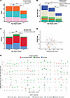 Figure 1
Adaptive functioning and psychopathology in our cohort of 54 individuals with KLEFS1 at different age categories. (A) Adaptive functioning (VABS) and its domains per age category. A total of 165 measurements were completed. *** P < 0.001. (B) Episodic psychiatric disorders measured with the mini PAS-ADD (DSM-categories). A total of 191 measurements were completed. (C) Total PANSS scores per domain per age category. A total of 93 measurements were completed. After the Bonferroni correction for multiple testing, differences were found between the age groups of patients aged 21–30 years and greater than or equal to 31 years on the positive, general pathology, and composite scales (significant on subscale general pathology). (D) Total individual PANSS scores. The horizontal line denotes the clinical cut-off of 28 points. A total of 93 measurements were completed, of which 33 measurements were on or above the cut-off point (35.5%). (E) Individual course over time of adaptive functioning and (olanzapine) treatment. Shows the individual course of adaptive functioning before and/or during treatment with psychiatric medication. Numbers corresponding to the Nijmegen cohort (e.g., 1= N1). The colors green and blue represent the target medicines olanzapine and clozapine, respectively. Red indicates that neither was administered. The form indicates whether other psychiatric medication was used. The category ‘other agents’ includes psychiatric medication from categories other than antipsychotics (antidepressants, lithium, antiepileptics for mood, and benzodiazepines). Treatment with olanzapine/clozapine was in accordance with Dutch guidelines regarding antipsychotic medication.
Psychopathology was assessed using the Mini Psychiatric Assessment Schedules for Adults with Developmental Disabilities Screening (mini-PAS-ADD) (Figure 1B) and Positive And Negative Syndrome Scale (PANSS) (Figure 1, C and D). Mean scores for several diagnostic categories were higher in the younger age groups compared with the oldest group in the mini-PAS-ADD results. Additionally, differences were observed between the 21–30 and 31+ age groups on the positive, general pathology, and composite scales of the PANSS with a peak in adolescent age.
Decline in adaptive functioning can be partially reversed by olanzapine
To determine whether there was a developmental regression in individuals, we assessed those with 3 or more adaptive measurements (n = 32) with no clinical suspicion for additional catatonia.
Sixteen of the participants experienced regression during the study and for this received olanzapine treatment (Figure 1E). Seven individuals clearly profited from olanzapine with recovery in functioning (numbers N1, N2, N3, N7, N9, N14, N21), 2 individuals stabilized but showed some recovery over time (N4, N23), 1 individual stabilized and showed some recovery after switch to clozapine (N19), 3 individuals stabilized for a while but continued to decline over time (N15, N17, N38), 1 individual initially declined but showed almost complete recovery 4 months later (N25), and 2 individuals further declined in functioning without any stabilization or minor recovery (N18, N28). So, in conclusion, 10 of 16 individuals did benefit in terms of improved functioning after an initial regressive period using olanzapine, and another 4 individuals stabilized at least temporarily. Extensive data points on the course of functionality that include data prior to initial regression phases were present for 7 of 10 individuals. They showed a decline in adaptive functioning during regression ranging from 25.6%– 87.2% of initial functioning, with greater range of decline in the higher functioning individuals. Recovery during olanzapine treatment in these individuals ranged from 78.4%–126% of the initial functioning. (Supplemental Figure 2). The latter indicates that the individual not only recovered to initial functioning but was able to even further improve. In 3 of 10 individuals (N2, N3, N14) data on their functioning before regression measures were lacking. Their functioning improved, respectively, 114%, 1434.9%, and 148.2% during olanzapine treatment. Dosage regimes and adverse reactions were comparable with those with idiopathic psychiatric illness (Supplemental Table 3).
EHMT1+/– human astrocytes show increased calcium response to glutamate and altered transcriptomic profile
To investigate the cellular mechanisms underlying the beneficial effects of olanzapine, we established a cellular model system to examine its impact. Based on transcriptomic data from the Brain RNA-seq database (15) and our own RNA-seq datasets (Supplemental Figure 3A), which both indicate a higher expression level of EHMT1 in astrocytes relative to neurons, we sought to explore its specific role in astrocytes, a cell type that has previously not been investigated in KLEFS1. We used a previously characterized EHMT1 loss-of-function isogenic pair engineered with CRISPR/Cas9 (EHMT1+/– and EHMT1+/+) (Figure 2A) (16). All iPS cells successfully differentiated into astrocytes, using a recently described protocol (34) and expressed typical astrocytic markers, including CD44, Vimentin, GFAP, and S100B (Supplemental Figure 3, B–E). As expected, EHMT1+/– astrocytes exhibited reduced EHMT1 expression compared with controls (Figure 2B).
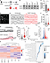 Figure 2
EHMT1+/– human astrocytes show increased calcium response to glutamate and altered transcriptomic profile. (A) IPS cells generated through CRIPSR/cas9 used in this study. (B) Western blot showing the EHMT1 protein levels in control and EHMT1+/– astrocytes. (C) Glutamate uptake ability of control and EHMT1+/– astrocytes, n = 4 independent cultures (D) Representative image of astrocytes for calcium imaging measure using Fluo8-AM. (E) Representative traces of control and EHMT1+/– astrocytes showing spontaneous calcium transients. (F) Quantification of AUC of spontaneous calcium transients. n = 139 for control and n = 197 for EHMT1+/–, 3 biological replicates (G) Representative traces of control and EHMT1+/– astrocytes showing evoked activity upon 50 μM glutamate stimulation. Thick black trace represents the average trace. (H) Quantification of AUC of evoked calcium events. n = 21 for control and n = 30 for EHMT1+/-, 3 biological replicates. (I) Principal component analysis (PCA) of RNA-seq data from control (Ctrl) and EHMT1+/– astrocytes, n = 3 independent cultures. (J) Volcano plots showing differentially expressed genes (DEGs) between EHMT1+/– and control human astrocytes. Relative to the control, significantly up- or downregulated genes are shown above the black dashed line in red and blue (FDR < 0.05; log2(FC) > 1; log2(FC) < –1).(K) Heatmap showing relative normalized gene expression across samples. Normalized counts were log2-transformed and scaled per gene to Z-scores, representing relative expression levels. Both genes and samples were hierarchically clustered based on Z-scores. (L) GO term analysis of molecular function. Data represent means ± SEM. *P < 0.05, **P < 0.01, ***P < 0.001. Unpaired Student’s t test (C, F and H).
A key functional property of astrocytes is their ability to support neuronal circuitry function by the uptake of extracellular glutamate. We observed that EHMT1+/– astrocytes exhibited significantly reduced glutamate uptake compared with control astrocytes (Figure 2C). Considering the crucial role of calcium signaling in astrocyte function, we next examined spontaneous cytosolic calcium dynamics in both control and EHMT1+/– astrocytes (Figure 2, D–F). While both genotypes displayed spontaneous calcium transients with no significant differences in frequency or amplitude (Figure 2F), glutamate stimulation elicited a markedly enhanced Ca²+ response in EHMT1+/– astrocytes compared with controls (Figure 2, G and H). These findings indicate that EHMT1+/– astrocytes exhibit altered calcium homeostasis and heightened sensitivity to glutamate-induced calcium signaling.
To investigate the molecular perturbations underlying altered astrocytic function in EHMT1-deficient astrocytes, we performed RNA sequencing on control and EHMT1+/– astrocytes. Differential expression analysis identified 1223 downregulated and 1427 significantly upregulated genes in EHMT1+/– astrocytes compared to controls (Figure 2, I and J, Supplemental Figure 4A, and Supplemental Table 7). The predominance of upregulated genes aligns with EHMT1’s known role as a transcriptional repressor (35). To obtain a broader perspective on affected pathways we performed a Gene Ontology (GO) analysis, which revealed significant enrichment in terms related to calcium ion binding, transmembrane signaling, ion transport and cytokines (Figure 2, K and L, and Supplemental Table 8).
To further gain insights into molecular underpinnings of the reduced glutamate uptake ability as well as increased calcium response to glutamate in EHMT1+/– astrocytes, we specifically examined genes involved in these processes. Astrocytic glutamate transporters, or excitatory amino acid transporters (EAATs), play a crucial role in maintaining extracellular glutamate homeostasis. Notably, our RNA-seq analysis revealed a significant downregulation of key glutamate transporter genes (SLC1A1, SLC1A3, and SLC1A4) in EHMT1+/– astrocytes (Supplemental Figure 4B). We independently validated the downregulation of these transporters via qPCR, providing a potential molecular explanation for the observed deficits in glutamate clearance (Supplemental Figure 4C).
Calcium homeostasis is regulated by a complex network of calcium channels, exchangers, binding proteins, and pumps. We identified multiple dysregulated genes associated with calcium homeostasis (Supplemental Table 8), including significant upregulation of store-operated calcium entry channel genes (TRPC3/4/6). Notably, some genes involved in calcium pathways, for example RyR1 and CACNA1H, exhibited dramatic increases of 626-fold and 53-fold, respectively, suggesting a major contribution to calcium signaling abnormalities in EHMT1+/– astrocytes. Additionally, several calcium-binding protein genes, such as CAMK2N1 and S100B, displayed altered expression (Supplemental Tables 7 and 8). Collectively, our findings indicate profound transcriptomic dysregulation in EHMT1+/– astrocytes, implicating disruptions in glutamate transport, calcium signaling, and broader cellular processes. Of interest, although the transcriptome of EHMT1+/– neurons (grown on control astrocytes) was more moderately affected (Supplemental Figure 4, D–H), GO term analysis revealed a similar enrichment in calcium ion binding (Supplemental Figure 4H and Supplemental Table 7).
Olanzapine rescues the neuronal network hyperactivity in EHMT1+/– astrocyte-neuron cocultures
To investigate whether astrocytes contribute to neuronal network dysfunction in KLEFS1, we cocultured human control or EHMT1+/– iPSC-derived Ngn2-induced glutamatergic neurons (iN) together with human astrocytes in an all-human model system. This allowed us to generate networks in which neurons and astrocytes of different genotypes can be cocultured in different combinations (i.e., iNCtrl+ACtrl, iNCtrl+AEhmt1+/–, iNEHMT1+/–+ACtrl and iNEHMT1+/–+AEHMT1+/–, Figure 3A). After 3 weeks of development in vitro, we measured the neuronal activity on MEAs (Figure 3B). iNEHMT1+/–+AEHMT1+/– networks showed a significant increase in their global activity level compared with iNCtrl+ACtrl networks (Figure 3C). Specifically, we found that the mean firing rate (MFR) of EHMT1+/– neurons was exacerbated when grown on EHMT1+/– astrocytes (iNEHMT1+/–+AEHMT1+/–, yellow) compared with culturing EHMT1+/– neurons on control astrocytes (iNEHMT1+/–+ACtrl, orange). Importantly, we found no difference in the density of astrocytes between the different conditions, nor was the ratio between astrocytes (GFAP) and neurons (MAP2) different between conditions (Supplemental Figure 3F).
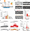 Figure 3
EHMT1+/– human astrocytes lead to neuronal network hyperactivity in EHMT1+/– neurons, which can be rescued by olanzapine. (A) Schematic overview of human astrocyte and human neuron cocultures. (B) Representative raster plot and (C) quantification of mean firing rate exhibited by control or EHMT1+/– astrocyte coculture with control or EHMT1+/– human neurons on MEA at DIV21. iNCtrl+ACtrl n = 12, iNCtrl+AEHMT1+/– n = 12, iNEHMT1+/–+ACtrl n = 11 and iNEHMT1+/–+AEHMT1+/– n = 12. Data represent means ± SEM. *P < 0.05, **P < 0.01, ***P < 0.001. One-way ANOVA test and post hoc Bonferroni correction (D) Representative raster plots and (E) quantification of mean firing rate exhibited by EHMT1+/– astrocyte-neuron cocultures on MEAs treated with 10 μM olanzapine for 1 week. Dashed line represents mean of untreated control cells, n = 6. Data represent means ± SEM. *P < 0.05, **P < 0.01, ***P < 0.001. Unpaired Student’s t test (E). (F) Representative traces and (G) quantification of calcium wave from control and EHMT1+/– astrocytes treated with/without olanzapine upon 50 μM glutamate stimulation. Control n = 94, Control + olanzapine n = 54, EHMT1+/– n = 104, EHMT1+/– + olanzapine n = 81 Data represent means ± SEM. *P < 0.05, **P < 0.01, ***P < 0.001. One-way ANOVA test and post hoc Bonferroni correction (G).
To corroborate these data, we also generated networks composed of human iN and mouse cortical astrocytes isolated from control or Ehmt1+/– mice, as rodent astrocytes are considered the gold standard in this type of in vitro models (Supplemental Figure 5, A and B). Networks consisting of mouse astrocytes showed more distinct synchronization of spontaneous network activity — represented by network bursts — after 3 weeks, compared with all-human cultures. We first recapitulated the glutamatergic neuron–specific network phenotype in iNEHMT+/–+ mouseACtrl, as we described before (16) (orange), which included a lower network burst rate and a longer burst duration compared with iNCtrl+mouseACtrl networks (dark grey) (Supplemental Figure 5, C–F). Similar to the all-human cultures, we found that the network phenotype of EHMT1+/– neurons was exacerbated when grown on mouse Ehmt1+/– astrocytes (iNEHMT+/–+mouseAEhmt1+/–, yellow) compared with culturing EHMT1+/– neurons on control astrocytes (Supplemental Figure 5C). Indeed, iNEHMT+/–+mouseAEhmt1+/– networks showed a significant increase in MFR compared with iNEHMT+/–+mouseACtrl networks (Supplemental Figure 5D). Together, these results confirm that astrocytes significantly contribute to neuronal network dysfunction in KLEFS1. Notably, when control neurons were grown with EHMT1-deficient astrocytes (iNCtrl+mouseAEhmt1+/–), we did not observe any significant differences in neuronal network activity compared with iNCtrl+mouseACtrl networks. These results suggest that EHMT1 deficiency in astrocytes alone is not sufficient to induce a major neuronal network phenotype. Moreover, these findings suggest that EHMT1+/– neurons are more sensitive than control neurons to the presence of EHMT1-deficient astrocytes, resulting in the observed cumulative effect on the network phenotype. Consistently, when measuring spontaneous excitatory postsynaptic currents (sEPSCs) using whole-cell patch-clamp recordings, we found that mouse Ehmt1+/– astrocytes aggravated the single-cell phenotype of EHMT1+/– neurons by means of an increased sEPSC frequency but not amplitude (Supplemental Figure 5, G–I).
Having established an all-human model system exhibiting a clear EHMT1 haploinsufficiency phenotype, we next investigated whether olanzapine could rescue the associated neuronal hyperactivity. To this end, we treated EHMT1+/– astrocyte-neuron cocultures with olanzapine for a week and compared them with untreated controls. olanzapine effectively reduced overall network activity to levels comparable with those of untreated control networks (Figure 3, D and E). This effect seemed to be specific for olanzapine, as other antipsychotics such as quetiapine and aripiprazole did not reduce overall network activity in EHMT1+/– astrocyte-neuron cocultures (Supplemental Figure 6).
Given our previous results showing that EHMT1 haploinsufficient astrocytes exhibited enhanced calcium response to glutamate stimulation and considering evidence that astrocytic calcium activity can influence neuronal excitability along with the previous reported impact of olanzapine on calcium signaling (36), we tested whether olanzapine could also normalize calcium signaling in EHMT1+/– astrocytes. Indeed, olanzapine treatment reduced the heightened calcium response to glutamate stimulation in EHMT1+/– astrocytes (Figure 3, F and G). These findings suggest that olanzapine can mitigate the hyperactive neuronal network phenotype associated with EHMT1 deficiency, potentially through its stabilizing effect on aberrant astrocytic calcium signaling.
To corroborate our results in an independent isogenic KS patient line, we generated iNeurons and astrocytes from previously characterized isogenic iPSCs obtained from a female who has a mosaic microdeletion on chromosome 9q34 (233 kb), including EHMT1 (KSmos and Cmos, Supplemental Figure 6, A and B) (16). KSmos neuronal networks were hyperactive compared with control Cmos networks (Supplemental Figure 7, C and D). This was accompanied by increased calcium response to glutamate. Importantly, these phenotypes could be rescued by treating the cells with olanzapine (Supplemental Figure 7E). Together, these data indicate a more general mechanism at play in KLEFS1.
Because EHMT1 is also expressed in other cell types, including dopaminergic and serotonergic neurons, and olanzapine is also known to exert some of its effects by modulating dopaminergic (D2) and serotonergic (5-HT2A) receptors (37), we explored the role of EHMT1 in these cells. To this end, we generated human iPSC-derived dopaminergic and serotonergic neurons (Supplemental Figure 8) and grew them on MEAs to measure changes in neuronal network activity. We performed a detailed analysis of neuronal network properties during development, but we did not observe any neuronal network phenotype between control and EHMT1-haploinsufficient dopaminergic or serotonergic neurons (Supplemental Figure 8). This suggests that these cell types are not directly affected by EHMT1 haploinsufficiency.
EHMT1+/– human astrocytes show increased S100B, which can be reduced by olanzapine treatment
We next sought to understand the underlying mechanisms by which olanzapine rescues abnormal calcium activity in EHMT1+/– astrocytes and attenuates hyperactive neuronal activity. Examining our astrocytic transcriptomic data, we were particularly interested in S100B, a key regulator of calcium wave propagation. Our RNA-seq data revealed an increased expression of S100B mRNA in EHMT1+/– astrocytes. Notably, previous studies have reported that olanzapine can reduce S100B levels (38). Based on this, we hypothesized that the elevated S100B expression in EHMT1+/– astrocytes could contribute to the observed calcium dysregulation and hyperactive neuronal network phenotype, and that olanzapine potentially exerts its rescue effect by downregulating S100B expression.
To test this hypothesis, we first confirmed, in independent experiments, that EHMT1 haploinsufficiency leads to increased S100B transcript and extracellular protein expression from human astrocytes. We performed both qPCR and ELISA for control and EHMT1+/– astrocytes at DIV 28. The results showed that, at both mRNA and protein levels, EHMT1 haploinsufficiency resulted in a significant increase of S100B (Figure 4, A and B). Further, using CUT&RUN-qPCR we measured reduced H3K9me2 occupancy at S100B promoter in EHMT1+/– astrocytes (Supplemental Figure 9A). In addition, we observed that 4 out of 8 CpG sites showed prominently decreased DNA methylation in EHMT1+/– astrocytes compared with control astrocytes (Supplemental Figure 9B). These results suggest that EHMT1 deficiency directly or indirectly results in reduced H3K9me2 and DNA methylation at the S100B promoter in astrocytes, possibly responsible for the increased expression of S100B.
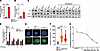 Figure 4
EHMT1+/– human astrocytes show increased S100B, and the level of S100B can be reduced by olanzapine treatment. (A) Relative expression of S100B in control and EHMT1+/– astrocytes using qPCR. Control n = 6, EHMT1+/– n = 6. (B) Relative protein level of S100B in the culture medium of control and EHMT1+/– astrocytes using ELISA. Control n = 4, EHMT1+/– n = 4. (C) Western blot showing the JAK/STAT3 protein levels in astrocytes in the absence or presence of olanzapine (D) quantification of Western Blot, normalized to control. Control n = 4, EHMT1+/– n = 4. (E) Representative images and (F) quantification of CellROX fluorescent intensity in control, control + Rotenone and EHMT1+/– astrocytes. Control n = 34, Control + 100 nM Rotenone n = 34, EHMT1+/– n = 41. (G) olanzapine (100 nM, 1 μM, 10 μM, 100 μM) treatment on EHMT1+/– astrocytes for 1 week could reduce S100B level in the culture medium in a dose-dependent manner as tested by ELISA. n = 3. Data represent means ± SEM. *P < 0.05, **P < 0.01, ***P < 0.001. One-way ANOVA test and post hoc Bonferroni correction (F) and unpaired Student’s t test (A, B, and D).
At nanomolar concentrations, S100B has been shown to activate the Ras/ERK/NF-kB pathway to promote neurotrophic functions (39, 40). However, when released at high concentrations (micromolar range) (41), S100B is suggested to act as a Damage-Associated Molecular Pattern (DAMP) molecule that triggers several stress response pathways (42). These include, among others, the JNK/p38 and the Janus Kinase (JAK)/signal transducer and activator of transcription (STAT) pathways, which are known to serve as downstream effectors potentially relevant to reactive oxygen species (ROS) production and reactive gliosis (42–45). This raised the question whether EHMT1+/– astrocytes showed aberrant activation of JAK/STAT signaling and accumulated higher ROS levels. To test this, we performed immunoblotting of JAK and STAT3. EHMT1+/– astrocytes showed significantly higher levels of total JAK and STAT3 (Figure 4, C and D). Strikingly, the higher levels of total JAK and STAT3 were abolished when cells were pretreated with olanzapine (Figure 4, C and D). Next, we assessed ROS levels using CellROX Green in EHMT1+/– astrocytes. As a positive control, we included control astrocytes treated with Rotenone, a mitochondrial complex one inhibitor, to induce oxidative stress (46). EHMT1+/– human astrocytes produced significantly higher ROS than control astrocytes (Figure 4, E and F). Consistent with the data collected from human astrocytes, Ehmt1+/– mouse astrocytes showed a similar increase in S100b (Supplemental Figure 10, A, C–-E, and H–J) and produced more ROS than control astrocytes in primary cultures (Supplemental Figure 10B). In line with these in vitro data, we also observed increased S100B, GFAP expression, and GFAP area in brain slices of the visual cortex and hippocampus of Ehmt1+/– mice, suggesting increased astrocyte reactivity and signs of neuroinflammation ex vivo (Supplemental Figure 10, C, F–H, K, and L). This further emphasizes the cross-species changes in S100B expression and increased oxidative stress in astrocytes with EHMT1 deficiency.
Treatment of olanzapine to individuals with schizophrenia (SCZ) for 2 weeks has been shown to significantly reduce plasma S100B level (38). Considering that astrocytes are the main cell type that could secrete S100B, we further explored whether, in our human astrocyte model, olanzapine could reduce S100B expression. To test this, we treated EHMT1+/– astrocytes with different doses of olanzapine for 1 week, after which we measured S100B protein level. We found that olanzapine reduced the extracellular S100B level in a dose-dependent manner (Figure 4G). Taken together, this indicates that EHMT1+/– astrocytes display an upregulation in S100B, which can be attenuated by the administration of olanzapine.
S100B inhibition in EHMT1+/– astrocytes rescues the neuronal network hyperactivity in EHMT1+/– astrocyte-neuron cocultures.
S100B has been reported as one of the important regulators in calcium signaling (44). We next investigated whether reducing S100B expression would be sufficient to revert the increased Ca2+ responses and neuronal network hyperactivity phenotypes. To pharmacologically reduce S100B expression, we treated EHMT1+/– astrocytes with the S100B synthesis inhibitor, ONO2506, for 24 hours. We observed that ONO2506 significantly reduced the increased calcium response to glutamate of EHMT1+/– astrocytes (Figure 5, A and B). Considering that calcium signaling in astrocytes could potentially influence neuronal excitability, we next tested whether, by pharmacologically inhibiting S100B, the hyperactive network phenotype in iNEHMT1+/–+AEHMT1+/– could be rescued. After 24 hours ONO2506 treatment (100 μM), we found that the global activity of iNEHMT1+/–+AEHMT1+/– cultures was significantly reduced (Figure 5, C and D). Next to pharmacological inhibition, we also used short hairpin RNA (shRNA) to genetically knock down S100B (Figure 5E). qPCR data confirmed that S100B shRNA significantly decreased S100B expression (Figure 5F). Consistently, both increased glutamate-evoked calcium response in EHMT1+/– astrocytes and the hyperactive network phenotype in astrocyte-neuron cocultures were normalized to control levels (Figure 5, G–J). Thus, we conclude that the hyperactivity of EHMT1+/– network, at least partially, is caused by increased S100B from EHMT1+/– astrocytes; and olanzapine could rescue the EHMT1+/– network phenotype via reducing S100B. Collectively, these data offers a new mechanistic perspective on how olanzapine can impact EHMT1 haploinsufficient neuronal networks.
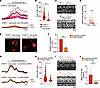 Figure 5
Pharmacological inhibition and genetic knockdown of S100B in EHMT1+/– astrocytes rescue the neuronal network hyperactivity in EHMT1+/– astrocyte-neuron cocultures. (A) Representative traces and (B) quantification of EHMT1+/– astrocytes with ONO2506 showing reduced evoked activity upon 50 μM glutamate stimulation. n = 57 for EHMT1+/– and n = 63 for EHMT1+/– with ONO2506. (C) Representative raster plots and (D) quantification of mean firing rate exhibited by EHMT1+/– astrocytes cocultured with EHMT1+/– neurons on MEAs at DIV21 after 24-hour treatment of 100 μM ONO2506. n = 12. (E) EHMT1+/– astrocytes infected lentivirus expressing mcherry and scrambled or S100B shRNA. (F) Relative expression of mRNA S100B in EHMT1+/– astrocytes infected with S100B shRNA compared with astrocytes transfected with scrambled shRNA. n = 6 for each group. (G) Representative traces and (H) quantification of calcium wave from EHMT1+/– astrocytes transfected with scrambled shRNA and S100B shRNA upon 50 μM glutamate stimulation. n = 29 for EHMT1+/– + scrambled shRNA and n = 21 for EHMT1+/– + S100B shRNA. (I) Representative raster plots and (J) quantification of mean firing rate exhibited by EHMT1+/– neurons cocultured with EHMT1+/– astrocytes transfected with scrambled shRNA or S100B shRNA on MEAs. Dashed line represents mean of untreated control cells, n = 6 for scrambled shRNA. n = 15 for S100B shRNA. Data represent means ± SEM. *P < 0.05, **P < 0.01, ***P < 0.001. Unpaired Student’s t test (B, F, H, and J) and paired Student’s t test (D).


Copyright © 2026 American Society for Clinical Investigation
ISSN: 0021-9738 (print), 1558-8238 (online)