
Advertisement
Research ArticleCell biologyOncology
Open Access |  10.1172/JCI189034
Targeting N-acetyltransferase 10 reprograms Treg function to enhance anti–PD-1 immunotherapy sensitivity in murine lung tumors
Xuewei Qi,1 Dan Cui,2 Xiao Li,3 Wenfan Fu,4 Handong Ma,1 Wenting Guo,5 Jiabao Wang,1 Jiaxi Song,1 Jiaxin Ma,1 Jing Yang,1 Haoyu Yu,1 Yuting Hong,1 Qian Wang,6 Susu Luo,7 Jia Wang,7 Baixing Wu,5 Shanshan Li,3 Hao Cheng,8 Min Cheng,9 Bin Li,8 Kaiming Zhang,3 Jia Li,2,10 and Fengyin Li1,11
1State Key Laboratory of Immune Response and Immunotherapy, School of Basic Medical Sciences, Division of Life Sciences and Medicine, University of Science and Technology of China, Hefei, China.
2Guangzhou National Laboratory, Xing Dao Huan Bei Road, Guangzhou International Bio Island, Guangzhou, China.
3Ministry of Education Key Laboratory for Cellular Dynamics, Center for Advanced Interdisciplinary Science and Biomedicine of Institute of Health and Medicine (IHM), Division of Life Sciences and Medicine, University of Science and Technology of China, Hefei, China.
4Department of Chest Surgery, Affiliated Cancer Hospital and Institute of Guangzhou Medical University, Guangzhou, China.
5Guangdong Provincial Key Laboratory of Malignant Tumor Epigenetics and Gene Regulation, Guangdong-Hong Kong Joint Laboratory for RNA Medicine, Medical Research Center, Sun Yat-Sen Memorial Hospital, Sun Yat-Sen University, Guangzhou, China.
6Affiliated Cancer Hospital and Institute of Guangzhou Medical University, Guangzhou, China.
7Guangzhou Medical University–Guangzhou Institutes of Biomedicine and Health Joint School of Life Sciences, Guangdong-Hong Kong-Macau Joint Laboratory for Cell Fate Regulation and Diseases, Guangzhou Medical University, Guangzhou, China.
8Center for Immune-related Disease at Shanghai Institute of Immunology, Department of Respiratory and Critical Care Medicine of Ruijin Hospital, Shanghai Jiao Tong University School of Medicine, Shanghai, China.
9Department of Geriatrics, Gerontology Institute of Anhui Province, The First Affiliated Hospital of University of Science and Technology of China, Division of Life Sciences and Medicine, University of Science and Technology of China, Hefei, China.
10State Key Laboratory of Respiratory Disease, National Clinical Research Center for Respiratory Disease, Guangzhou Institute of Respiratory Health, The First Affiliated Hospital of Guangzhou Medical University, Guangzhou, China.
11Center for Advanced Interdisciplinary Science and Biomedicine of IHM, Division of Life Sciences and Medicine, University of Science and Technology of China, Hefei, China.
Address for correspondence. Fengyin Li, 443 Huangshan Road, University of Science and Technology of China, Anhui, China. Phone: 86.0551.63600939; Email: fyli88@ustc.edu.cn. Or to: Jia Li, 151 Yanjiang West Road, The First Affiliated Hospital of Guangzhou Medical University, Guangdong, China. Phone: 86.19878341601; Email: jiali@gzhmu.edu.cn. Or to: Kaiming Zhang, 443 Huangshan Road, University of Science and Technology of China, Anhui, China. Phone: 86.13694415677; Email: kmzhang@ustc.edu.cn. Or to: Bin Li, 280 South Chongqing Road, Shanghai Jiao Tong University School of Medicine, Shanghai, China. Phone: 86.021.6384.6590.776783; Email: binli@shsmu.edu.cn. Or to: Min Cheng, 443 Huangshan Road, University of Science and Technology of China, Anhui, China. Phone: 86.0551.62282371; Email: chengmin@ustc.edu.cn.
Authorship note: XQ, DC, XL, WF, and HM are co–first authors.
Find articles by Qi, X. in: PubMed | Google Scholar
1State Key Laboratory of Immune Response and Immunotherapy, School of Basic Medical Sciences, Division of Life Sciences and Medicine, University of Science and Technology of China, Hefei, China.
2Guangzhou National Laboratory, Xing Dao Huan Bei Road, Guangzhou International Bio Island, Guangzhou, China.
3Ministry of Education Key Laboratory for Cellular Dynamics, Center for Advanced Interdisciplinary Science and Biomedicine of Institute of Health and Medicine (IHM), Division of Life Sciences and Medicine, University of Science and Technology of China, Hefei, China.
4Department of Chest Surgery, Affiliated Cancer Hospital and Institute of Guangzhou Medical University, Guangzhou, China.
5Guangdong Provincial Key Laboratory of Malignant Tumor Epigenetics and Gene Regulation, Guangdong-Hong Kong Joint Laboratory for RNA Medicine, Medical Research Center, Sun Yat-Sen Memorial Hospital, Sun Yat-Sen University, Guangzhou, China.
6Affiliated Cancer Hospital and Institute of Guangzhou Medical University, Guangzhou, China.
7Guangzhou Medical University–Guangzhou Institutes of Biomedicine and Health Joint School of Life Sciences, Guangdong-Hong Kong-Macau Joint Laboratory for Cell Fate Regulation and Diseases, Guangzhou Medical University, Guangzhou, China.
8Center for Immune-related Disease at Shanghai Institute of Immunology, Department of Respiratory and Critical Care Medicine of Ruijin Hospital, Shanghai Jiao Tong University School of Medicine, Shanghai, China.
9Department of Geriatrics, Gerontology Institute of Anhui Province, The First Affiliated Hospital of University of Science and Technology of China, Division of Life Sciences and Medicine, University of Science and Technology of China, Hefei, China.
10State Key Laboratory of Respiratory Disease, National Clinical Research Center for Respiratory Disease, Guangzhou Institute of Respiratory Health, The First Affiliated Hospital of Guangzhou Medical University, Guangzhou, China.
11Center for Advanced Interdisciplinary Science and Biomedicine of IHM, Division of Life Sciences and Medicine, University of Science and Technology of China, Hefei, China.
Address for correspondence. Fengyin Li, 443 Huangshan Road, University of Science and Technology of China, Anhui, China. Phone: 86.0551.63600939; Email: fyli88@ustc.edu.cn. Or to: Jia Li, 151 Yanjiang West Road, The First Affiliated Hospital of Guangzhou Medical University, Guangdong, China. Phone: 86.19878341601; Email: jiali@gzhmu.edu.cn. Or to: Kaiming Zhang, 443 Huangshan Road, University of Science and Technology of China, Anhui, China. Phone: 86.13694415677; Email: kmzhang@ustc.edu.cn. Or to: Bin Li, 280 South Chongqing Road, Shanghai Jiao Tong University School of Medicine, Shanghai, China. Phone: 86.021.6384.6590.776783; Email: binli@shsmu.edu.cn. Or to: Min Cheng, 443 Huangshan Road, University of Science and Technology of China, Anhui, China. Phone: 86.0551.62282371; Email: chengmin@ustc.edu.cn.
Authorship note: XQ, DC, XL, WF, and HM are co–first authors.
Find articles by Cui, D. in: PubMed | Google Scholar
1State Key Laboratory of Immune Response and Immunotherapy, School of Basic Medical Sciences, Division of Life Sciences and Medicine, University of Science and Technology of China, Hefei, China.
2Guangzhou National Laboratory, Xing Dao Huan Bei Road, Guangzhou International Bio Island, Guangzhou, China.
3Ministry of Education Key Laboratory for Cellular Dynamics, Center for Advanced Interdisciplinary Science and Biomedicine of Institute of Health and Medicine (IHM), Division of Life Sciences and Medicine, University of Science and Technology of China, Hefei, China.
4Department of Chest Surgery, Affiliated Cancer Hospital and Institute of Guangzhou Medical University, Guangzhou, China.
5Guangdong Provincial Key Laboratory of Malignant Tumor Epigenetics and Gene Regulation, Guangdong-Hong Kong Joint Laboratory for RNA Medicine, Medical Research Center, Sun Yat-Sen Memorial Hospital, Sun Yat-Sen University, Guangzhou, China.
6Affiliated Cancer Hospital and Institute of Guangzhou Medical University, Guangzhou, China.
7Guangzhou Medical University–Guangzhou Institutes of Biomedicine and Health Joint School of Life Sciences, Guangdong-Hong Kong-Macau Joint Laboratory for Cell Fate Regulation and Diseases, Guangzhou Medical University, Guangzhou, China.
8Center for Immune-related Disease at Shanghai Institute of Immunology, Department of Respiratory and Critical Care Medicine of Ruijin Hospital, Shanghai Jiao Tong University School of Medicine, Shanghai, China.
9Department of Geriatrics, Gerontology Institute of Anhui Province, The First Affiliated Hospital of University of Science and Technology of China, Division of Life Sciences and Medicine, University of Science and Technology of China, Hefei, China.
10State Key Laboratory of Respiratory Disease, National Clinical Research Center for Respiratory Disease, Guangzhou Institute of Respiratory Health, The First Affiliated Hospital of Guangzhou Medical University, Guangzhou, China.
11Center for Advanced Interdisciplinary Science and Biomedicine of IHM, Division of Life Sciences and Medicine, University of Science and Technology of China, Hefei, China.
Address for correspondence. Fengyin Li, 443 Huangshan Road, University of Science and Technology of China, Anhui, China. Phone: 86.0551.63600939; Email: fyli88@ustc.edu.cn. Or to: Jia Li, 151 Yanjiang West Road, The First Affiliated Hospital of Guangzhou Medical University, Guangdong, China. Phone: 86.19878341601; Email: jiali@gzhmu.edu.cn. Or to: Kaiming Zhang, 443 Huangshan Road, University of Science and Technology of China, Anhui, China. Phone: 86.13694415677; Email: kmzhang@ustc.edu.cn. Or to: Bin Li, 280 South Chongqing Road, Shanghai Jiao Tong University School of Medicine, Shanghai, China. Phone: 86.021.6384.6590.776783; Email: binli@shsmu.edu.cn. Or to: Min Cheng, 443 Huangshan Road, University of Science and Technology of China, Anhui, China. Phone: 86.0551.62282371; Email: chengmin@ustc.edu.cn.
Authorship note: XQ, DC, XL, WF, and HM are co–first authors.
Find articles by Li, X. in: PubMed | Google Scholar
1State Key Laboratory of Immune Response and Immunotherapy, School of Basic Medical Sciences, Division of Life Sciences and Medicine, University of Science and Technology of China, Hefei, China.
2Guangzhou National Laboratory, Xing Dao Huan Bei Road, Guangzhou International Bio Island, Guangzhou, China.
3Ministry of Education Key Laboratory for Cellular Dynamics, Center for Advanced Interdisciplinary Science and Biomedicine of Institute of Health and Medicine (IHM), Division of Life Sciences and Medicine, University of Science and Technology of China, Hefei, China.
4Department of Chest Surgery, Affiliated Cancer Hospital and Institute of Guangzhou Medical University, Guangzhou, China.
5Guangdong Provincial Key Laboratory of Malignant Tumor Epigenetics and Gene Regulation, Guangdong-Hong Kong Joint Laboratory for RNA Medicine, Medical Research Center, Sun Yat-Sen Memorial Hospital, Sun Yat-Sen University, Guangzhou, China.
6Affiliated Cancer Hospital and Institute of Guangzhou Medical University, Guangzhou, China.
7Guangzhou Medical University–Guangzhou Institutes of Biomedicine and Health Joint School of Life Sciences, Guangdong-Hong Kong-Macau Joint Laboratory for Cell Fate Regulation and Diseases, Guangzhou Medical University, Guangzhou, China.
8Center for Immune-related Disease at Shanghai Institute of Immunology, Department of Respiratory and Critical Care Medicine of Ruijin Hospital, Shanghai Jiao Tong University School of Medicine, Shanghai, China.
9Department of Geriatrics, Gerontology Institute of Anhui Province, The First Affiliated Hospital of University of Science and Technology of China, Division of Life Sciences and Medicine, University of Science and Technology of China, Hefei, China.
10State Key Laboratory of Respiratory Disease, National Clinical Research Center for Respiratory Disease, Guangzhou Institute of Respiratory Health, The First Affiliated Hospital of Guangzhou Medical University, Guangzhou, China.
11Center for Advanced Interdisciplinary Science and Biomedicine of IHM, Division of Life Sciences and Medicine, University of Science and Technology of China, Hefei, China.
Address for correspondence. Fengyin Li, 443 Huangshan Road, University of Science and Technology of China, Anhui, China. Phone: 86.0551.63600939; Email: fyli88@ustc.edu.cn. Or to: Jia Li, 151 Yanjiang West Road, The First Affiliated Hospital of Guangzhou Medical University, Guangdong, China. Phone: 86.19878341601; Email: jiali@gzhmu.edu.cn. Or to: Kaiming Zhang, 443 Huangshan Road, University of Science and Technology of China, Anhui, China. Phone: 86.13694415677; Email: kmzhang@ustc.edu.cn. Or to: Bin Li, 280 South Chongqing Road, Shanghai Jiao Tong University School of Medicine, Shanghai, China. Phone: 86.021.6384.6590.776783; Email: binli@shsmu.edu.cn. Or to: Min Cheng, 443 Huangshan Road, University of Science and Technology of China, Anhui, China. Phone: 86.0551.62282371; Email: chengmin@ustc.edu.cn.
Authorship note: XQ, DC, XL, WF, and HM are co–first authors.
Find articles by Fu, W. in: PubMed | Google Scholar
1State Key Laboratory of Immune Response and Immunotherapy, School of Basic Medical Sciences, Division of Life Sciences and Medicine, University of Science and Technology of China, Hefei, China.
2Guangzhou National Laboratory, Xing Dao Huan Bei Road, Guangzhou International Bio Island, Guangzhou, China.
3Ministry of Education Key Laboratory for Cellular Dynamics, Center for Advanced Interdisciplinary Science and Biomedicine of Institute of Health and Medicine (IHM), Division of Life Sciences and Medicine, University of Science and Technology of China, Hefei, China.
4Department of Chest Surgery, Affiliated Cancer Hospital and Institute of Guangzhou Medical University, Guangzhou, China.
5Guangdong Provincial Key Laboratory of Malignant Tumor Epigenetics and Gene Regulation, Guangdong-Hong Kong Joint Laboratory for RNA Medicine, Medical Research Center, Sun Yat-Sen Memorial Hospital, Sun Yat-Sen University, Guangzhou, China.
6Affiliated Cancer Hospital and Institute of Guangzhou Medical University, Guangzhou, China.
7Guangzhou Medical University–Guangzhou Institutes of Biomedicine and Health Joint School of Life Sciences, Guangdong-Hong Kong-Macau Joint Laboratory for Cell Fate Regulation and Diseases, Guangzhou Medical University, Guangzhou, China.
8Center for Immune-related Disease at Shanghai Institute of Immunology, Department of Respiratory and Critical Care Medicine of Ruijin Hospital, Shanghai Jiao Tong University School of Medicine, Shanghai, China.
9Department of Geriatrics, Gerontology Institute of Anhui Province, The First Affiliated Hospital of University of Science and Technology of China, Division of Life Sciences and Medicine, University of Science and Technology of China, Hefei, China.
10State Key Laboratory of Respiratory Disease, National Clinical Research Center for Respiratory Disease, Guangzhou Institute of Respiratory Health, The First Affiliated Hospital of Guangzhou Medical University, Guangzhou, China.
11Center for Advanced Interdisciplinary Science and Biomedicine of IHM, Division of Life Sciences and Medicine, University of Science and Technology of China, Hefei, China.
Address for correspondence. Fengyin Li, 443 Huangshan Road, University of Science and Technology of China, Anhui, China. Phone: 86.0551.63600939; Email: fyli88@ustc.edu.cn. Or to: Jia Li, 151 Yanjiang West Road, The First Affiliated Hospital of Guangzhou Medical University, Guangdong, China. Phone: 86.19878341601; Email: jiali@gzhmu.edu.cn. Or to: Kaiming Zhang, 443 Huangshan Road, University of Science and Technology of China, Anhui, China. Phone: 86.13694415677; Email: kmzhang@ustc.edu.cn. Or to: Bin Li, 280 South Chongqing Road, Shanghai Jiao Tong University School of Medicine, Shanghai, China. Phone: 86.021.6384.6590.776783; Email: binli@shsmu.edu.cn. Or to: Min Cheng, 443 Huangshan Road, University of Science and Technology of China, Anhui, China. Phone: 86.0551.62282371; Email: chengmin@ustc.edu.cn.
Authorship note: XQ, DC, XL, WF, and HM are co–first authors.
Find articles by Ma, H. in: PubMed | Google Scholar
1State Key Laboratory of Immune Response and Immunotherapy, School of Basic Medical Sciences, Division of Life Sciences and Medicine, University of Science and Technology of China, Hefei, China.
2Guangzhou National Laboratory, Xing Dao Huan Bei Road, Guangzhou International Bio Island, Guangzhou, China.
3Ministry of Education Key Laboratory for Cellular Dynamics, Center for Advanced Interdisciplinary Science and Biomedicine of Institute of Health and Medicine (IHM), Division of Life Sciences and Medicine, University of Science and Technology of China, Hefei, China.
4Department of Chest Surgery, Affiliated Cancer Hospital and Institute of Guangzhou Medical University, Guangzhou, China.
5Guangdong Provincial Key Laboratory of Malignant Tumor Epigenetics and Gene Regulation, Guangdong-Hong Kong Joint Laboratory for RNA Medicine, Medical Research Center, Sun Yat-Sen Memorial Hospital, Sun Yat-Sen University, Guangzhou, China.
6Affiliated Cancer Hospital and Institute of Guangzhou Medical University, Guangzhou, China.
7Guangzhou Medical University–Guangzhou Institutes of Biomedicine and Health Joint School of Life Sciences, Guangdong-Hong Kong-Macau Joint Laboratory for Cell Fate Regulation and Diseases, Guangzhou Medical University, Guangzhou, China.
8Center for Immune-related Disease at Shanghai Institute of Immunology, Department of Respiratory and Critical Care Medicine of Ruijin Hospital, Shanghai Jiao Tong University School of Medicine, Shanghai, China.
9Department of Geriatrics, Gerontology Institute of Anhui Province, The First Affiliated Hospital of University of Science and Technology of China, Division of Life Sciences and Medicine, University of Science and Technology of China, Hefei, China.
10State Key Laboratory of Respiratory Disease, National Clinical Research Center for Respiratory Disease, Guangzhou Institute of Respiratory Health, The First Affiliated Hospital of Guangzhou Medical University, Guangzhou, China.
11Center for Advanced Interdisciplinary Science and Biomedicine of IHM, Division of Life Sciences and Medicine, University of Science and Technology of China, Hefei, China.
Address for correspondence. Fengyin Li, 443 Huangshan Road, University of Science and Technology of China, Anhui, China. Phone: 86.0551.63600939; Email: fyli88@ustc.edu.cn. Or to: Jia Li, 151 Yanjiang West Road, The First Affiliated Hospital of Guangzhou Medical University, Guangdong, China. Phone: 86.19878341601; Email: jiali@gzhmu.edu.cn. Or to: Kaiming Zhang, 443 Huangshan Road, University of Science and Technology of China, Anhui, China. Phone: 86.13694415677; Email: kmzhang@ustc.edu.cn. Or to: Bin Li, 280 South Chongqing Road, Shanghai Jiao Tong University School of Medicine, Shanghai, China. Phone: 86.021.6384.6590.776783; Email: binli@shsmu.edu.cn. Or to: Min Cheng, 443 Huangshan Road, University of Science and Technology of China, Anhui, China. Phone: 86.0551.62282371; Email: chengmin@ustc.edu.cn.
Authorship note: XQ, DC, XL, WF, and HM are co–first authors.
Find articles by Guo, W. in: PubMed | Google Scholar
1State Key Laboratory of Immune Response and Immunotherapy, School of Basic Medical Sciences, Division of Life Sciences and Medicine, University of Science and Technology of China, Hefei, China.
2Guangzhou National Laboratory, Xing Dao Huan Bei Road, Guangzhou International Bio Island, Guangzhou, China.
3Ministry of Education Key Laboratory for Cellular Dynamics, Center for Advanced Interdisciplinary Science and Biomedicine of Institute of Health and Medicine (IHM), Division of Life Sciences and Medicine, University of Science and Technology of China, Hefei, China.
4Department of Chest Surgery, Affiliated Cancer Hospital and Institute of Guangzhou Medical University, Guangzhou, China.
5Guangdong Provincial Key Laboratory of Malignant Tumor Epigenetics and Gene Regulation, Guangdong-Hong Kong Joint Laboratory for RNA Medicine, Medical Research Center, Sun Yat-Sen Memorial Hospital, Sun Yat-Sen University, Guangzhou, China.
6Affiliated Cancer Hospital and Institute of Guangzhou Medical University, Guangzhou, China.
7Guangzhou Medical University–Guangzhou Institutes of Biomedicine and Health Joint School of Life Sciences, Guangdong-Hong Kong-Macau Joint Laboratory for Cell Fate Regulation and Diseases, Guangzhou Medical University, Guangzhou, China.
8Center for Immune-related Disease at Shanghai Institute of Immunology, Department of Respiratory and Critical Care Medicine of Ruijin Hospital, Shanghai Jiao Tong University School of Medicine, Shanghai, China.
9Department of Geriatrics, Gerontology Institute of Anhui Province, The First Affiliated Hospital of University of Science and Technology of China, Division of Life Sciences and Medicine, University of Science and Technology of China, Hefei, China.
10State Key Laboratory of Respiratory Disease, National Clinical Research Center for Respiratory Disease, Guangzhou Institute of Respiratory Health, The First Affiliated Hospital of Guangzhou Medical University, Guangzhou, China.
11Center for Advanced Interdisciplinary Science and Biomedicine of IHM, Division of Life Sciences and Medicine, University of Science and Technology of China, Hefei, China.
Address for correspondence. Fengyin Li, 443 Huangshan Road, University of Science and Technology of China, Anhui, China. Phone: 86.0551.63600939; Email: fyli88@ustc.edu.cn. Or to: Jia Li, 151 Yanjiang West Road, The First Affiliated Hospital of Guangzhou Medical University, Guangdong, China. Phone: 86.19878341601; Email: jiali@gzhmu.edu.cn. Or to: Kaiming Zhang, 443 Huangshan Road, University of Science and Technology of China, Anhui, China. Phone: 86.13694415677; Email: kmzhang@ustc.edu.cn. Or to: Bin Li, 280 South Chongqing Road, Shanghai Jiao Tong University School of Medicine, Shanghai, China. Phone: 86.021.6384.6590.776783; Email: binli@shsmu.edu.cn. Or to: Min Cheng, 443 Huangshan Road, University of Science and Technology of China, Anhui, China. Phone: 86.0551.62282371; Email: chengmin@ustc.edu.cn.
Authorship note: XQ, DC, XL, WF, and HM are co–first authors.
Find articles by Wang, J. in: PubMed | Google Scholar
1State Key Laboratory of Immune Response and Immunotherapy, School of Basic Medical Sciences, Division of Life Sciences and Medicine, University of Science and Technology of China, Hefei, China.
2Guangzhou National Laboratory, Xing Dao Huan Bei Road, Guangzhou International Bio Island, Guangzhou, China.
3Ministry of Education Key Laboratory for Cellular Dynamics, Center for Advanced Interdisciplinary Science and Biomedicine of Institute of Health and Medicine (IHM), Division of Life Sciences and Medicine, University of Science and Technology of China, Hefei, China.
4Department of Chest Surgery, Affiliated Cancer Hospital and Institute of Guangzhou Medical University, Guangzhou, China.
5Guangdong Provincial Key Laboratory of Malignant Tumor Epigenetics and Gene Regulation, Guangdong-Hong Kong Joint Laboratory for RNA Medicine, Medical Research Center, Sun Yat-Sen Memorial Hospital, Sun Yat-Sen University, Guangzhou, China.
6Affiliated Cancer Hospital and Institute of Guangzhou Medical University, Guangzhou, China.
7Guangzhou Medical University–Guangzhou Institutes of Biomedicine and Health Joint School of Life Sciences, Guangdong-Hong Kong-Macau Joint Laboratory for Cell Fate Regulation and Diseases, Guangzhou Medical University, Guangzhou, China.
8Center for Immune-related Disease at Shanghai Institute of Immunology, Department of Respiratory and Critical Care Medicine of Ruijin Hospital, Shanghai Jiao Tong University School of Medicine, Shanghai, China.
9Department of Geriatrics, Gerontology Institute of Anhui Province, The First Affiliated Hospital of University of Science and Technology of China, Division of Life Sciences and Medicine, University of Science and Technology of China, Hefei, China.
10State Key Laboratory of Respiratory Disease, National Clinical Research Center for Respiratory Disease, Guangzhou Institute of Respiratory Health, The First Affiliated Hospital of Guangzhou Medical University, Guangzhou, China.
11Center for Advanced Interdisciplinary Science and Biomedicine of IHM, Division of Life Sciences and Medicine, University of Science and Technology of China, Hefei, China.
Address for correspondence. Fengyin Li, 443 Huangshan Road, University of Science and Technology of China, Anhui, China. Phone: 86.0551.63600939; Email: fyli88@ustc.edu.cn. Or to: Jia Li, 151 Yanjiang West Road, The First Affiliated Hospital of Guangzhou Medical University, Guangdong, China. Phone: 86.19878341601; Email: jiali@gzhmu.edu.cn. Or to: Kaiming Zhang, 443 Huangshan Road, University of Science and Technology of China, Anhui, China. Phone: 86.13694415677; Email: kmzhang@ustc.edu.cn. Or to: Bin Li, 280 South Chongqing Road, Shanghai Jiao Tong University School of Medicine, Shanghai, China. Phone: 86.021.6384.6590.776783; Email: binli@shsmu.edu.cn. Or to: Min Cheng, 443 Huangshan Road, University of Science and Technology of China, Anhui, China. Phone: 86.0551.62282371; Email: chengmin@ustc.edu.cn.
Authorship note: XQ, DC, XL, WF, and HM are co–first authors.
Find articles by Song, J. in: PubMed | Google Scholar
1State Key Laboratory of Immune Response and Immunotherapy, School of Basic Medical Sciences, Division of Life Sciences and Medicine, University of Science and Technology of China, Hefei, China.
2Guangzhou National Laboratory, Xing Dao Huan Bei Road, Guangzhou International Bio Island, Guangzhou, China.
3Ministry of Education Key Laboratory for Cellular Dynamics, Center for Advanced Interdisciplinary Science and Biomedicine of Institute of Health and Medicine (IHM), Division of Life Sciences and Medicine, University of Science and Technology of China, Hefei, China.
4Department of Chest Surgery, Affiliated Cancer Hospital and Institute of Guangzhou Medical University, Guangzhou, China.
5Guangdong Provincial Key Laboratory of Malignant Tumor Epigenetics and Gene Regulation, Guangdong-Hong Kong Joint Laboratory for RNA Medicine, Medical Research Center, Sun Yat-Sen Memorial Hospital, Sun Yat-Sen University, Guangzhou, China.
6Affiliated Cancer Hospital and Institute of Guangzhou Medical University, Guangzhou, China.
7Guangzhou Medical University–Guangzhou Institutes of Biomedicine and Health Joint School of Life Sciences, Guangdong-Hong Kong-Macau Joint Laboratory for Cell Fate Regulation and Diseases, Guangzhou Medical University, Guangzhou, China.
8Center for Immune-related Disease at Shanghai Institute of Immunology, Department of Respiratory and Critical Care Medicine of Ruijin Hospital, Shanghai Jiao Tong University School of Medicine, Shanghai, China.
9Department of Geriatrics, Gerontology Institute of Anhui Province, The First Affiliated Hospital of University of Science and Technology of China, Division of Life Sciences and Medicine, University of Science and Technology of China, Hefei, China.
10State Key Laboratory of Respiratory Disease, National Clinical Research Center for Respiratory Disease, Guangzhou Institute of Respiratory Health, The First Affiliated Hospital of Guangzhou Medical University, Guangzhou, China.
11Center for Advanced Interdisciplinary Science and Biomedicine of IHM, Division of Life Sciences and Medicine, University of Science and Technology of China, Hefei, China.
Address for correspondence. Fengyin Li, 443 Huangshan Road, University of Science and Technology of China, Anhui, China. Phone: 86.0551.63600939; Email: fyli88@ustc.edu.cn. Or to: Jia Li, 151 Yanjiang West Road, The First Affiliated Hospital of Guangzhou Medical University, Guangdong, China. Phone: 86.19878341601; Email: jiali@gzhmu.edu.cn. Or to: Kaiming Zhang, 443 Huangshan Road, University of Science and Technology of China, Anhui, China. Phone: 86.13694415677; Email: kmzhang@ustc.edu.cn. Or to: Bin Li, 280 South Chongqing Road, Shanghai Jiao Tong University School of Medicine, Shanghai, China. Phone: 86.021.6384.6590.776783; Email: binli@shsmu.edu.cn. Or to: Min Cheng, 443 Huangshan Road, University of Science and Technology of China, Anhui, China. Phone: 86.0551.62282371; Email: chengmin@ustc.edu.cn.
Authorship note: XQ, DC, XL, WF, and HM are co–first authors.
Find articles by Ma, J. in: PubMed | Google Scholar
1State Key Laboratory of Immune Response and Immunotherapy, School of Basic Medical Sciences, Division of Life Sciences and Medicine, University of Science and Technology of China, Hefei, China.
2Guangzhou National Laboratory, Xing Dao Huan Bei Road, Guangzhou International Bio Island, Guangzhou, China.
3Ministry of Education Key Laboratory for Cellular Dynamics, Center for Advanced Interdisciplinary Science and Biomedicine of Institute of Health and Medicine (IHM), Division of Life Sciences and Medicine, University of Science and Technology of China, Hefei, China.
4Department of Chest Surgery, Affiliated Cancer Hospital and Institute of Guangzhou Medical University, Guangzhou, China.
5Guangdong Provincial Key Laboratory of Malignant Tumor Epigenetics and Gene Regulation, Guangdong-Hong Kong Joint Laboratory for RNA Medicine, Medical Research Center, Sun Yat-Sen Memorial Hospital, Sun Yat-Sen University, Guangzhou, China.
6Affiliated Cancer Hospital and Institute of Guangzhou Medical University, Guangzhou, China.
7Guangzhou Medical University–Guangzhou Institutes of Biomedicine and Health Joint School of Life Sciences, Guangdong-Hong Kong-Macau Joint Laboratory for Cell Fate Regulation and Diseases, Guangzhou Medical University, Guangzhou, China.
8Center for Immune-related Disease at Shanghai Institute of Immunology, Department of Respiratory and Critical Care Medicine of Ruijin Hospital, Shanghai Jiao Tong University School of Medicine, Shanghai, China.
9Department of Geriatrics, Gerontology Institute of Anhui Province, The First Affiliated Hospital of University of Science and Technology of China, Division of Life Sciences and Medicine, University of Science and Technology of China, Hefei, China.
10State Key Laboratory of Respiratory Disease, National Clinical Research Center for Respiratory Disease, Guangzhou Institute of Respiratory Health, The First Affiliated Hospital of Guangzhou Medical University, Guangzhou, China.
11Center for Advanced Interdisciplinary Science and Biomedicine of IHM, Division of Life Sciences and Medicine, University of Science and Technology of China, Hefei, China.
Address for correspondence. Fengyin Li, 443 Huangshan Road, University of Science and Technology of China, Anhui, China. Phone: 86.0551.63600939; Email: fyli88@ustc.edu.cn. Or to: Jia Li, 151 Yanjiang West Road, The First Affiliated Hospital of Guangzhou Medical University, Guangdong, China. Phone: 86.19878341601; Email: jiali@gzhmu.edu.cn. Or to: Kaiming Zhang, 443 Huangshan Road, University of Science and Technology of China, Anhui, China. Phone: 86.13694415677; Email: kmzhang@ustc.edu.cn. Or to: Bin Li, 280 South Chongqing Road, Shanghai Jiao Tong University School of Medicine, Shanghai, China. Phone: 86.021.6384.6590.776783; Email: binli@shsmu.edu.cn. Or to: Min Cheng, 443 Huangshan Road, University of Science and Technology of China, Anhui, China. Phone: 86.0551.62282371; Email: chengmin@ustc.edu.cn.
Authorship note: XQ, DC, XL, WF, and HM are co–first authors.
Find articles by Yang, J. in: PubMed | Google Scholar
1State Key Laboratory of Immune Response and Immunotherapy, School of Basic Medical Sciences, Division of Life Sciences and Medicine, University of Science and Technology of China, Hefei, China.
2Guangzhou National Laboratory, Xing Dao Huan Bei Road, Guangzhou International Bio Island, Guangzhou, China.
3Ministry of Education Key Laboratory for Cellular Dynamics, Center for Advanced Interdisciplinary Science and Biomedicine of Institute of Health and Medicine (IHM), Division of Life Sciences and Medicine, University of Science and Technology of China, Hefei, China.
4Department of Chest Surgery, Affiliated Cancer Hospital and Institute of Guangzhou Medical University, Guangzhou, China.
5Guangdong Provincial Key Laboratory of Malignant Tumor Epigenetics and Gene Regulation, Guangdong-Hong Kong Joint Laboratory for RNA Medicine, Medical Research Center, Sun Yat-Sen Memorial Hospital, Sun Yat-Sen University, Guangzhou, China.
6Affiliated Cancer Hospital and Institute of Guangzhou Medical University, Guangzhou, China.
7Guangzhou Medical University–Guangzhou Institutes of Biomedicine and Health Joint School of Life Sciences, Guangdong-Hong Kong-Macau Joint Laboratory for Cell Fate Regulation and Diseases, Guangzhou Medical University, Guangzhou, China.
8Center for Immune-related Disease at Shanghai Institute of Immunology, Department of Respiratory and Critical Care Medicine of Ruijin Hospital, Shanghai Jiao Tong University School of Medicine, Shanghai, China.
9Department of Geriatrics, Gerontology Institute of Anhui Province, The First Affiliated Hospital of University of Science and Technology of China, Division of Life Sciences and Medicine, University of Science and Technology of China, Hefei, China.
10State Key Laboratory of Respiratory Disease, National Clinical Research Center for Respiratory Disease, Guangzhou Institute of Respiratory Health, The First Affiliated Hospital of Guangzhou Medical University, Guangzhou, China.
11Center for Advanced Interdisciplinary Science and Biomedicine of IHM, Division of Life Sciences and Medicine, University of Science and Technology of China, Hefei, China.
Address for correspondence. Fengyin Li, 443 Huangshan Road, University of Science and Technology of China, Anhui, China. Phone: 86.0551.63600939; Email: fyli88@ustc.edu.cn. Or to: Jia Li, 151 Yanjiang West Road, The First Affiliated Hospital of Guangzhou Medical University, Guangdong, China. Phone: 86.19878341601; Email: jiali@gzhmu.edu.cn. Or to: Kaiming Zhang, 443 Huangshan Road, University of Science and Technology of China, Anhui, China. Phone: 86.13694415677; Email: kmzhang@ustc.edu.cn. Or to: Bin Li, 280 South Chongqing Road, Shanghai Jiao Tong University School of Medicine, Shanghai, China. Phone: 86.021.6384.6590.776783; Email: binli@shsmu.edu.cn. Or to: Min Cheng, 443 Huangshan Road, University of Science and Technology of China, Anhui, China. Phone: 86.0551.62282371; Email: chengmin@ustc.edu.cn.
Authorship note: XQ, DC, XL, WF, and HM are co–first authors.
Find articles by Yu, H. in: PubMed | Google Scholar
1State Key Laboratory of Immune Response and Immunotherapy, School of Basic Medical Sciences, Division of Life Sciences and Medicine, University of Science and Technology of China, Hefei, China.
2Guangzhou National Laboratory, Xing Dao Huan Bei Road, Guangzhou International Bio Island, Guangzhou, China.
3Ministry of Education Key Laboratory for Cellular Dynamics, Center for Advanced Interdisciplinary Science and Biomedicine of Institute of Health and Medicine (IHM), Division of Life Sciences and Medicine, University of Science and Technology of China, Hefei, China.
4Department of Chest Surgery, Affiliated Cancer Hospital and Institute of Guangzhou Medical University, Guangzhou, China.
5Guangdong Provincial Key Laboratory of Malignant Tumor Epigenetics and Gene Regulation, Guangdong-Hong Kong Joint Laboratory for RNA Medicine, Medical Research Center, Sun Yat-Sen Memorial Hospital, Sun Yat-Sen University, Guangzhou, China.
6Affiliated Cancer Hospital and Institute of Guangzhou Medical University, Guangzhou, China.
7Guangzhou Medical University–Guangzhou Institutes of Biomedicine and Health Joint School of Life Sciences, Guangdong-Hong Kong-Macau Joint Laboratory for Cell Fate Regulation and Diseases, Guangzhou Medical University, Guangzhou, China.
8Center for Immune-related Disease at Shanghai Institute of Immunology, Department of Respiratory and Critical Care Medicine of Ruijin Hospital, Shanghai Jiao Tong University School of Medicine, Shanghai, China.
9Department of Geriatrics, Gerontology Institute of Anhui Province, The First Affiliated Hospital of University of Science and Technology of China, Division of Life Sciences and Medicine, University of Science and Technology of China, Hefei, China.
10State Key Laboratory of Respiratory Disease, National Clinical Research Center for Respiratory Disease, Guangzhou Institute of Respiratory Health, The First Affiliated Hospital of Guangzhou Medical University, Guangzhou, China.
11Center for Advanced Interdisciplinary Science and Biomedicine of IHM, Division of Life Sciences and Medicine, University of Science and Technology of China, Hefei, China.
Address for correspondence. Fengyin Li, 443 Huangshan Road, University of Science and Technology of China, Anhui, China. Phone: 86.0551.63600939; Email: fyli88@ustc.edu.cn. Or to: Jia Li, 151 Yanjiang West Road, The First Affiliated Hospital of Guangzhou Medical University, Guangdong, China. Phone: 86.19878341601; Email: jiali@gzhmu.edu.cn. Or to: Kaiming Zhang, 443 Huangshan Road, University of Science and Technology of China, Anhui, China. Phone: 86.13694415677; Email: kmzhang@ustc.edu.cn. Or to: Bin Li, 280 South Chongqing Road, Shanghai Jiao Tong University School of Medicine, Shanghai, China. Phone: 86.021.6384.6590.776783; Email: binli@shsmu.edu.cn. Or to: Min Cheng, 443 Huangshan Road, University of Science and Technology of China, Anhui, China. Phone: 86.0551.62282371; Email: chengmin@ustc.edu.cn.
Authorship note: XQ, DC, XL, WF, and HM are co–first authors.
Find articles by Hong, Y. in: PubMed | Google Scholar
1State Key Laboratory of Immune Response and Immunotherapy, School of Basic Medical Sciences, Division of Life Sciences and Medicine, University of Science and Technology of China, Hefei, China.
2Guangzhou National Laboratory, Xing Dao Huan Bei Road, Guangzhou International Bio Island, Guangzhou, China.
3Ministry of Education Key Laboratory for Cellular Dynamics, Center for Advanced Interdisciplinary Science and Biomedicine of Institute of Health and Medicine (IHM), Division of Life Sciences and Medicine, University of Science and Technology of China, Hefei, China.
4Department of Chest Surgery, Affiliated Cancer Hospital and Institute of Guangzhou Medical University, Guangzhou, China.
5Guangdong Provincial Key Laboratory of Malignant Tumor Epigenetics and Gene Regulation, Guangdong-Hong Kong Joint Laboratory for RNA Medicine, Medical Research Center, Sun Yat-Sen Memorial Hospital, Sun Yat-Sen University, Guangzhou, China.
6Affiliated Cancer Hospital and Institute of Guangzhou Medical University, Guangzhou, China.
7Guangzhou Medical University–Guangzhou Institutes of Biomedicine and Health Joint School of Life Sciences, Guangdong-Hong Kong-Macau Joint Laboratory for Cell Fate Regulation and Diseases, Guangzhou Medical University, Guangzhou, China.
8Center for Immune-related Disease at Shanghai Institute of Immunology, Department of Respiratory and Critical Care Medicine of Ruijin Hospital, Shanghai Jiao Tong University School of Medicine, Shanghai, China.
9Department of Geriatrics, Gerontology Institute of Anhui Province, The First Affiliated Hospital of University of Science and Technology of China, Division of Life Sciences and Medicine, University of Science and Technology of China, Hefei, China.
10State Key Laboratory of Respiratory Disease, National Clinical Research Center for Respiratory Disease, Guangzhou Institute of Respiratory Health, The First Affiliated Hospital of Guangzhou Medical University, Guangzhou, China.
11Center for Advanced Interdisciplinary Science and Biomedicine of IHM, Division of Life Sciences and Medicine, University of Science and Technology of China, Hefei, China.
Address for correspondence. Fengyin Li, 443 Huangshan Road, University of Science and Technology of China, Anhui, China. Phone: 86.0551.63600939; Email: fyli88@ustc.edu.cn. Or to: Jia Li, 151 Yanjiang West Road, The First Affiliated Hospital of Guangzhou Medical University, Guangdong, China. Phone: 86.19878341601; Email: jiali@gzhmu.edu.cn. Or to: Kaiming Zhang, 443 Huangshan Road, University of Science and Technology of China, Anhui, China. Phone: 86.13694415677; Email: kmzhang@ustc.edu.cn. Or to: Bin Li, 280 South Chongqing Road, Shanghai Jiao Tong University School of Medicine, Shanghai, China. Phone: 86.021.6384.6590.776783; Email: binli@shsmu.edu.cn. Or to: Min Cheng, 443 Huangshan Road, University of Science and Technology of China, Anhui, China. Phone: 86.0551.62282371; Email: chengmin@ustc.edu.cn.
Authorship note: XQ, DC, XL, WF, and HM are co–first authors.
Find articles by Wang, Q. in: PubMed | Google Scholar
1State Key Laboratory of Immune Response and Immunotherapy, School of Basic Medical Sciences, Division of Life Sciences and Medicine, University of Science and Technology of China, Hefei, China.
2Guangzhou National Laboratory, Xing Dao Huan Bei Road, Guangzhou International Bio Island, Guangzhou, China.
3Ministry of Education Key Laboratory for Cellular Dynamics, Center for Advanced Interdisciplinary Science and Biomedicine of Institute of Health and Medicine (IHM), Division of Life Sciences and Medicine, University of Science and Technology of China, Hefei, China.
4Department of Chest Surgery, Affiliated Cancer Hospital and Institute of Guangzhou Medical University, Guangzhou, China.
5Guangdong Provincial Key Laboratory of Malignant Tumor Epigenetics and Gene Regulation, Guangdong-Hong Kong Joint Laboratory for RNA Medicine, Medical Research Center, Sun Yat-Sen Memorial Hospital, Sun Yat-Sen University, Guangzhou, China.
6Affiliated Cancer Hospital and Institute of Guangzhou Medical University, Guangzhou, China.
7Guangzhou Medical University–Guangzhou Institutes of Biomedicine and Health Joint School of Life Sciences, Guangdong-Hong Kong-Macau Joint Laboratory for Cell Fate Regulation and Diseases, Guangzhou Medical University, Guangzhou, China.
8Center for Immune-related Disease at Shanghai Institute of Immunology, Department of Respiratory and Critical Care Medicine of Ruijin Hospital, Shanghai Jiao Tong University School of Medicine, Shanghai, China.
9Department of Geriatrics, Gerontology Institute of Anhui Province, The First Affiliated Hospital of University of Science and Technology of China, Division of Life Sciences and Medicine, University of Science and Technology of China, Hefei, China.
10State Key Laboratory of Respiratory Disease, National Clinical Research Center for Respiratory Disease, Guangzhou Institute of Respiratory Health, The First Affiliated Hospital of Guangzhou Medical University, Guangzhou, China.
11Center for Advanced Interdisciplinary Science and Biomedicine of IHM, Division of Life Sciences and Medicine, University of Science and Technology of China, Hefei, China.
Address for correspondence. Fengyin Li, 443 Huangshan Road, University of Science and Technology of China, Anhui, China. Phone: 86.0551.63600939; Email: fyli88@ustc.edu.cn. Or to: Jia Li, 151 Yanjiang West Road, The First Affiliated Hospital of Guangzhou Medical University, Guangdong, China. Phone: 86.19878341601; Email: jiali@gzhmu.edu.cn. Or to: Kaiming Zhang, 443 Huangshan Road, University of Science and Technology of China, Anhui, China. Phone: 86.13694415677; Email: kmzhang@ustc.edu.cn. Or to: Bin Li, 280 South Chongqing Road, Shanghai Jiao Tong University School of Medicine, Shanghai, China. Phone: 86.021.6384.6590.776783; Email: binli@shsmu.edu.cn. Or to: Min Cheng, 443 Huangshan Road, University of Science and Technology of China, Anhui, China. Phone: 86.0551.62282371; Email: chengmin@ustc.edu.cn.
Authorship note: XQ, DC, XL, WF, and HM are co–first authors.
Find articles by Luo, S. in: PubMed | Google Scholar
1State Key Laboratory of Immune Response and Immunotherapy, School of Basic Medical Sciences, Division of Life Sciences and Medicine, University of Science and Technology of China, Hefei, China.
2Guangzhou National Laboratory, Xing Dao Huan Bei Road, Guangzhou International Bio Island, Guangzhou, China.
3Ministry of Education Key Laboratory for Cellular Dynamics, Center for Advanced Interdisciplinary Science and Biomedicine of Institute of Health and Medicine (IHM), Division of Life Sciences and Medicine, University of Science and Technology of China, Hefei, China.
4Department of Chest Surgery, Affiliated Cancer Hospital and Institute of Guangzhou Medical University, Guangzhou, China.
5Guangdong Provincial Key Laboratory of Malignant Tumor Epigenetics and Gene Regulation, Guangdong-Hong Kong Joint Laboratory for RNA Medicine, Medical Research Center, Sun Yat-Sen Memorial Hospital, Sun Yat-Sen University, Guangzhou, China.
6Affiliated Cancer Hospital and Institute of Guangzhou Medical University, Guangzhou, China.
7Guangzhou Medical University–Guangzhou Institutes of Biomedicine and Health Joint School of Life Sciences, Guangdong-Hong Kong-Macau Joint Laboratory for Cell Fate Regulation and Diseases, Guangzhou Medical University, Guangzhou, China.
8Center for Immune-related Disease at Shanghai Institute of Immunology, Department of Respiratory and Critical Care Medicine of Ruijin Hospital, Shanghai Jiao Tong University School of Medicine, Shanghai, China.
9Department of Geriatrics, Gerontology Institute of Anhui Province, The First Affiliated Hospital of University of Science and Technology of China, Division of Life Sciences and Medicine, University of Science and Technology of China, Hefei, China.
10State Key Laboratory of Respiratory Disease, National Clinical Research Center for Respiratory Disease, Guangzhou Institute of Respiratory Health, The First Affiliated Hospital of Guangzhou Medical University, Guangzhou, China.
11Center for Advanced Interdisciplinary Science and Biomedicine of IHM, Division of Life Sciences and Medicine, University of Science and Technology of China, Hefei, China.
Address for correspondence. Fengyin Li, 443 Huangshan Road, University of Science and Technology of China, Anhui, China. Phone: 86.0551.63600939; Email: fyli88@ustc.edu.cn. Or to: Jia Li, 151 Yanjiang West Road, The First Affiliated Hospital of Guangzhou Medical University, Guangdong, China. Phone: 86.19878341601; Email: jiali@gzhmu.edu.cn. Or to: Kaiming Zhang, 443 Huangshan Road, University of Science and Technology of China, Anhui, China. Phone: 86.13694415677; Email: kmzhang@ustc.edu.cn. Or to: Bin Li, 280 South Chongqing Road, Shanghai Jiao Tong University School of Medicine, Shanghai, China. Phone: 86.021.6384.6590.776783; Email: binli@shsmu.edu.cn. Or to: Min Cheng, 443 Huangshan Road, University of Science and Technology of China, Anhui, China. Phone: 86.0551.62282371; Email: chengmin@ustc.edu.cn.
Authorship note: XQ, DC, XL, WF, and HM are co–first authors.
Find articles by Wang, J. in: PubMed | Google Scholar
1State Key Laboratory of Immune Response and Immunotherapy, School of Basic Medical Sciences, Division of Life Sciences and Medicine, University of Science and Technology of China, Hefei, China.
2Guangzhou National Laboratory, Xing Dao Huan Bei Road, Guangzhou International Bio Island, Guangzhou, China.
3Ministry of Education Key Laboratory for Cellular Dynamics, Center for Advanced Interdisciplinary Science and Biomedicine of Institute of Health and Medicine (IHM), Division of Life Sciences and Medicine, University of Science and Technology of China, Hefei, China.
4Department of Chest Surgery, Affiliated Cancer Hospital and Institute of Guangzhou Medical University, Guangzhou, China.
5Guangdong Provincial Key Laboratory of Malignant Tumor Epigenetics and Gene Regulation, Guangdong-Hong Kong Joint Laboratory for RNA Medicine, Medical Research Center, Sun Yat-Sen Memorial Hospital, Sun Yat-Sen University, Guangzhou, China.
6Affiliated Cancer Hospital and Institute of Guangzhou Medical University, Guangzhou, China.
7Guangzhou Medical University–Guangzhou Institutes of Biomedicine and Health Joint School of Life Sciences, Guangdong-Hong Kong-Macau Joint Laboratory for Cell Fate Regulation and Diseases, Guangzhou Medical University, Guangzhou, China.
8Center for Immune-related Disease at Shanghai Institute of Immunology, Department of Respiratory and Critical Care Medicine of Ruijin Hospital, Shanghai Jiao Tong University School of Medicine, Shanghai, China.
9Department of Geriatrics, Gerontology Institute of Anhui Province, The First Affiliated Hospital of University of Science and Technology of China, Division of Life Sciences and Medicine, University of Science and Technology of China, Hefei, China.
10State Key Laboratory of Respiratory Disease, National Clinical Research Center for Respiratory Disease, Guangzhou Institute of Respiratory Health, The First Affiliated Hospital of Guangzhou Medical University, Guangzhou, China.
11Center for Advanced Interdisciplinary Science and Biomedicine of IHM, Division of Life Sciences and Medicine, University of Science and Technology of China, Hefei, China.
Address for correspondence. Fengyin Li, 443 Huangshan Road, University of Science and Technology of China, Anhui, China. Phone: 86.0551.63600939; Email: fyli88@ustc.edu.cn. Or to: Jia Li, 151 Yanjiang West Road, The First Affiliated Hospital of Guangzhou Medical University, Guangdong, China. Phone: 86.19878341601; Email: jiali@gzhmu.edu.cn. Or to: Kaiming Zhang, 443 Huangshan Road, University of Science and Technology of China, Anhui, China. Phone: 86.13694415677; Email: kmzhang@ustc.edu.cn. Or to: Bin Li, 280 South Chongqing Road, Shanghai Jiao Tong University School of Medicine, Shanghai, China. Phone: 86.021.6384.6590.776783; Email: binli@shsmu.edu.cn. Or to: Min Cheng, 443 Huangshan Road, University of Science and Technology of China, Anhui, China. Phone: 86.0551.62282371; Email: chengmin@ustc.edu.cn.
Authorship note: XQ, DC, XL, WF, and HM are co–first authors.
Find articles by Wu, B. in: PubMed | Google Scholar
1State Key Laboratory of Immune Response and Immunotherapy, School of Basic Medical Sciences, Division of Life Sciences and Medicine, University of Science and Technology of China, Hefei, China.
2Guangzhou National Laboratory, Xing Dao Huan Bei Road, Guangzhou International Bio Island, Guangzhou, China.
3Ministry of Education Key Laboratory for Cellular Dynamics, Center for Advanced Interdisciplinary Science and Biomedicine of Institute of Health and Medicine (IHM), Division of Life Sciences and Medicine, University of Science and Technology of China, Hefei, China.
4Department of Chest Surgery, Affiliated Cancer Hospital and Institute of Guangzhou Medical University, Guangzhou, China.
5Guangdong Provincial Key Laboratory of Malignant Tumor Epigenetics and Gene Regulation, Guangdong-Hong Kong Joint Laboratory for RNA Medicine, Medical Research Center, Sun Yat-Sen Memorial Hospital, Sun Yat-Sen University, Guangzhou, China.
6Affiliated Cancer Hospital and Institute of Guangzhou Medical University, Guangzhou, China.
7Guangzhou Medical University–Guangzhou Institutes of Biomedicine and Health Joint School of Life Sciences, Guangdong-Hong Kong-Macau Joint Laboratory for Cell Fate Regulation and Diseases, Guangzhou Medical University, Guangzhou, China.
8Center for Immune-related Disease at Shanghai Institute of Immunology, Department of Respiratory and Critical Care Medicine of Ruijin Hospital, Shanghai Jiao Tong University School of Medicine, Shanghai, China.
9Department of Geriatrics, Gerontology Institute of Anhui Province, The First Affiliated Hospital of University of Science and Technology of China, Division of Life Sciences and Medicine, University of Science and Technology of China, Hefei, China.
10State Key Laboratory of Respiratory Disease, National Clinical Research Center for Respiratory Disease, Guangzhou Institute of Respiratory Health, The First Affiliated Hospital of Guangzhou Medical University, Guangzhou, China.
11Center for Advanced Interdisciplinary Science and Biomedicine of IHM, Division of Life Sciences and Medicine, University of Science and Technology of China, Hefei, China.
Address for correspondence. Fengyin Li, 443 Huangshan Road, University of Science and Technology of China, Anhui, China. Phone: 86.0551.63600939; Email: fyli88@ustc.edu.cn. Or to: Jia Li, 151 Yanjiang West Road, The First Affiliated Hospital of Guangzhou Medical University, Guangdong, China. Phone: 86.19878341601; Email: jiali@gzhmu.edu.cn. Or to: Kaiming Zhang, 443 Huangshan Road, University of Science and Technology of China, Anhui, China. Phone: 86.13694415677; Email: kmzhang@ustc.edu.cn. Or to: Bin Li, 280 South Chongqing Road, Shanghai Jiao Tong University School of Medicine, Shanghai, China. Phone: 86.021.6384.6590.776783; Email: binli@shsmu.edu.cn. Or to: Min Cheng, 443 Huangshan Road, University of Science and Technology of China, Anhui, China. Phone: 86.0551.62282371; Email: chengmin@ustc.edu.cn.
Authorship note: XQ, DC, XL, WF, and HM are co–first authors.
Find articles by Li, S. in: PubMed | Google Scholar
1State Key Laboratory of Immune Response and Immunotherapy, School of Basic Medical Sciences, Division of Life Sciences and Medicine, University of Science and Technology of China, Hefei, China.
2Guangzhou National Laboratory, Xing Dao Huan Bei Road, Guangzhou International Bio Island, Guangzhou, China.
3Ministry of Education Key Laboratory for Cellular Dynamics, Center for Advanced Interdisciplinary Science and Biomedicine of Institute of Health and Medicine (IHM), Division of Life Sciences and Medicine, University of Science and Technology of China, Hefei, China.
4Department of Chest Surgery, Affiliated Cancer Hospital and Institute of Guangzhou Medical University, Guangzhou, China.
5Guangdong Provincial Key Laboratory of Malignant Tumor Epigenetics and Gene Regulation, Guangdong-Hong Kong Joint Laboratory for RNA Medicine, Medical Research Center, Sun Yat-Sen Memorial Hospital, Sun Yat-Sen University, Guangzhou, China.
6Affiliated Cancer Hospital and Institute of Guangzhou Medical University, Guangzhou, China.
7Guangzhou Medical University–Guangzhou Institutes of Biomedicine and Health Joint School of Life Sciences, Guangdong-Hong Kong-Macau Joint Laboratory for Cell Fate Regulation and Diseases, Guangzhou Medical University, Guangzhou, China.
8Center for Immune-related Disease at Shanghai Institute of Immunology, Department of Respiratory and Critical Care Medicine of Ruijin Hospital, Shanghai Jiao Tong University School of Medicine, Shanghai, China.
9Department of Geriatrics, Gerontology Institute of Anhui Province, The First Affiliated Hospital of University of Science and Technology of China, Division of Life Sciences and Medicine, University of Science and Technology of China, Hefei, China.
10State Key Laboratory of Respiratory Disease, National Clinical Research Center for Respiratory Disease, Guangzhou Institute of Respiratory Health, The First Affiliated Hospital of Guangzhou Medical University, Guangzhou, China.
11Center for Advanced Interdisciplinary Science and Biomedicine of IHM, Division of Life Sciences and Medicine, University of Science and Technology of China, Hefei, China.
Address for correspondence. Fengyin Li, 443 Huangshan Road, University of Science and Technology of China, Anhui, China. Phone: 86.0551.63600939; Email: fyli88@ustc.edu.cn. Or to: Jia Li, 151 Yanjiang West Road, The First Affiliated Hospital of Guangzhou Medical University, Guangdong, China. Phone: 86.19878341601; Email: jiali@gzhmu.edu.cn. Or to: Kaiming Zhang, 443 Huangshan Road, University of Science and Technology of China, Anhui, China. Phone: 86.13694415677; Email: kmzhang@ustc.edu.cn. Or to: Bin Li, 280 South Chongqing Road, Shanghai Jiao Tong University School of Medicine, Shanghai, China. Phone: 86.021.6384.6590.776783; Email: binli@shsmu.edu.cn. Or to: Min Cheng, 443 Huangshan Road, University of Science and Technology of China, Anhui, China. Phone: 86.0551.62282371; Email: chengmin@ustc.edu.cn.
Authorship note: XQ, DC, XL, WF, and HM are co–first authors.
Find articles by Cheng, H. in: PubMed | Google Scholar
1State Key Laboratory of Immune Response and Immunotherapy, School of Basic Medical Sciences, Division of Life Sciences and Medicine, University of Science and Technology of China, Hefei, China.
2Guangzhou National Laboratory, Xing Dao Huan Bei Road, Guangzhou International Bio Island, Guangzhou, China.
3Ministry of Education Key Laboratory for Cellular Dynamics, Center for Advanced Interdisciplinary Science and Biomedicine of Institute of Health and Medicine (IHM), Division of Life Sciences and Medicine, University of Science and Technology of China, Hefei, China.
4Department of Chest Surgery, Affiliated Cancer Hospital and Institute of Guangzhou Medical University, Guangzhou, China.
5Guangdong Provincial Key Laboratory of Malignant Tumor Epigenetics and Gene Regulation, Guangdong-Hong Kong Joint Laboratory for RNA Medicine, Medical Research Center, Sun Yat-Sen Memorial Hospital, Sun Yat-Sen University, Guangzhou, China.
6Affiliated Cancer Hospital and Institute of Guangzhou Medical University, Guangzhou, China.
7Guangzhou Medical University–Guangzhou Institutes of Biomedicine and Health Joint School of Life Sciences, Guangdong-Hong Kong-Macau Joint Laboratory for Cell Fate Regulation and Diseases, Guangzhou Medical University, Guangzhou, China.
8Center for Immune-related Disease at Shanghai Institute of Immunology, Department of Respiratory and Critical Care Medicine of Ruijin Hospital, Shanghai Jiao Tong University School of Medicine, Shanghai, China.
9Department of Geriatrics, Gerontology Institute of Anhui Province, The First Affiliated Hospital of University of Science and Technology of China, Division of Life Sciences and Medicine, University of Science and Technology of China, Hefei, China.
10State Key Laboratory of Respiratory Disease, National Clinical Research Center for Respiratory Disease, Guangzhou Institute of Respiratory Health, The First Affiliated Hospital of Guangzhou Medical University, Guangzhou, China.
11Center for Advanced Interdisciplinary Science and Biomedicine of IHM, Division of Life Sciences and Medicine, University of Science and Technology of China, Hefei, China.
Address for correspondence. Fengyin Li, 443 Huangshan Road, University of Science and Technology of China, Anhui, China. Phone: 86.0551.63600939; Email: fyli88@ustc.edu.cn. Or to: Jia Li, 151 Yanjiang West Road, The First Affiliated Hospital of Guangzhou Medical University, Guangdong, China. Phone: 86.19878341601; Email: jiali@gzhmu.edu.cn. Or to: Kaiming Zhang, 443 Huangshan Road, University of Science and Technology of China, Anhui, China. Phone: 86.13694415677; Email: kmzhang@ustc.edu.cn. Or to: Bin Li, 280 South Chongqing Road, Shanghai Jiao Tong University School of Medicine, Shanghai, China. Phone: 86.021.6384.6590.776783; Email: binli@shsmu.edu.cn. Or to: Min Cheng, 443 Huangshan Road, University of Science and Technology of China, Anhui, China. Phone: 86.0551.62282371; Email: chengmin@ustc.edu.cn.
Authorship note: XQ, DC, XL, WF, and HM are co–first authors.
Find articles by Cheng, M. in: PubMed | Google Scholar
1State Key Laboratory of Immune Response and Immunotherapy, School of Basic Medical Sciences, Division of Life Sciences and Medicine, University of Science and Technology of China, Hefei, China.
2Guangzhou National Laboratory, Xing Dao Huan Bei Road, Guangzhou International Bio Island, Guangzhou, China.
3Ministry of Education Key Laboratory for Cellular Dynamics, Center for Advanced Interdisciplinary Science and Biomedicine of Institute of Health and Medicine (IHM), Division of Life Sciences and Medicine, University of Science and Technology of China, Hefei, China.
4Department of Chest Surgery, Affiliated Cancer Hospital and Institute of Guangzhou Medical University, Guangzhou, China.
5Guangdong Provincial Key Laboratory of Malignant Tumor Epigenetics and Gene Regulation, Guangdong-Hong Kong Joint Laboratory for RNA Medicine, Medical Research Center, Sun Yat-Sen Memorial Hospital, Sun Yat-Sen University, Guangzhou, China.
6Affiliated Cancer Hospital and Institute of Guangzhou Medical University, Guangzhou, China.
7Guangzhou Medical University–Guangzhou Institutes of Biomedicine and Health Joint School of Life Sciences, Guangdong-Hong Kong-Macau Joint Laboratory for Cell Fate Regulation and Diseases, Guangzhou Medical University, Guangzhou, China.
8Center for Immune-related Disease at Shanghai Institute of Immunology, Department of Respiratory and Critical Care Medicine of Ruijin Hospital, Shanghai Jiao Tong University School of Medicine, Shanghai, China.
9Department of Geriatrics, Gerontology Institute of Anhui Province, The First Affiliated Hospital of University of Science and Technology of China, Division of Life Sciences and Medicine, University of Science and Technology of China, Hefei, China.
10State Key Laboratory of Respiratory Disease, National Clinical Research Center for Respiratory Disease, Guangzhou Institute of Respiratory Health, The First Affiliated Hospital of Guangzhou Medical University, Guangzhou, China.
11Center for Advanced Interdisciplinary Science and Biomedicine of IHM, Division of Life Sciences and Medicine, University of Science and Technology of China, Hefei, China.
Address for correspondence. Fengyin Li, 443 Huangshan Road, University of Science and Technology of China, Anhui, China. Phone: 86.0551.63600939; Email: fyli88@ustc.edu.cn. Or to: Jia Li, 151 Yanjiang West Road, The First Affiliated Hospital of Guangzhou Medical University, Guangdong, China. Phone: 86.19878341601; Email: jiali@gzhmu.edu.cn. Or to: Kaiming Zhang, 443 Huangshan Road, University of Science and Technology of China, Anhui, China. Phone: 86.13694415677; Email: kmzhang@ustc.edu.cn. Or to: Bin Li, 280 South Chongqing Road, Shanghai Jiao Tong University School of Medicine, Shanghai, China. Phone: 86.021.6384.6590.776783; Email: binli@shsmu.edu.cn. Or to: Min Cheng, 443 Huangshan Road, University of Science and Technology of China, Anhui, China. Phone: 86.0551.62282371; Email: chengmin@ustc.edu.cn.
Authorship note: XQ, DC, XL, WF, and HM are co–first authors.
Find articles by
Li, B.
in:
PubMed
|
Google Scholar
|

1State Key Laboratory of Immune Response and Immunotherapy, School of Basic Medical Sciences, Division of Life Sciences and Medicine, University of Science and Technology of China, Hefei, China.
2Guangzhou National Laboratory, Xing Dao Huan Bei Road, Guangzhou International Bio Island, Guangzhou, China.
3Ministry of Education Key Laboratory for Cellular Dynamics, Center for Advanced Interdisciplinary Science and Biomedicine of Institute of Health and Medicine (IHM), Division of Life Sciences and Medicine, University of Science and Technology of China, Hefei, China.
4Department of Chest Surgery, Affiliated Cancer Hospital and Institute of Guangzhou Medical University, Guangzhou, China.
5Guangdong Provincial Key Laboratory of Malignant Tumor Epigenetics and Gene Regulation, Guangdong-Hong Kong Joint Laboratory for RNA Medicine, Medical Research Center, Sun Yat-Sen Memorial Hospital, Sun Yat-Sen University, Guangzhou, China.
6Affiliated Cancer Hospital and Institute of Guangzhou Medical University, Guangzhou, China.
7Guangzhou Medical University–Guangzhou Institutes of Biomedicine and Health Joint School of Life Sciences, Guangdong-Hong Kong-Macau Joint Laboratory for Cell Fate Regulation and Diseases, Guangzhou Medical University, Guangzhou, China.
8Center for Immune-related Disease at Shanghai Institute of Immunology, Department of Respiratory and Critical Care Medicine of Ruijin Hospital, Shanghai Jiao Tong University School of Medicine, Shanghai, China.
9Department of Geriatrics, Gerontology Institute of Anhui Province, The First Affiliated Hospital of University of Science and Technology of China, Division of Life Sciences and Medicine, University of Science and Technology of China, Hefei, China.
10State Key Laboratory of Respiratory Disease, National Clinical Research Center for Respiratory Disease, Guangzhou Institute of Respiratory Health, The First Affiliated Hospital of Guangzhou Medical University, Guangzhou, China.
11Center for Advanced Interdisciplinary Science and Biomedicine of IHM, Division of Life Sciences and Medicine, University of Science and Technology of China, Hefei, China.
Address for correspondence. Fengyin Li, 443 Huangshan Road, University of Science and Technology of China, Anhui, China. Phone: 86.0551.63600939; Email: fyli88@ustc.edu.cn. Or to: Jia Li, 151 Yanjiang West Road, The First Affiliated Hospital of Guangzhou Medical University, Guangdong, China. Phone: 86.19878341601; Email: jiali@gzhmu.edu.cn. Or to: Kaiming Zhang, 443 Huangshan Road, University of Science and Technology of China, Anhui, China. Phone: 86.13694415677; Email: kmzhang@ustc.edu.cn. Or to: Bin Li, 280 South Chongqing Road, Shanghai Jiao Tong University School of Medicine, Shanghai, China. Phone: 86.021.6384.6590.776783; Email: binli@shsmu.edu.cn. Or to: Min Cheng, 443 Huangshan Road, University of Science and Technology of China, Anhui, China. Phone: 86.0551.62282371; Email: chengmin@ustc.edu.cn.
Authorship note: XQ, DC, XL, WF, and HM are co–first authors.
Find articles by Zhang, K. in: PubMed | Google Scholar
1State Key Laboratory of Immune Response and Immunotherapy, School of Basic Medical Sciences, Division of Life Sciences and Medicine, University of Science and Technology of China, Hefei, China.
2Guangzhou National Laboratory, Xing Dao Huan Bei Road, Guangzhou International Bio Island, Guangzhou, China.
3Ministry of Education Key Laboratory for Cellular Dynamics, Center for Advanced Interdisciplinary Science and Biomedicine of Institute of Health and Medicine (IHM), Division of Life Sciences and Medicine, University of Science and Technology of China, Hefei, China.
4Department of Chest Surgery, Affiliated Cancer Hospital and Institute of Guangzhou Medical University, Guangzhou, China.
5Guangdong Provincial Key Laboratory of Malignant Tumor Epigenetics and Gene Regulation, Guangdong-Hong Kong Joint Laboratory for RNA Medicine, Medical Research Center, Sun Yat-Sen Memorial Hospital, Sun Yat-Sen University, Guangzhou, China.
6Affiliated Cancer Hospital and Institute of Guangzhou Medical University, Guangzhou, China.
7Guangzhou Medical University–Guangzhou Institutes of Biomedicine and Health Joint School of Life Sciences, Guangdong-Hong Kong-Macau Joint Laboratory for Cell Fate Regulation and Diseases, Guangzhou Medical University, Guangzhou, China.
8Center for Immune-related Disease at Shanghai Institute of Immunology, Department of Respiratory and Critical Care Medicine of Ruijin Hospital, Shanghai Jiao Tong University School of Medicine, Shanghai, China.
9Department of Geriatrics, Gerontology Institute of Anhui Province, The First Affiliated Hospital of University of Science and Technology of China, Division of Life Sciences and Medicine, University of Science and Technology of China, Hefei, China.
10State Key Laboratory of Respiratory Disease, National Clinical Research Center for Respiratory Disease, Guangzhou Institute of Respiratory Health, The First Affiliated Hospital of Guangzhou Medical University, Guangzhou, China.
11Center for Advanced Interdisciplinary Science and Biomedicine of IHM, Division of Life Sciences and Medicine, University of Science and Technology of China, Hefei, China.
Address for correspondence. Fengyin Li, 443 Huangshan Road, University of Science and Technology of China, Anhui, China. Phone: 86.0551.63600939; Email: fyli88@ustc.edu.cn. Or to: Jia Li, 151 Yanjiang West Road, The First Affiliated Hospital of Guangzhou Medical University, Guangdong, China. Phone: 86.19878341601; Email: jiali@gzhmu.edu.cn. Or to: Kaiming Zhang, 443 Huangshan Road, University of Science and Technology of China, Anhui, China. Phone: 86.13694415677; Email: kmzhang@ustc.edu.cn. Or to: Bin Li, 280 South Chongqing Road, Shanghai Jiao Tong University School of Medicine, Shanghai, China. Phone: 86.021.6384.6590.776783; Email: binli@shsmu.edu.cn. Or to: Min Cheng, 443 Huangshan Road, University of Science and Technology of China, Anhui, China. Phone: 86.0551.62282371; Email: chengmin@ustc.edu.cn.
Authorship note: XQ, DC, XL, WF, and HM are co–first authors.
Find articles by Li, J. in: PubMed | Google Scholar
1State Key Laboratory of Immune Response and Immunotherapy, School of Basic Medical Sciences, Division of Life Sciences and Medicine, University of Science and Technology of China, Hefei, China.
2Guangzhou National Laboratory, Xing Dao Huan Bei Road, Guangzhou International Bio Island, Guangzhou, China.
3Ministry of Education Key Laboratory for Cellular Dynamics, Center for Advanced Interdisciplinary Science and Biomedicine of Institute of Health and Medicine (IHM), Division of Life Sciences and Medicine, University of Science and Technology of China, Hefei, China.
4Department of Chest Surgery, Affiliated Cancer Hospital and Institute of Guangzhou Medical University, Guangzhou, China.
5Guangdong Provincial Key Laboratory of Malignant Tumor Epigenetics and Gene Regulation, Guangdong-Hong Kong Joint Laboratory for RNA Medicine, Medical Research Center, Sun Yat-Sen Memorial Hospital, Sun Yat-Sen University, Guangzhou, China.
6Affiliated Cancer Hospital and Institute of Guangzhou Medical University, Guangzhou, China.
7Guangzhou Medical University–Guangzhou Institutes of Biomedicine and Health Joint School of Life Sciences, Guangdong-Hong Kong-Macau Joint Laboratory for Cell Fate Regulation and Diseases, Guangzhou Medical University, Guangzhou, China.
8Center for Immune-related Disease at Shanghai Institute of Immunology, Department of Respiratory and Critical Care Medicine of Ruijin Hospital, Shanghai Jiao Tong University School of Medicine, Shanghai, China.
9Department of Geriatrics, Gerontology Institute of Anhui Province, The First Affiliated Hospital of University of Science and Technology of China, Division of Life Sciences and Medicine, University of Science and Technology of China, Hefei, China.
10State Key Laboratory of Respiratory Disease, National Clinical Research Center for Respiratory Disease, Guangzhou Institute of Respiratory Health, The First Affiliated Hospital of Guangzhou Medical University, Guangzhou, China.
11Center for Advanced Interdisciplinary Science and Biomedicine of IHM, Division of Life Sciences and Medicine, University of Science and Technology of China, Hefei, China.
Address for correspondence. Fengyin Li, 443 Huangshan Road, University of Science and Technology of China, Anhui, China. Phone: 86.0551.63600939; Email: fyli88@ustc.edu.cn. Or to: Jia Li, 151 Yanjiang West Road, The First Affiliated Hospital of Guangzhou Medical University, Guangdong, China. Phone: 86.19878341601; Email: jiali@gzhmu.edu.cn. Or to: Kaiming Zhang, 443 Huangshan Road, University of Science and Technology of China, Anhui, China. Phone: 86.13694415677; Email: kmzhang@ustc.edu.cn. Or to: Bin Li, 280 South Chongqing Road, Shanghai Jiao Tong University School of Medicine, Shanghai, China. Phone: 86.021.6384.6590.776783; Email: binli@shsmu.edu.cn. Or to: Min Cheng, 443 Huangshan Road, University of Science and Technology of China, Anhui, China. Phone: 86.0551.62282371; Email: chengmin@ustc.edu.cn.
Authorship note: XQ, DC, XL, WF, and HM are co–first authors.
Find articles by Li, F. in: PubMed | Google Scholar
Published November 2, 2026 - More info
J Clin Invest. 2026;136(21):e189034. https://doi.org/10.1172/JCI189034.
© 2026 Qi et al. This work is licensed under the Creative Commons Attribution 4.0 International License. To view a copy of this license, visit http://creativecommons.org/licenses/by/4.0/.
Received: November 11, 2024; Accepted: September 10, 2026

-
Results
NAT10 is essential for the activation of intratumoral Tregs. To investigate the role of RNA epigenetic modifications in intratumoral Tregs, we analyzed single-cell RNA-seq (scRNA-seq) data from patients with non–small cell lung cancer (NSCLC) (GSE99254), identifying 11,769 TI lymphocytes (TILs). From this original dataset, we first identified 5,547 CD4+ T cells and 2,612 Tregs (Supplemental Figure 1, A and B; supplemental material available online with this article; https://doi.org/10.1172/JCI189034DS1). Given the enrichment of CD4+FOXP3+CTLA4+ Tregs in tumors (30), we further focused on this subset (1,146 cells) and computed Treg activation scores based on canonical markers (LAG3, PDCD1, CXCR3, TNFRSF18, TNFRSF4, TNFRSF9, CCR8, CTLA4, ICOS, ENTPD1, LAYN, IL2RA, and FOXP3). Clusters 1 and 3 showed markedly higher activation scores than clusters 0 and 2 (Figure 1, A and B). Differential expression analysis between these groups revealed distinct gene expression patterns (Figure 1C). To identify regulators of Treg activation, we calculated Pearson correlation coefficients between Treg activation scores and the expression of RNA epigenetic modifiers. Genes that were both highly expressed in clusters 1 and 3 and showed strong positive correlations were considered potential activators of these clusters. Among these, NAT10 was most significantly upregulated (FDR = 0.01) and displayed the strongest correlation with Treg activation (r = 0.33) (Figure 1D), outperforming other modifiers such as METTL3, METTL4, METTL6, METTL8, and METTL14 (Supplemental Table 1).
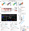 Figure 1
Increased NAT10 expression in Tregs in human and mouse cancer. (A) UMAP representation of the cells subclustered by CD4–FOXP3–CTLA4 Tregs (1,146 cells) expressing NAT10 in the GSE99254 dataset. (B) Calculating the Treg activation score for each cell using classical Treg activation markers (left: UMAP, right: Violin plot). (C) Dot plot showing the classical Treg activation markers in the NAT10 differentiated expression clusters. (D) Comparison of the correlation coefficient between the expression of each RNA epigenetic modification gene and its Treg activation score. (E) Immunofluorescence staining of human NSCLC tumor sections used the markers CD4, NAT10, FOXP3, and EpCAM to identify CD4+ T cells, CD4+FOXP3+ Tregs, NAT10+CD4+FOXP3+ Tregs, and EpCAM+ tumor cells. Scale bar: 2 μm. (F) Percentage per field of CD4+FOXP3+ Tregs among NAT10+ cells or NAT10+ Tregs among CD4+FOXP3+ Tregs (n = 19). (G and H) Kaplan-Meier plot of survival analysis using NAT10 expression in anti–PD-L1 immunotherapy (G) and paclitaxel chemotherapy (H) of the NSCLC cohort. (I) Real-time PCR showing relative mRNA expression of Nat10 in TI Tregs after anti–PD-L1, cisplatin, or paclitaxel treatment (n = 12). Data represent 2–3 independent experiments; mean ± SEM. P values were calculated using 1-way ANOVA (I). ***P < 0.001.
These findings were corroborated using Tregs from treatment-naive patients with lung cancer (GSE235500) (Supplemental Figure 1, C–H). Immunofluorescence staining confirmed the presence of NAT10+CD4+FOXP3+ Tregs infiltrating NSCLC tumor tissues (Figure 1E, Supplemental Figure 2A, Supplemental Figure 3, and Supplemental Table 2), with some tumor cells also expressing NAT10, consistent with previous reports (31). Quantitative immunofluorescence analysis revealed that, on average, 20% of NAT10+ cells were CD4+FOXP3+ Tregs, while 45% of CD4+FOXP3+ Tregs were NAT10+ (Figure 1F).
To evaluate the clinical relevance of NAT10 expression, we analyzed transcriptomic data from 439 patients with NSCLC treated with atezolizumab (anti–PD-L1) (32, 33). Focusing on samples with FOXP3 ≥2 transcripts per million (TPM) (n = 269), we calculated the NAT10/FOXP3 ratio and stratified patients into high and low groups based on the median value (n = 134 vs. 135) (34). Kaplan-Meier and Cox regression analyses revealed that a higher NAT10/FOXP3 ratio was significantly associated with poorer survival (P = 0.0029) (Figure 1G). A similar study in the paclitaxel-treated chemotherapy cohort (n = 372) showed a marginal association (P = 0.052) (Figure 1H). Multivariate analysis with available data for the NAT10/FOXP3 TPM ratio, PD-L1 expression, immune infiltration score, and histological subtype showed that the NAT10/FOXP3 TPM ratio remained significantly associated with survival in atezolizumab-treated (n = 269, P = 0.049) and chemotherapy (n = 61, P = 0.021) patients with NSCLC. Detailed patient information is provided in the Supplemental Data Set 1 (Excel for multivariant survival). Together, these results suggest a correlation between elevated NAT10 signal in FOXP3+‑rich tumor regions and poorer clinical outcomes. The bulk‑derived NAT10/FOXP3 TPM ratio exhibits an independent statistical association with survival among patients with NSCLC treated with anti–PD-L1 or chemotherapy.
In mice lung tumor model, immunofluorescence analysis confirmed the presence of NAT10+ FOXP3+ Tregs (Supplemental Figure 2B). Given that TI Tregs may arise from CD4+ T cells activated by antigen presentation via MHC molecules on dendritic or tumor cells, we examined whether Nat10 expression in TI Tregs, or MHC molecule expression in antigen-presenting cells, was induced during anti–PD-1, cisplatin, or paclitaxel therapy (Supplemental Figure 4A). These treatments markedly upregulated Nat10 expression in TI Tregs (Figure 1I). Additionally, anti–PD-1 and paclitaxel treatments upregulated MHC-II (mH2-A and mH2-E) and MHC-I (mH2-D and mH2-K) expression in tumor cells (Supplemental Figure 4, B and C) but not in dendritic cells (Supplemental Figure 4, D and E). These data suggest that drug therapy–induced MHC-II expression on tumor cells may facilitate CD4+ T cell activation and promote iTreg (iTreg) differentiation in the TME.
Nat10 ablation in TI Tregs suppresses tumor progression. To assess the requirement of NAT10 in Tregs, we designed single guide RNAs (sgRNAs) targeting exons 4–5, which are critical for protein function (35), and generated Nat10flox mice via CRISPR. LoxP sites were confirmed by genomic sequencing (Supplemental Figure 5A). To investigate whether Nat10 ablation destabilizes intratumoral Tregs, we generated Foxp3GFP-cre-ERT2 × Nat10fl/fl mice in which tamoxifen administration triggers FOXP3+ Treg-specific deletion of Nat10 and simultaneous GFP expression. As expected, GFP was observed exclusively in CD4+FOXP3+ Tregs from both the spleen and TILs of B16F10-bearing mice but not in CD4+FOXP3– non-Tregs or CD8+ T cells (Supplemental Figure 5, B and C).
Subsequent evaluation revealed that NAT10 expression was diminished in Tregs from tamoxifen-treated Nat10fl/fl Foxp3GFP-cre-ERT2 mice (Figure 2, A and B). These mice exhibited markedly delayed tumor growth and prolonged survival compared with Nat10+/+ Foxp3GFP-cre-ERT2 controls (Figure 2, C–G, and Supplemental Figure 5, D–I). Flow cytometry analysis of TI Tregs showed a significant reduction in both frequency and number in Nat10-cKO mice relative to controls, regardless of tumor models (Figure 2H and Supplemental Figure 6, A and C), whereas FOXP3 protein levels remained unchanged (Figure 2I and Supplemental Figure 6B). In contrast, the frequency and number of Tregs in the spleen were comparable between Nat10-Ctrl and Nat10-cKO mice (Supplemental Figure 7, A and B), suggesting that Nat10 deletion selectively affects intratumoral but not systemic Treg populations. These findings indicate that Nat10 ablation may restrict Treg infiltration or iTreg differentiation specifically within the TME.
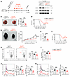 Figure 2
Nat10 deficiency in TI Tregs shows delayed tumor growth and prolonged survival. (A) Schematic of the experiment. (B) Western blot for the NAT10 protein level in Tregs from Nat10+/+ Foxp3cre-ERT2 and Nat10fl/fl Foxp3cre-ERT2 mice treated with tamoxifen. (C) Quantification of tumor nodules in the lungs of Nat10+/+ Foxp3cre-ERT2 and Nat10fl/fl Foxp3cre-ERT2 mice at day 14 after intravenous injection with B16F10 cells (n = 10). Scale bar: 1 cm. (D) Survival curve of Nat10+/+ Foxp3cre-ERT2 and Nat10fl/fl Foxp3cre-ERT2 mice intravenously injected with B16F10 cells (n = 12). (E and F) Tumor growth rate (E) and tumor mass (F) in Nat10+/+ Foxp3cre-ERT2 and Nat10fl/fl Foxp3cre-ERT2 mice at day 14 after subcutaneous injection with B16F10 cells (n = 10). Scale bar: 1 cm. (G) Survival curve of Nat10+/+ Foxp3cre-ERT2 and Nat10fl/fl Foxp3cre-ERT2 mice subcutaneously injected with B16F10 cells (n = 13). (H and I) Representative flow cytometry plots (left) and quantification of the percentage (right) of TI Tregs (H) and MFI of FOXP3 (I) in Nat10+/+ Foxp3cre-ERT2 and Nat10fl/fl Foxp3cre-ERT2 mice intravenously injected with B16F10 cells (n = 10). (J and K) Representative histograms (left) and quantification of the percentages (middle) and MFI (right) of PD-1 (J) and CD25 (K) in TI Tregs from Nat10+/+ Foxp3cre-ERT2 and Nat10fl/fl Foxp3cre-ERT2 mice intravenously injected with B16F10 cells (n = 10). Data represent 2–3 independent experiments; mean ± SEM. P values were calculated using 2-tailed unpaired Student’s t test (C, F, and H–K), 2-way ANOVA (E), and log-rank test (D and G). **P < 0.01, ****P < 0.0001.
In both lung and subcutaneous tumor models, Nat10-cKO mice exhibited significantly reduced ratios of TI Tregs to CD8+ T cells and TI PD-1+ Tregs to PD-1+ CD8+ T cells compared with controls (Supplemental Figure 7, C and D). Frequency and expression of key activation and immunosuppressive markers, including PD-1, CD25, CXCR3, KLRG1, CTLA4, NRP1, and ICOS, were markedly downregulated in TI Tregs from Nat10-cKO mice (Figure 2, J and K, Supplemental Figure 6, D–J, and Supplemental Figure 7, E–I). Notably, although the number of TI CD8+ T cells remained unchanged, their frequency significantly increased in the Nat10-cKO group (Supplemental Figure 8A). Both the frequency and number of CD44hiCD62L– effector CD8+ T cells were significantly higher in Nat10-cKO mice (Supplemental Figure 8B), along with a marked elevation in CD44 protein levels (Supplemental Figure 8C). Together, these results demonstrate that conditional Nat10 deletion in Tregs reduces their accumulation and suppressive phenotype within tumors, excluding FOXP3 expression, while enhancing the activation of cytotoxic CD8+ T cells. This suggests that NAT10 plays a key role in maintaining intratumoral Treg function and phenotype.
Nat10 ablation in TI Tregs enhances antitumor immunity. IFN-γ is known to induce intratumoral Treg functional fragility, impairing their suppressive capacity without disrupting FOXP3 expression (36). Similarly, deletion of Nrp1, Pdcd1, or Jmjd1c enhances IFN-γ production and promotes Treg fragility, thereby improving antitumor immunity (20, 37, 38). In our experiments, TI Tregs from Nat10-cKO mice secreted significantly more IFN-γ following PMA/ionomycin (Figure 3A) or anti-CD3/anti-CD28 (Supplemental Figure 8D) stimulation, indicating increased functional fragility. To further investigate whether Nat10 deficiency influences Treg fragility, we performed RNA-seq on TI Tregs from control and Nat10-cKO mice and analyzed the data by using both curated gene set enrichment analysis (39). The MSigDB GSE14308 Th1-versus-natural Treg signatures showed that the GSE14308_TH1_VS_NATURAL_Treg_UP gene set was enriched in Nat10-deficient TI Tregs (normalized enrichment score [NES] = 1.92; FDR q = 6.69 × 10⁻⁷) (Supplemental Figure 8E), whereas the reciprocal GSE14308_TH1_VS_NATURAL_Treg_Down gene set was enriched in control TI Tregs (NES = –1.68; FDR q = 4.75 × 10⁻⁵) (Supplemental Figure 8F). Together, these data indicate that Nat10-deficient TI Tregs display a Th1-skewed transcriptional profile with attenuation of multiple Treg-associated genes, consistent with increased Treg fragility.
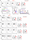 Figure 3
NAT10 is essential for preventing the fragility and suppressive function of Tregs. (A) Representative flow cytometry plots (left) and quantification of the percentages (middle) and MFI (right) of IFN-γ in TI Tregs after ex vivo stimulation with PMA and ionomycin from Nat10+/+ Foxp3cre-ERT2 and Nat10fl/fl Foxp3cre-ERT2 mice intravenously injected with B16F10 cells (n = 10). (B) Body weight changes in Rag1−/− recipient mice transferred with naive conventional CD4+ cells isolated from the spleens of CD45.1 mice alone or with Tregs isolated from the spleens of Nat10+/+ Foxp3cre-ERT2 and Nat10fl/fl Foxp3cre-ERT2 mice (n = 6). Rag1−/− mice were intraperitoneally injected with tamoxifen as indicated. (C) Representative histogram of CTV (left) and quantification of the percentages (right) of each division in naive CD8+ T cells isolated from the spleens of CD45.1 mice. Cells were cultured for 72 hours alone or with TI Tregs isolated from Nat10+/+ Foxp3cre-ERT2 and Nat10fl/fl Foxp3cre-ERT2 mice in the presence or absence of anti-CD3 and anti-CD28 antibodies (n = 3). (D–G) Representative flow cytometry plots (left) and quantification of the percentages (middle) and MFI (right) of granzyme B (D), IFN-γ (E), perforin (F), and TNF-α (G) in TI CD8+ cells after ex vivo stimulation from Nat10+/+ Foxp3cre-ERT2 and Nat10fl/fl Foxp3cre-ERT2 mice intravenously injected with B16F10 cells (n = 10). Data represent 2–3 independent experiments; mean ± SEM. P values were calculated using 2-tailed unpaired Student’s t test (A and C–G) and 2-way ANOVA (B). **P < 0.01, ***P < 0.001, ****P < 0.0001.
To further examine the suppressive capacity of Nat10-deficient Tregs, we performed both in vivo and in vitro assays. In vivo, Rag1−/− mice were adoptively transferred with CD45.1+ naive CD4+ T cells alone or with WT or Nat10-deficient Tregs, followed by tamoxifen. Mice receiving Nat10-deficient Tregs were less protected from weight loss compared with those receiving WT Tregs, indicating impaired immunoregulatory function (Figure 3B). In vitro, CellTrace Violet–labeled (CTV-labeled) CD45.1+CD8+ T cells were cocultured with WT or Nat10-deficient TI Tregs for 72 hours. CD8+ T cells cocultured with Nat10-deficient Tregs proliferated significantly more than those cocultured with WT Tregs (Figure 3C), confirming that loss of Nat10 weakens Treg-mediated suppression of CD8+ T cell activation. Moreover, TI CD8+ T cells from mice with Nat10-deficient Tregs secreted higher levels of granzyme B, IFN-γ, perforin, and TNF-α (Figure 3, D–G), indicating an enhanced cytotoxic potential. Taken together, these findings demonstrate that Nat10 deletion increases Treg fragility and diminishes their suppressive function on conventional CD4+ and CD8+ T cells, thereby enhancing antitumor immunity. This highlights NAT10 as a key regulator of Treg fragility and immunosuppression within the TME.
NAT10 is essential for the differentiation of iTregs in tumors. To assess whether NAT10 is intrinsically required for iTreg development, we generated Nat10fl/fl Ox40cre mice. Since OX40 is expressed in activated but not naive CD4+ T cells, this model avoided confounding effects on T cell homeostasis. CTV-labeled naive CD4+ T cells were cultured with TGF-β and IL-2 for 96 hours to induce iTreg differentiation (Supplemental Figure 9A). Nat10-deficient CD4+ T cells showed a significantly reduced frequency of iTregs compared with controls (Supplemental Figure 9B). Both control and Nat10-deficient CD4+ cells divided up to 6 times (Supplemental Figure 9D) but a greater proportion of Nat10-deficient iTregs stalled at the first division (Supplemental Figure 9C). When Nat10 was deleted in naive CD4+ T cells via Cd4cre, cells failed to divide beyond the first round (Supplemental Figure 9E), suggesting that NAT10 is essential for initiating iTreg differentiation.
To determine whether NAT10 is required for iTreg differentiation in vivo, we conditionally deleted Nat10 in TI Tregs using tamoxifen in B16F10-bearing mice (Supplemental Figure 10A). After 14 days, the frequency and number of Tregs from Nat10-deficient mice were significantly reduced compared with controls (Supplemental Figure 10B), despite unaltered FOXP3 protein expression (Supplemental Figure 10C). Analysis of the Treg-specific demethylated region revealed an approximately 20% reduction in CpG demethylation in Nat10-deficient Tregs compared with controls (Supplemental Figure 10D), suggesting compromised epigenetic commitment. To confirm this, we adoptively transferred naive CD4+ T cells from Nat10fl/fl Ox40cre and control mice into CD45.1+ recipients bearing B16F10 tumors (Supplemental Figure 10E). After 14 days, the frequency and number of Tregs derived from Nat10-deficient donors were markedly reduced compared with controls (Supplemental Figure 10F), although FOXP3 expression was unchanged (Supplemental Figure 10G). These results suggest that NAT10 is potentially required for iTreg differentiation from activated CD4+ T cells within the TME.
Indeed, in tumors from Nat10 cKO mice bearing B16F10, the frequency and number of CD44hiCD62L– effector and CD44+CD69+ activated Tregs were significantly reduced compared with controls (Supplemental Figure 11, A, B, and D). Concomitantly, the frequency of naive-like Tregs were increased, although their numbers remained unchanged (Supplemental Figure 11C). Consistently, Nat10-deficient Tregs exhibited markedly lower CD44 and CD69 expression (Supplemental Figure 11, E and F). To evaluate proliferative capacity, Tregs were sorted and labeled with CTV (Supplemental Figure 11G). Flow cytometry data revealed that Nat10-deficient Tregs displayed a significantly lower rate of cell division compared with controls (Supplemental Figure 11H). Collectively, these results demonstrate that NAT10 is critical not only for iTreg differentiation but also for maintaining activated and effector Treg populations within the TME.
NAT10 interacts with FOXP3 to promote iTreg differentiation in the TME. To explore the mechanisms underlying the impaired Treg upon Nat10 deletion, we examined the localization of the NAT10 protein. Western blot revealed that NAT10 was predominantly nuclear in Tregs, with no detectable cytoplasmic expression (Supplemental Figure 12A). TI Tregs from lung tumor model were sorted for ac4C RNA immunoprecipitation sequencing (RIP-seq) analysis. The ac4C signal intensity was comparable between Nat10-deficient and control TI Tregs (Supplemental Figure 12B). Furthermore, stratifying genes based on high or low ac4C signals showed no marked downregulation in Nat10-deficient cells regardless of ac4C enrichment (Supplemental Figure 12C). These results suggest that NAT10’s role in TI Treg differentiation is largely independent of its RNA acetyltransferase activity via ac4C modification.
Next, we performed CUT&Tag profiling on TI Tregs sorted from lung tumor model. Comparative analysis of NAT10 and FOXP3 CUT&Tag datasets revealed that 34.9% of NAT10 peaks overlapped with FOXP3 binding sites (clusters 1 and 2), while 65.1% represented NAT10-only regions lacking FOXP3 enrichment (cluster 3) (Figure 4A). Cobound regions exhibited elevated H3K27ac and assay for transposase-accessible chromatin with high-throughput sequencing (ATAC-seq) signals, along with reduced DNA methylation (Figure 4, B–D, red lines), indicating increased chromatin accessibility and transcriptionally activity. Whereas NAT10-only sites were depleted of activation marks and showed high DNA methylation (Figure 4, B–D, blue lines). Importantly, NAT10-FOXP3 cobound regions were enriched in promoters of Treg signature genes (Figure 4, E and F), whereas NAT10-only binding sites were enriched in conventional CD4+ T cell genes (Supplemental Figure 12D). These findings suggest that NAT10 cooperates with FOXP3 to regulate Treg-specific gene expression and iTreg differentiation.
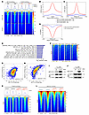 Figure 4
NAT10 interacts with FOXP3 to directly regulate the activation of TI Tregs. (A) Heatmaps showing NAT10 and FOXP3 CUT&Tag peaks resolved into 3 clusters by K-means clustering. (B) Curve plot showing the H3K27ac CUT&Tag signals in clusters 1 and 2 (red) and cluster 3 (blue) peak region. (C) Curve plot displaying the chromosome accessibility signals in clusters 1 and 2 (red) and cluster 3 (blue) peak region. (D) Curve plot displaying the mean methylated cytosine signals in clusters 1 and 2 (red) and cluster 3 (blue) peak region. (E) Gene set enrichment analysis of the candidate genes within 2,000 of clusters 1 and 2 peak regions. (F) Heatmaps showing the NAT10 and FOXP3 CUT&Tag signals enriched in the promoter regions of Treg signature genes. (G) Scatterplot showing the correlation between the NAT10 and FOXP3 enrichment signals on H3K27ac peak regions. (H) Scatterplot showing the correlation between the NAT10 and H3K27ac genome-wide enrichment signals. (I) FOXP3 pull-down NAT10 in TI Tregs by the co-IP experiment. (J) NAT10 pull-down FOXP3 in TI Tregs by the co-IP experiment. (K) Heatmaps exhibiting reduced FOXP3 peaks in Nat10fl/fl Foxp3cre-ERT2 TI Tregs. (L) Heatmaps showing reduced H3K27ac enrichment in Nat10fl/fl Foxp3cre-ERT2 TI Tregs.
Consistent with prior reports of FOXP3 binding at H3K27ac-enriched regions, we observed cooccupancy of NAT10 and H3K27ac (Supplemental Figure 12, E and F). Correlation analyses demonstrated strong associations between NAT10 and FOXP3 as well as between NAT10 and H3K27ac (Spearman coefficients = 0.52 and 0.76, respectively; Figure 4, G and H), suggesting synergistic regulation of Treg activation in the TME. Protein co-IP confirmed the physical interaction between NAT10 and FOXP3 in TI Tregs (Figure 4, I and J). FOXP3 CUT&Tag analysis revealed a marked loss of peaks within clusters 1 and 2 in Nat10-deficient TI Tregs (Figure 4K). In contrast, regions lacking FOXP3 in cluster 3 remained unaffected (Supplemental Figure 12G). H3K27ac CUT&Tag analysis identified 2,898 peaks with reduced H3K27ac enrichment in Nat10-deficient Tregs (Figure 4L). These peaks were associated with genes involved in T cell aggregation, activation, differentiation, and immune regulation (Supplemental Figure 12, H and I). In summary, our data demonstrate that NAT10 cooperates with FOXP3 to direct the transcriptional and epigenetic programs essential for iTreg differentiation and function in the TME. Loss of Nat10 disrupts FOXP3 binding and chromatin activation, thereby impairing TI iTreg generation and maintenance.
NAT10 facilitates natural Treg recruitment and iTreg activation via direct DNA binding, independent of RNA acetylation. To further investigate the impaired suppressive capacity of TI Tregs following Nat10 ablation, we integrated Treg activation and function markers with NAT10 and H3K27ac CUT&Tag data. Our analysis revealed strong enrichment of enhancers and superenhancers at gene loci critical for Treg costimulation and activation in TI Tregs, including Tnfrsf18 (GITR), Tnfrsf4 (OX40), Pdcd1 (PD-1), Tnfrsf9 (4-1BB), Icos, and Cxcr4 (Figure 5A). Analysis of our 3D genome data showed that NAT10, together with FOXP3, participates in loop formation at loci such as Cxcr4, which promotes Treg recruitment to the TME (40). Furthermore, the chromosome loops with anchors cobinding with NAT10 and FOXP3 were impaired in Nat10-deficient Tregs (Figure 5B). CUT&Tag and ATAC-seq analyses demonstrated a marked reduction in H3K27ac signal and chromatin accessibility at Cxcr4 loci in Nat10-deficient TI Tregs, accompanied by a marked decrease in FOXP3 binding (Figure 5C). FOXP3 CUT&Tag-qPCR confirmed that FOXP3 enrichment at the Cxcr4 locus was substantially reduced in Nat10-deficient Tregs (Figure 5D). Flow cytometry further demonstrated a significant decrease in CXCR4 protein expression in Nat10-deficient TI Tregs (Figure 5E). As CXCR4 is a key homing receptor for immune cells migrating to the bone marrow, a known reservoir for tumor antigen–specific and TI Tregs in patients with cancer (41), we examined CXCR4+ Treg populations in the bone marrow of mice bearing B16F10 lung tumors. The frequency of CXCR4+ cells and CXCR4 protein levels were significantly reduced in Nat10-deficient Tregs compared with controls (Supplemental Figure 13A). To further assess NAT10’s role in Treg accumulation within tumors, we cotransferred Nat10-cKO Tregs (CD45.2+) and control Tregs (CD45.1+ CD45.2+) into tumor-bearing mice (Figure 5F). Flow cytometry data revealed significantly lower frequencies and numbers of CD45.2+ Nat10 cKO Tregs compared with CD45.1+CD45.2+ control Tregs within tumors (Figure 5G). These data collectively suggest that NAT10 promotes Treg recruitment to the TME, potentially through modulating local chromatin‑loop interactions and establishing Treg-specific superenhancer landscapes.
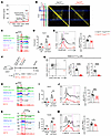 Figure 5
NAT10 interacts with FOXP3 to facilitate the Treg superenhancer establishment. (A) Analysis of TI Tregs in Figure 4. Treg-specific super-enhancers are associated with Treg signature genes, including Tnfrsf18 (GITR), Tnfrsf4 (OX40), Cxcr4, Tnfrsf9 (4-1BB), Pdcd1 (PD-1), and Icos. (B) Chromosome loops with anchors cobinding with NAT10 and FOXP3 in WT and Nat10-deficient Tregs. Representative Hi-C contact maps at the Cxcr4-containing region in WT and Nat10-deficient Tregs at 5 kb resolution. Blue boxes highlight the local region used for comparison. (C) NAT10-CUT&Tag, FOXP3-CUT&Tag, H3K27ac-CUT&Tag, and ATAC-seq tracks at the Cxcr4 gene locus. (D) qPCR showing the enrichment of FOXP3 at the promoter of Cxcr4 in TI Tregs (n = 4). (E) Representative histogram (left) and quantification of the MFI (right) of CXCR4 in TI Tregs (n = 10). (F) Schematic of the experiment. (G) Representative flow cytometry plot (left) and quantification of the percentages (middle) and number (right) of TI Tregs in lung tumors of the CD45.1 mice adoptively transferred with WT or Nat10-deficient Tregs (n = 8). (H) NAT10-CUT&Tag, FOXP3-CUT&Tag, H3K27ac-CUT&Tag, and ATAC-seq tracks at the Pdcd1 gene locus. (I) qPCR showing the enrichment of FOXP3 at the promoter of Pdcd1 in TI Tregs (n = 4). (J) Representative histogram (left) and quantification of the MFI (right) of PD-1 in TI Tregs (n = 10). (K) NAT10-CUT&Tag, FOXP3-CUT&Tag, H3K27ac-CUT&Tag, and ATAC-seq tracks at the Tnfrsf9 gene locus. (L) qPCR showing the enrichment of FOXP3 at the promoter of Tnfrsf9 in TI Tregs (n = 4). (M) Representative histogram (left) and quantification of the MFI (right) of 4-1BB in TI Tregs (n = 10). Data represent 2–3 independent experiments; mean ± SEM. P values were calculated using 2-tailed unpaired Student’s t test (D, E, G, I, J, L, and M). *P < 0.05, **P < 0.01, ***P < 0.001, ****P < 0.0001.
Nat10 ablation also markedly reduced H3K27ac enrichment and chromatin accessibility at Pdcd1, Tnfrsf9, Tnfrsf4, Tnfrsf18, and Icos loci (Figure 5, H and K, and Supplemental Figure 13, B and E). FOXP3 CUT&Tag-qPCR confirmed a marked reduction in FOXP3 occupancy at these loci in Nat10-deficient TI Tregs (Figure 5, I and L, and Supplemental Figure 13G). Correspondingly, flow cytometry data revealed reduced protein expression of PD-1, 4-1BB, OX40, GITR, and ICOS in Nat10-deficient TI Tregs (Figure 5, J and M, and Supplemental Figure 13, C, D and F), indicating that NAT10 is essential for sustaining the expression of key Treg activation and immunosuppressive markers in the TME.
To examine whether NAT10 exerts a similar role in humans, we performed CUT&Tag on healthy human CD4+CD25– T cells and in vitro iTregs (Supplemental Figure 14A). iTregs showed increased enrichment of NAT10, FOXP3, and H3K27ac signals compared with CD4+CD25– T cells (Supplemental Figure 14, B–D). Comparative analysis of NAT10 and FOXP3 profiles revealed that genomic regions cooccupied by both factors in iTregs exhibited higher signal intensity following the differentiation of CD4+CD25– T cells (Supplemental Figure 14E). Regions cooccupied by NAT10 and FOXP3 exhibited higher signal intensity in iTregs, particularly near genes involved in T cell costimulation and activation, such as TNFRSF9, PDCD1, ICOS, TNFRSF18, TNFRSF4, and CXCR4, which were also identified as Nat10-target genes in murine TI Tregs (Supplemental Figure 14, F–J). These results suggest that NAT10 and FOXP3 may play conserved roles in shaping chromatin accessibility at activation- and immunosuppression-associated loci during human iTreg differentiation.
To further determine whether NAT10 was required for the expression of these activation- and immunosuppression-associated markers during CD4+ T cell activation, we utilized Ox40cre mice to delete Nat10 in activated CD4+ T cells. Naive CD4+ T cells from Nat10+/+ Ox40cre and Nat10fl/fl Ox40cre mice were sorted and activated in vitro (Supplemental Figure 15A). Surprisingly, Nat10-deficient activated CD4+ T cells expressed comparable levels of PD-1, 4-1BB, ICOS, OX40, GITR, and CXCR4 to controls (Supplemental Figure 15, B–H), indicating that NAT10 is dispensable for the expression of these markers in activated CD4+ T cells.
NAT10 was required for activation- and immunosuppression-associated gene expression in iTregs. To confirm NAT10’s DNA-binding capacity, we first purified the NAT10 protein (Figure 6A) and performed electrophoretic mobility shift assays (EMSAs) with 8 candidate DNA sequences (Supplemental Table 3). Among these, DNA2, DNA5, and DNA8 showed strong binding affinity for NAT10, as evidenced by pronounced band shifts (Supplemental Figure 16, A–H). Sequence alignment of DNA2, DNA5, and DNA8 identified a conserved 46-nucleotide motif (termed DNA9), which was used in binding analyses and confirmed NAT10’s DNA-binding capability (Figure 6B).
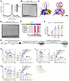 Figure 6
NAT10 promotes iTreg differentiation by positively transcriptional regulation of activation-associated genes. (A) Size-exclusion chromatography analysis of mouse NAT10. (B) EMSA analysis of WT NAT10 binding to DNA. (C) Left: Three-dimensional structure of dimerized NAT10 predicted by AlphaFold3. Two monomers of NAT10 are shown in blue and orange. Right: Surface electrostatic potential of the NAT10 dimer. The positively charged residues and acetyl-CoA-binding residues are colored in pink and green, respectively. (D) EMSA analysis of mutant NAT10 (exon 4–5 deletion) binding to DNA. (E) Sequence conservation analysis. The sequence logo, which depicts the relative frequency of amino acids at each position in a multiple sequence alignment, includes color-coded residues based on their chemical properties. (F) Compared with the WT NAT10 protein, the EMSA analysis of mutant NAT10 (H537A/Y538A) binding to DNA. The binding rate was determined by calculating the proportion of remaining free DNA, as measured by a grayscale value obtained using ImageJ (NIH) (n = 3). (G) The schematic of the experiment. (H–J) Representative histograms (left) and quantification of the MFI (right) of PD-1 (H), 4-1BB (I), and ICOS (J) in iTregs of Nat10+/+ Ox40cre and Nat10fl/fl Ox40cre mice (n = 3). Data represent 2–3 independent experiments; mean ± SEM. P values were calculated using 2-tailed unpaired Student’s t test (F) and 1-way ANOVA (H–J). **P < 0.01, ***P < 0.001, ****P < 0.0001.
Although NAT10 is an RNA acetyltransferase, it lacks canonical DNA-binding motifs such as zinc fingers or helix-turn-helix domains. Instead, it appears to bind DNA via electrostatic interactions mediated by positively charged residues within exons 4–5 (pink region), distinct from the its RNA acetyltransferase catalytic residues H537/Y538 (green region) (42) (Figure 6C). Deletion of exons 4–5 markedly impaired DNA-binding ability (Figure 6D), whereas mutation of H537A/Y538A had no effect (Figure 6, E and F), indicating that NAT10 can directly bind DNA independent of its acetyltransferase activity. To determine whether NAT10 directly regulates downstream target gene expression via its genomic DNA-binding activity, we constructed a luciferase reporter plasmid incorporating the promoter region of a NAT10-bound activation-associated gene, including the 46 bp conserved motif (Supplemental Figure 16I). Cotransfection of this reporter with a Nat10 overexpression plasmid into HEK293T cells significantly increased luciferase activity compared with the empty vector (Supplemental Figure 16J). These data suggest that NAT10 can directly promote the transcription of activation- and immunosuppression-related genes in iTregs via genomic DNA binding.
To further elucidate the role of NAT10 in iTreg differentiation, we expressed WT Nat10, a catalytically inactive mutant (mutNat10H537A/Y538A), and a DNA-binding mutant (exon 4–5 deletion, mutNat10exon4–5) in differentiating iTregs (Figure 6G). The iTregs from Nat10fl/fl Ox40cre mice showed significantly reduced expression of PD-1, 4-1BB, ICOS, OX40, GITR, and CXCR4 compared with Nat10+/+ Ox40cre controls. Remarkably, ectopic expression of WT Nat10 or mutNat10H537A/Y538A restored expression of these proteins, whereas mutNat10exon4–5 did not (Figure 6, H–J, and Supplemental Figure 16, L–N). In addition, the purified mutNAT10H537A/Y538A protein showed a marked reduction of its acetyltransferase activity compared with WT NAT10 protein (Supplemental Figure 17, A and B). The molecular size peak chart and electrophoresis results demonstrated that the integrity and stability of mutNAT10exon4–5 was similar with mutNAT10H537A/Y538A and WT NAT10 (Supplemental Figure 17, A and C). The AlphaFold3 structure prediction showed high confidence of mutNAT10exon4–5, compared with WT NAT10 (Supplemental Figure 17D). Meanwhile, compared with control iTregs with pMigR1 and Nat10-deficient iTregs with WT Nat10, we observed that both the expressed mutants did not alter its protein localization in the differentiated iTregs (Supplemental Figure 16K). These results indicate that exon 4–5 deletion does not affect NAT10 protein structure, integrity, and localization in cells. Collectively, these findings suggest that NAT10 regulates the expression of iTreg activation- and suppression-associated genes through direct DNA binding, independent of its RNA acetyltransferase activity.
To evaluate whether NAT10 regulates antigen-specific activated CD4+ T cell differentiation, we crossed Nat10fl/fl Ox40cre mice with Smarta TCR transgenic mice (specific for LCMV GP61-80), generating Smarta/Nat10fl/fl and Smarta/Nat10+/+ Ox40cre controls. CD45.2+ Smarta CD4+ T cells from these mice were transferred into CD45.1 recipient mice, followed by chronic LCMV-Cl13 infection to simulate a tumor-like microenvironment (Supplemental Figure 18A). At day 30 after infection, donor-derived CD4+ T cells showed no significant differences in the proportions of CXCR5–SLAMhi Th1 cells and CXCR5+ SLAMlo T follicular helper cells between the two groups (Supplemental Figure 18B). However, the frequency of FOXP3+ Tregs was markedly decreased in the Nat10-deficient group, despite no change in FOXP3 protein expression levels (Supplemental Figure 18, C and D). Western blot further revealed reduced phosphorylation of TCR signaling mediators ZAP70 and AKT in activated CD4+ T cells from the Nat10-deficient group, while total protein levels remained unchanged (Supplemental Figure 18E). Collectively, these findings suggest that Nat10 deletion in OX40+ activated CD4+ T cells impairs iTreg differentiation in response to antigen stimulation, without affecting the expression of FOXP3 itself.
NAT10 was induced in Treg subsets during iTreg differentiation (Figure 1D, clusters 1 and 3) and was essential for iTreg generation in tumor-bearing (Supplemental Figure 10, E–G) and chronic infection hosts (Supplemental Figure 18, A–D). Mechanistically, NAT10 promotes the expression of costimulatory molecules and members of the TNFR superfamily, thereby enhancing TCR signal transduction required for Treg activation and immunosuppressive function (Supplemental Figure 18F). Furthermore, anti–PD-1 immunotherapy and chemotherapy induce Nat10 expression in TI Tregs (Figure 1I), accompanied by increased MHC-II expression in tumor cells (Supplemental Figure 4B). Together, these data suggest that NAT10 facilitates the differentiation of tumor antigen–activated CD4+ T cells into iTregs within the TME in response to immunotherapy or chemotherapy.
Intratumoral Treg activation and immunosuppressive function are critically dependent on the NAT10 protein. To evaluate NAT10’s role in TI Treg function, we treated B16F10-bearing C57BL/6 mice daily with the NAT10 inhibitor remodelin from day 4 after inoculation for 6 days (Supplemental Figure 19A). Remodelin did not affect body weight at either dosage (Supplemental Figure 19B) but markedly reduced NAT10 protein levels without altering FOXP3 expression versus the vehicle group (Supplemental Figure 19C), consistent with previous reports (43, 44). Remodelin dose-dependently delayed tumor growth and extended survival (Supplemental Figure 19, D–F). By day 14 after inoculation, the frequency and number of TI Tregs were markedly lower in groups treated with high doses (200 mg/Kg) of remodelin compared with those treated with low doses (100 mg/Kg) or vehicle (Supplemental Figure 19G). Additionally, the expression of key Treg markers, such as CD25 and PD-1, was reduced in a dose-dependent manner (Supplemental Figure 19, H and I). The frequency ratios of Tregs to CD8+ T cells and PD-1+ Tregs to PD-1+ CD8+ T cells were also diminished (Supplemental Figure 19, J and K).
To assess potential autoimmune effects of systemic Nat10 deletion in Tregs, we administered tamoxifen to tumor-free Nat10+/+Foxp3cre-ERT2 and Nat10fl/flFoxp3cre-ERT2 mice, and neither group showed signs of autoimmunity (Supplemental Figure 20, A–E). Moreover, remodelin treatment did not affect lung metastasis or subcutaneous tumor growth in Rag1–/– mice (Supplemental Figure 20, F–I), indicating that remodelin’s antitumor effects are lymphocyte-dependent. Collectively, these results indicate that remodelin selectively and dose-dependently impairs TI Tregs, supporting the 200 mg/Kg dose for further studies.
To further dissect the pharmacological effects of NAT10 inhibition on TI Treg phenotype and function, we treated Nat10+/+Foxp3cre-ERT2 and Nat10fl/flFoxp3cre-ERT2 mice with tamoxifen, with or without remodelin (Supplemental Figure 21A). Compared with the Nat10+/+Foxp3cre-ERT2 and vehicle group, Nat10+/+Foxp3cre-ERT2 and remodelin mice exhibited significantly fewer tumor nodules (Supplemental Figure 21B), reduced frequency and number of Tregs (Supplemental Figure 21C), and lower CD25 and PD-1 expression (Supplemental Figure 21, D and E). However, in Nat10fl/flFoxp3cre-ERT2 mice, remodelin did not further reduce tumor burden, Treg frequency or number, or Treg-associated marker expression compared with vehicle (Supplemental Figure 21, B–E), suggesting that Nat10 loss and pharmacological inhibition may converge on overlapping pathways. Importantly, cytotoxic CD8+ T cells from Nat10+/+Foxp3cre-ERT2 and remodelin, Nat10fl/flFoxp3cre-ERT2 and vehicle, and Nat10fl/flFoxp3cre-ERT2 and remodelin groups showed comparably elevated levels of effector molecules, including granzyme B, IFN-γ, perforin, and TNF-α, relative to the Nat10+/+Foxp3cre-ERT2 and vehicle control groups (Supplemental Figure 22, A–D). In summary, these findings demonstrate that NAT10 is essential for TI Treg expansion, activation, and immunosuppressive function. Pharmacological inhibition of NAT10 with remodelin effectively suppresses TI Treg activity and enhances antitumor CD8+ T cell responses, underscoring its therapeutic potential to overcome immune suppression in the TME.
Pharmacological inhibition of NAT10 enhances anti–PD-1 immunotherapy by selectively targeting TI Tregs. To assess the synergistic efficacy of NAT10 inhibition and anti–PD-1 immunotherapy in enhancing antitumor responses, B16F10-bearing C57BL/6 mice were treated with remodelin (200 mg/Kg) and/or anti–PD-1 antibody (200 μg) at indicated time points, starting on day 7 after tumor inoculation (Figure 7A). Anti–PD-1 alone moderately controlled tumor progression and prolong survival compared with vehicle. In contrast, both remodelin alone and its combination with anti–PD-1 significantly suppressed tumor growth and extended survival, with the combination treatment demonstrating the greatest efficacy (Figure 7, B and C). These results suggest that targeting NAT10 in Tregs can potentiate the effectiveness of immune checkpoint therapy in cancer. Flow cytometry analysis revealed that both remodelin alone and its combination with anti–PD-1 significantly reduced the frequency and number of TI Tregs, compared with vehicle or anti–PD-1 alone (Figure 7D). Immunofluorescence staining showed accumulation of NAT10+FOXP3+ Tregs surrounding tumor cells in lung tumors (Figure 7E). Notably, the frequency of NAT10+FOXP3+ Tregs was significantly lower in the remodelin-treated group than in the vehicle or anti–PD-1-treated groups, whereas anti–PD-1 alone had no effect on this population (Figure 7F). TI Tregs from combination-treated mice exhibited the lowest CD25 and PD-1 expression (Figure 7, G and H). Additionally, remodelin markedly reduced the ratio of Tregs to CD8+ T cells and the ratio of PD-1+ Tregs to PD-1+ CD8+ T cells (Figure 7, I and J), indicating enhanced CD8+ T cell activation. Furthermore, CD8+ T cells from combination-treated mice produced the highest levels of cytotoxic effectors, such as granzyme B, IFN-γ, perforin, and TNF-α (Supplemental Figure 23, A–D). This highlights the critical role of NAT10 inhibition in reversing Treg-mediated suppression of CD8+ T cell activity.
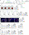 Figure 7
Inhibition of NAT10 synergizes with anti–PD-1 therapy to enhance control of tumor growth. (A) Schematic of the experiment. (B) Quantification of tumor nodules in the lungs of C57BL/6 mice treated with vehicle, anti–PD-1, remodelin, and anti–PD-1 and remodelin at day 14 after intravenous injection with B16F10 cells (n = 7). Scale bar: 1 cm. (C) Survival curve of B16F10 tumor-bearing C57BL/6 mice treated with vehicle, anti–PD-1, remodelin, and anti–PD-1 and remodelin (n = 15). (D) Representative flow cytometry plots (left) and quantification of the percentage (middle) and number (right) of TI Tregs in B16F10 tumor-bearing C57BL/6 mice treated with vehicle, anti–PD-1, remodelin, and anti–PD-1 and remodelin (n = 7). (E and F) Representative immunofluorescence staining (E) and quantification of the percentage (F) of intratumoral NAT10+ FOXP3+ Tregs (n = 6). P, peri-tumoral tissue; T, tumor tissue. (G and H) Representative histograms (left) and quantification of the percentages (middle) and MFI (right) of CD25 (G) and PD-1 (H) in TI Tregs from B16F10 tumor-bearing C57BL/6 mice treated with vehicle, anti–PD-1, remodelin, and anti–PD-1 and remodelin (n = 7). (I and J) Ratio of Tregs to CD8+ T cells (I) and PD-1+ Tregs to CD8+ T cells (J) in B16F10 tumor-bearing C57BL/6 mice treated with vehicle, anti–PD-1, remodelin, and anti–PD-1 and remodelin (n = 7). Data represent 2–3 independent experiments; mean ± SEM. P values were calculated using 1-way ANOVA (B, D, and F–J) and log-rank test (C). *P < 0.05, **P < 0.01, ***P < 0.001, ****P < 0.0001.
To further elucidate whether remodelin-induced TI Treg impairment depends on its direct effect on these cells, we treated Nat10+/+Foxp3cre-ERT2 and Nat10fl/flFoxp3cre-ERT2 mice with tamoxifen, with or without anti–PD-1 (Supplemental Figure 24, A and B). The Nat10fl/flFoxp3cre-ERT2 and anti–PD-1 groups showed a markedly reduced lung tumor burden compared with groups treated with monotherapies or controls (Supplemental Figure 24C), accompanied by decreased TI Treg frequency and number (Supplemental Figure 24D) and attenuated CD25 and PD-1 expression (Supplemental Figure 24, E and F). Combination therapy also led to a robust increase in functional CD8+ T cells, with the highest frequencies and protein levels of granzyme B, IFN-γ, perforin, and TNF-α compared with other treatment groups (Supplemental Figure 25, A–D), indicating reinvigoration of the antitumor CD8+ T cell compartment. In conclusion, NAT10 inhibition in TI Tregs is essential for overcoming their suppressive influence on cytotoxic CD8+ T cells. When combined with anti–PD-1 therapy, NAT10 blockade enhances antitumor immunity by suppressing Treg function and restoring CD8+ T cell activity. Thus, NAT10 represents a promising target to synergize with checkpoint blockade therapies in the TME.


Copyright © 2026 American Society for Clinical Investigation
ISSN: 0021-9738 (print), 1558-8238 (online)