
Advertisement
Research ArticleAIDS/HIVInfectious diseaseVirology
Open Access |  10.1172/JCI187961
Smac mimetic combined with eCD4-Ig reverses latency without reducing SHIV reservoirs in rhesus macaques
Lars Pache,1 John K. Bui,2,3 Lindsay M. Klouser,4 Christine M. Fennessey,5 Alexander C. Noyola,2 Teresa Einhaus,2 Haiying Zhu,4 Laurence Stensland,4 Isai Leguizamo,6 Abubakarr A. Koroma,6 Peter Teriete,1 W.L. William Chang,7,8 Ollivier Hyrien,9 Natasha N. Duggan,10 Dominik Heimann,1 Ailyn C. Pérez-Osorio,4 Katharine J. Bar,11 Nicholas D.P. Cosford,1 Brandon F. Keele,5 Dennis J. Hartigan-O’Connor,7,11,12 Michael Farzan,13,14 Matthew R. Gardner,6,15 Keith R. Jerome,4,16 Sumit K. Chanda,10 Hans-Peter Kiem,2,3 and Christopher W. Peterson2,3
1Center for Therapeutics Discovery, NCI Designated Cancer Center, Sanford Burnham Prebys Medical Discovery Institute, La Jolla, California, USA.
2Translational Science and Therapeutics Division, Fred Hutchinson Cancer Center, Seattle, Washington, USA.
3Department of Medicine and
4Department of Laboratory Medicine and Pathology, University of Washington, Seattle, Washington, USA.
5AIDS and Cancer Virus Program, Frederick National Laboratory for Cancer Research, Frederick, Maryland, USA.
6Division of Microbiology and Immunology, Emory National Primate Research Center, Emory University, Atlanta, Georgia, USA.
7California National Primate Research Center, University of California, Davis, Davis, California, USA.
8Department of Medical Microbiology and Immunology, School of Medicine, University of California, Davis, Davis, California, USA.
9Biostatistics, Bioinformatics and Epidemiology Program, Vaccine and Infectious Disease Division, Fred Hutchinson Cancer Center, Seattle, Washington, USA.
10Department of Immunology and Microbiology, Scripps Research, La Jolla, California, USA.
11Department of Medicine, Perelman School of Medicine, University of Pennsylvania, Philadelphia, Pennsylvania, USA.
12Division of Experimental Medicine, Department of Medicine, University of California, San Francisco, San Francisco, California, USA.
13Department of Pediatrics, Boston Children’s Hospital, Harvard Medical School, Boston, Massachusetts, USA.
14The Center for Integrated Solutions to Infectious Diseases (CISID), The Broad Institute of MIT and Harvard, Cambridge, Massachusetts, USA.
15Department of Medicine, Division of Infectious Diseases, Emory University, Atlanta, Georgia, USA.
16Vaccine and Infectious Disease Division, Fred Hutchinson Cancer Center, Seattle, Washington, USA.
Address correspondence to: Christopher W. Peterson or Hans-Peter Kiem, Fred Hutchinson Cancer Center, PO Box 19024, Mail Stop D1-100, 1100 Fairview Ave. N., Seattle, Washington 98109-1024, USA. Phone: 206.667.6646; Email: cwpeters@fredhutch.org (CWP). Phone: 206.667.4425; Email: hkiem@fredhutch.org (HPK). Or to: Sumit K. Chanda, Scripps Research, 10550 N. Torrey Pines Rd., La Jolla, California 92037, USA. Phone: 858.784.1000; Email: schanda@scripps.edu. Or to: Lars Pache, Sanford Burnham Prebys Medical Discovery Institute, 10901 N. Torrey Pines Rd., La Jolla, California 92037, USA. Phone: 858.646.3100; Email: lpache@sbpdiscovery.org.
Authorship note: LP, SKC, HPK, and CWP are co–senior authors.
Find articles by Pache, L. in: PubMed | Google Scholar
1Center for Therapeutics Discovery, NCI Designated Cancer Center, Sanford Burnham Prebys Medical Discovery Institute, La Jolla, California, USA.
2Translational Science and Therapeutics Division, Fred Hutchinson Cancer Center, Seattle, Washington, USA.
3Department of Medicine and
4Department of Laboratory Medicine and Pathology, University of Washington, Seattle, Washington, USA.
5AIDS and Cancer Virus Program, Frederick National Laboratory for Cancer Research, Frederick, Maryland, USA.
6Division of Microbiology and Immunology, Emory National Primate Research Center, Emory University, Atlanta, Georgia, USA.
7California National Primate Research Center, University of California, Davis, Davis, California, USA.
8Department of Medical Microbiology and Immunology, School of Medicine, University of California, Davis, Davis, California, USA.
9Biostatistics, Bioinformatics and Epidemiology Program, Vaccine and Infectious Disease Division, Fred Hutchinson Cancer Center, Seattle, Washington, USA.
10Department of Immunology and Microbiology, Scripps Research, La Jolla, California, USA.
11Department of Medicine, Perelman School of Medicine, University of Pennsylvania, Philadelphia, Pennsylvania, USA.
12Division of Experimental Medicine, Department of Medicine, University of California, San Francisco, San Francisco, California, USA.
13Department of Pediatrics, Boston Children’s Hospital, Harvard Medical School, Boston, Massachusetts, USA.
14The Center for Integrated Solutions to Infectious Diseases (CISID), The Broad Institute of MIT and Harvard, Cambridge, Massachusetts, USA.
15Department of Medicine, Division of Infectious Diseases, Emory University, Atlanta, Georgia, USA.
16Vaccine and Infectious Disease Division, Fred Hutchinson Cancer Center, Seattle, Washington, USA.
Address correspondence to: Christopher W. Peterson or Hans-Peter Kiem, Fred Hutchinson Cancer Center, PO Box 19024, Mail Stop D1-100, 1100 Fairview Ave. N., Seattle, Washington 98109-1024, USA. Phone: 206.667.6646; Email: cwpeters@fredhutch.org (CWP). Phone: 206.667.4425; Email: hkiem@fredhutch.org (HPK). Or to: Sumit K. Chanda, Scripps Research, 10550 N. Torrey Pines Rd., La Jolla, California 92037, USA. Phone: 858.784.1000; Email: schanda@scripps.edu. Or to: Lars Pache, Sanford Burnham Prebys Medical Discovery Institute, 10901 N. Torrey Pines Rd., La Jolla, California 92037, USA. Phone: 858.646.3100; Email: lpache@sbpdiscovery.org.
Authorship note: LP, SKC, HPK, and CWP are co–senior authors.
Find articles by Bui, J. in: PubMed | Google Scholar
1Center for Therapeutics Discovery, NCI Designated Cancer Center, Sanford Burnham Prebys Medical Discovery Institute, La Jolla, California, USA.
2Translational Science and Therapeutics Division, Fred Hutchinson Cancer Center, Seattle, Washington, USA.
3Department of Medicine and
4Department of Laboratory Medicine and Pathology, University of Washington, Seattle, Washington, USA.
5AIDS and Cancer Virus Program, Frederick National Laboratory for Cancer Research, Frederick, Maryland, USA.
6Division of Microbiology and Immunology, Emory National Primate Research Center, Emory University, Atlanta, Georgia, USA.
7California National Primate Research Center, University of California, Davis, Davis, California, USA.
8Department of Medical Microbiology and Immunology, School of Medicine, University of California, Davis, Davis, California, USA.
9Biostatistics, Bioinformatics and Epidemiology Program, Vaccine and Infectious Disease Division, Fred Hutchinson Cancer Center, Seattle, Washington, USA.
10Department of Immunology and Microbiology, Scripps Research, La Jolla, California, USA.
11Department of Medicine, Perelman School of Medicine, University of Pennsylvania, Philadelphia, Pennsylvania, USA.
12Division of Experimental Medicine, Department of Medicine, University of California, San Francisco, San Francisco, California, USA.
13Department of Pediatrics, Boston Children’s Hospital, Harvard Medical School, Boston, Massachusetts, USA.
14The Center for Integrated Solutions to Infectious Diseases (CISID), The Broad Institute of MIT and Harvard, Cambridge, Massachusetts, USA.
15Department of Medicine, Division of Infectious Diseases, Emory University, Atlanta, Georgia, USA.
16Vaccine and Infectious Disease Division, Fred Hutchinson Cancer Center, Seattle, Washington, USA.
Address correspondence to: Christopher W. Peterson or Hans-Peter Kiem, Fred Hutchinson Cancer Center, PO Box 19024, Mail Stop D1-100, 1100 Fairview Ave. N., Seattle, Washington 98109-1024, USA. Phone: 206.667.6646; Email: cwpeters@fredhutch.org (CWP). Phone: 206.667.4425; Email: hkiem@fredhutch.org (HPK). Or to: Sumit K. Chanda, Scripps Research, 10550 N. Torrey Pines Rd., La Jolla, California 92037, USA. Phone: 858.784.1000; Email: schanda@scripps.edu. Or to: Lars Pache, Sanford Burnham Prebys Medical Discovery Institute, 10901 N. Torrey Pines Rd., La Jolla, California 92037, USA. Phone: 858.646.3100; Email: lpache@sbpdiscovery.org.
Authorship note: LP, SKC, HPK, and CWP are co–senior authors.
Find articles by
Klouser, L.
in:
PubMed
|
Google Scholar
|

1Center for Therapeutics Discovery, NCI Designated Cancer Center, Sanford Burnham Prebys Medical Discovery Institute, La Jolla, California, USA.
2Translational Science and Therapeutics Division, Fred Hutchinson Cancer Center, Seattle, Washington, USA.
3Department of Medicine and
4Department of Laboratory Medicine and Pathology, University of Washington, Seattle, Washington, USA.
5AIDS and Cancer Virus Program, Frederick National Laboratory for Cancer Research, Frederick, Maryland, USA.
6Division of Microbiology and Immunology, Emory National Primate Research Center, Emory University, Atlanta, Georgia, USA.
7California National Primate Research Center, University of California, Davis, Davis, California, USA.
8Department of Medical Microbiology and Immunology, School of Medicine, University of California, Davis, Davis, California, USA.
9Biostatistics, Bioinformatics and Epidemiology Program, Vaccine and Infectious Disease Division, Fred Hutchinson Cancer Center, Seattle, Washington, USA.
10Department of Immunology and Microbiology, Scripps Research, La Jolla, California, USA.
11Department of Medicine, Perelman School of Medicine, University of Pennsylvania, Philadelphia, Pennsylvania, USA.
12Division of Experimental Medicine, Department of Medicine, University of California, San Francisco, San Francisco, California, USA.
13Department of Pediatrics, Boston Children’s Hospital, Harvard Medical School, Boston, Massachusetts, USA.
14The Center for Integrated Solutions to Infectious Diseases (CISID), The Broad Institute of MIT and Harvard, Cambridge, Massachusetts, USA.
15Department of Medicine, Division of Infectious Diseases, Emory University, Atlanta, Georgia, USA.
16Vaccine and Infectious Disease Division, Fred Hutchinson Cancer Center, Seattle, Washington, USA.
Address correspondence to: Christopher W. Peterson or Hans-Peter Kiem, Fred Hutchinson Cancer Center, PO Box 19024, Mail Stop D1-100, 1100 Fairview Ave. N., Seattle, Washington 98109-1024, USA. Phone: 206.667.6646; Email: cwpeters@fredhutch.org (CWP). Phone: 206.667.4425; Email: hkiem@fredhutch.org (HPK). Or to: Sumit K. Chanda, Scripps Research, 10550 N. Torrey Pines Rd., La Jolla, California 92037, USA. Phone: 858.784.1000; Email: schanda@scripps.edu. Or to: Lars Pache, Sanford Burnham Prebys Medical Discovery Institute, 10901 N. Torrey Pines Rd., La Jolla, California 92037, USA. Phone: 858.646.3100; Email: lpache@sbpdiscovery.org.
Authorship note: LP, SKC, HPK, and CWP are co–senior authors.
Find articles by
Fennessey, C.
in:
PubMed
|
Google Scholar
|
1Center for Therapeutics Discovery, NCI Designated Cancer Center, Sanford Burnham Prebys Medical Discovery Institute, La Jolla, California, USA.
2Translational Science and Therapeutics Division, Fred Hutchinson Cancer Center, Seattle, Washington, USA.
3Department of Medicine and
4Department of Laboratory Medicine and Pathology, University of Washington, Seattle, Washington, USA.
5AIDS and Cancer Virus Program, Frederick National Laboratory for Cancer Research, Frederick, Maryland, USA.
6Division of Microbiology and Immunology, Emory National Primate Research Center, Emory University, Atlanta, Georgia, USA.
7California National Primate Research Center, University of California, Davis, Davis, California, USA.
8Department of Medical Microbiology and Immunology, School of Medicine, University of California, Davis, Davis, California, USA.
9Biostatistics, Bioinformatics and Epidemiology Program, Vaccine and Infectious Disease Division, Fred Hutchinson Cancer Center, Seattle, Washington, USA.
10Department of Immunology and Microbiology, Scripps Research, La Jolla, California, USA.
11Department of Medicine, Perelman School of Medicine, University of Pennsylvania, Philadelphia, Pennsylvania, USA.
12Division of Experimental Medicine, Department of Medicine, University of California, San Francisco, San Francisco, California, USA.
13Department of Pediatrics, Boston Children’s Hospital, Harvard Medical School, Boston, Massachusetts, USA.
14The Center for Integrated Solutions to Infectious Diseases (CISID), The Broad Institute of MIT and Harvard, Cambridge, Massachusetts, USA.
15Department of Medicine, Division of Infectious Diseases, Emory University, Atlanta, Georgia, USA.
16Vaccine and Infectious Disease Division, Fred Hutchinson Cancer Center, Seattle, Washington, USA.
Address correspondence to: Christopher W. Peterson or Hans-Peter Kiem, Fred Hutchinson Cancer Center, PO Box 19024, Mail Stop D1-100, 1100 Fairview Ave. N., Seattle, Washington 98109-1024, USA. Phone: 206.667.6646; Email: cwpeters@fredhutch.org (CWP). Phone: 206.667.4425; Email: hkiem@fredhutch.org (HPK). Or to: Sumit K. Chanda, Scripps Research, 10550 N. Torrey Pines Rd., La Jolla, California 92037, USA. Phone: 858.784.1000; Email: schanda@scripps.edu. Or to: Lars Pache, Sanford Burnham Prebys Medical Discovery Institute, 10901 N. Torrey Pines Rd., La Jolla, California 92037, USA. Phone: 858.646.3100; Email: lpache@sbpdiscovery.org.
Authorship note: LP, SKC, HPK, and CWP are co–senior authors.
Find articles by Noyola, A. in: PubMed | Google Scholar
1Center for Therapeutics Discovery, NCI Designated Cancer Center, Sanford Burnham Prebys Medical Discovery Institute, La Jolla, California, USA.
2Translational Science and Therapeutics Division, Fred Hutchinson Cancer Center, Seattle, Washington, USA.
3Department of Medicine and
4Department of Laboratory Medicine and Pathology, University of Washington, Seattle, Washington, USA.
5AIDS and Cancer Virus Program, Frederick National Laboratory for Cancer Research, Frederick, Maryland, USA.
6Division of Microbiology and Immunology, Emory National Primate Research Center, Emory University, Atlanta, Georgia, USA.
7California National Primate Research Center, University of California, Davis, Davis, California, USA.
8Department of Medical Microbiology and Immunology, School of Medicine, University of California, Davis, Davis, California, USA.
9Biostatistics, Bioinformatics and Epidemiology Program, Vaccine and Infectious Disease Division, Fred Hutchinson Cancer Center, Seattle, Washington, USA.
10Department of Immunology and Microbiology, Scripps Research, La Jolla, California, USA.
11Department of Medicine, Perelman School of Medicine, University of Pennsylvania, Philadelphia, Pennsylvania, USA.
12Division of Experimental Medicine, Department of Medicine, University of California, San Francisco, San Francisco, California, USA.
13Department of Pediatrics, Boston Children’s Hospital, Harvard Medical School, Boston, Massachusetts, USA.
14The Center for Integrated Solutions to Infectious Diseases (CISID), The Broad Institute of MIT and Harvard, Cambridge, Massachusetts, USA.
15Department of Medicine, Division of Infectious Diseases, Emory University, Atlanta, Georgia, USA.
16Vaccine and Infectious Disease Division, Fred Hutchinson Cancer Center, Seattle, Washington, USA.
Address correspondence to: Christopher W. Peterson or Hans-Peter Kiem, Fred Hutchinson Cancer Center, PO Box 19024, Mail Stop D1-100, 1100 Fairview Ave. N., Seattle, Washington 98109-1024, USA. Phone: 206.667.6646; Email: cwpeters@fredhutch.org (CWP). Phone: 206.667.4425; Email: hkiem@fredhutch.org (HPK). Or to: Sumit K. Chanda, Scripps Research, 10550 N. Torrey Pines Rd., La Jolla, California 92037, USA. Phone: 858.784.1000; Email: schanda@scripps.edu. Or to: Lars Pache, Sanford Burnham Prebys Medical Discovery Institute, 10901 N. Torrey Pines Rd., La Jolla, California 92037, USA. Phone: 858.646.3100; Email: lpache@sbpdiscovery.org.
Authorship note: LP, SKC, HPK, and CWP are co–senior authors.
Find articles by Einhaus, T. in: PubMed | Google Scholar
1Center for Therapeutics Discovery, NCI Designated Cancer Center, Sanford Burnham Prebys Medical Discovery Institute, La Jolla, California, USA.
2Translational Science and Therapeutics Division, Fred Hutchinson Cancer Center, Seattle, Washington, USA.
3Department of Medicine and
4Department of Laboratory Medicine and Pathology, University of Washington, Seattle, Washington, USA.
5AIDS and Cancer Virus Program, Frederick National Laboratory for Cancer Research, Frederick, Maryland, USA.
6Division of Microbiology and Immunology, Emory National Primate Research Center, Emory University, Atlanta, Georgia, USA.
7California National Primate Research Center, University of California, Davis, Davis, California, USA.
8Department of Medical Microbiology and Immunology, School of Medicine, University of California, Davis, Davis, California, USA.
9Biostatistics, Bioinformatics and Epidemiology Program, Vaccine and Infectious Disease Division, Fred Hutchinson Cancer Center, Seattle, Washington, USA.
10Department of Immunology and Microbiology, Scripps Research, La Jolla, California, USA.
11Department of Medicine, Perelman School of Medicine, University of Pennsylvania, Philadelphia, Pennsylvania, USA.
12Division of Experimental Medicine, Department of Medicine, University of California, San Francisco, San Francisco, California, USA.
13Department of Pediatrics, Boston Children’s Hospital, Harvard Medical School, Boston, Massachusetts, USA.
14The Center for Integrated Solutions to Infectious Diseases (CISID), The Broad Institute of MIT and Harvard, Cambridge, Massachusetts, USA.
15Department of Medicine, Division of Infectious Diseases, Emory University, Atlanta, Georgia, USA.
16Vaccine and Infectious Disease Division, Fred Hutchinson Cancer Center, Seattle, Washington, USA.
Address correspondence to: Christopher W. Peterson or Hans-Peter Kiem, Fred Hutchinson Cancer Center, PO Box 19024, Mail Stop D1-100, 1100 Fairview Ave. N., Seattle, Washington 98109-1024, USA. Phone: 206.667.6646; Email: cwpeters@fredhutch.org (CWP). Phone: 206.667.4425; Email: hkiem@fredhutch.org (HPK). Or to: Sumit K. Chanda, Scripps Research, 10550 N. Torrey Pines Rd., La Jolla, California 92037, USA. Phone: 858.784.1000; Email: schanda@scripps.edu. Or to: Lars Pache, Sanford Burnham Prebys Medical Discovery Institute, 10901 N. Torrey Pines Rd., La Jolla, California 92037, USA. Phone: 858.646.3100; Email: lpache@sbpdiscovery.org.
Authorship note: LP, SKC, HPK, and CWP are co–senior authors.
Find articles by Zhu, H. in: PubMed | Google Scholar
1Center for Therapeutics Discovery, NCI Designated Cancer Center, Sanford Burnham Prebys Medical Discovery Institute, La Jolla, California, USA.
2Translational Science and Therapeutics Division, Fred Hutchinson Cancer Center, Seattle, Washington, USA.
3Department of Medicine and
4Department of Laboratory Medicine and Pathology, University of Washington, Seattle, Washington, USA.
5AIDS and Cancer Virus Program, Frederick National Laboratory for Cancer Research, Frederick, Maryland, USA.
6Division of Microbiology and Immunology, Emory National Primate Research Center, Emory University, Atlanta, Georgia, USA.
7California National Primate Research Center, University of California, Davis, Davis, California, USA.
8Department of Medical Microbiology and Immunology, School of Medicine, University of California, Davis, Davis, California, USA.
9Biostatistics, Bioinformatics and Epidemiology Program, Vaccine and Infectious Disease Division, Fred Hutchinson Cancer Center, Seattle, Washington, USA.
10Department of Immunology and Microbiology, Scripps Research, La Jolla, California, USA.
11Department of Medicine, Perelman School of Medicine, University of Pennsylvania, Philadelphia, Pennsylvania, USA.
12Division of Experimental Medicine, Department of Medicine, University of California, San Francisco, San Francisco, California, USA.
13Department of Pediatrics, Boston Children’s Hospital, Harvard Medical School, Boston, Massachusetts, USA.
14The Center for Integrated Solutions to Infectious Diseases (CISID), The Broad Institute of MIT and Harvard, Cambridge, Massachusetts, USA.
15Department of Medicine, Division of Infectious Diseases, Emory University, Atlanta, Georgia, USA.
16Vaccine and Infectious Disease Division, Fred Hutchinson Cancer Center, Seattle, Washington, USA.
Address correspondence to: Christopher W. Peterson or Hans-Peter Kiem, Fred Hutchinson Cancer Center, PO Box 19024, Mail Stop D1-100, 1100 Fairview Ave. N., Seattle, Washington 98109-1024, USA. Phone: 206.667.6646; Email: cwpeters@fredhutch.org (CWP). Phone: 206.667.4425; Email: hkiem@fredhutch.org (HPK). Or to: Sumit K. Chanda, Scripps Research, 10550 N. Torrey Pines Rd., La Jolla, California 92037, USA. Phone: 858.784.1000; Email: schanda@scripps.edu. Or to: Lars Pache, Sanford Burnham Prebys Medical Discovery Institute, 10901 N. Torrey Pines Rd., La Jolla, California 92037, USA. Phone: 858.646.3100; Email: lpache@sbpdiscovery.org.
Authorship note: LP, SKC, HPK, and CWP are co–senior authors.
Find articles by Stensland, L. in: PubMed | Google Scholar
1Center for Therapeutics Discovery, NCI Designated Cancer Center, Sanford Burnham Prebys Medical Discovery Institute, La Jolla, California, USA.
2Translational Science and Therapeutics Division, Fred Hutchinson Cancer Center, Seattle, Washington, USA.
3Department of Medicine and
4Department of Laboratory Medicine and Pathology, University of Washington, Seattle, Washington, USA.
5AIDS and Cancer Virus Program, Frederick National Laboratory for Cancer Research, Frederick, Maryland, USA.
6Division of Microbiology and Immunology, Emory National Primate Research Center, Emory University, Atlanta, Georgia, USA.
7California National Primate Research Center, University of California, Davis, Davis, California, USA.
8Department of Medical Microbiology and Immunology, School of Medicine, University of California, Davis, Davis, California, USA.
9Biostatistics, Bioinformatics and Epidemiology Program, Vaccine and Infectious Disease Division, Fred Hutchinson Cancer Center, Seattle, Washington, USA.
10Department of Immunology and Microbiology, Scripps Research, La Jolla, California, USA.
11Department of Medicine, Perelman School of Medicine, University of Pennsylvania, Philadelphia, Pennsylvania, USA.
12Division of Experimental Medicine, Department of Medicine, University of California, San Francisco, San Francisco, California, USA.
13Department of Pediatrics, Boston Children’s Hospital, Harvard Medical School, Boston, Massachusetts, USA.
14The Center for Integrated Solutions to Infectious Diseases (CISID), The Broad Institute of MIT and Harvard, Cambridge, Massachusetts, USA.
15Department of Medicine, Division of Infectious Diseases, Emory University, Atlanta, Georgia, USA.
16Vaccine and Infectious Disease Division, Fred Hutchinson Cancer Center, Seattle, Washington, USA.
Address correspondence to: Christopher W. Peterson or Hans-Peter Kiem, Fred Hutchinson Cancer Center, PO Box 19024, Mail Stop D1-100, 1100 Fairview Ave. N., Seattle, Washington 98109-1024, USA. Phone: 206.667.6646; Email: cwpeters@fredhutch.org (CWP). Phone: 206.667.4425; Email: hkiem@fredhutch.org (HPK). Or to: Sumit K. Chanda, Scripps Research, 10550 N. Torrey Pines Rd., La Jolla, California 92037, USA. Phone: 858.784.1000; Email: schanda@scripps.edu. Or to: Lars Pache, Sanford Burnham Prebys Medical Discovery Institute, 10901 N. Torrey Pines Rd., La Jolla, California 92037, USA. Phone: 858.646.3100; Email: lpache@sbpdiscovery.org.
Authorship note: LP, SKC, HPK, and CWP are co–senior authors.
Find articles by Leguizamo, I. in: PubMed | Google Scholar
1Center for Therapeutics Discovery, NCI Designated Cancer Center, Sanford Burnham Prebys Medical Discovery Institute, La Jolla, California, USA.
2Translational Science and Therapeutics Division, Fred Hutchinson Cancer Center, Seattle, Washington, USA.
3Department of Medicine and
4Department of Laboratory Medicine and Pathology, University of Washington, Seattle, Washington, USA.
5AIDS and Cancer Virus Program, Frederick National Laboratory for Cancer Research, Frederick, Maryland, USA.
6Division of Microbiology and Immunology, Emory National Primate Research Center, Emory University, Atlanta, Georgia, USA.
7California National Primate Research Center, University of California, Davis, Davis, California, USA.
8Department of Medical Microbiology and Immunology, School of Medicine, University of California, Davis, Davis, California, USA.
9Biostatistics, Bioinformatics and Epidemiology Program, Vaccine and Infectious Disease Division, Fred Hutchinson Cancer Center, Seattle, Washington, USA.
10Department of Immunology and Microbiology, Scripps Research, La Jolla, California, USA.
11Department of Medicine, Perelman School of Medicine, University of Pennsylvania, Philadelphia, Pennsylvania, USA.
12Division of Experimental Medicine, Department of Medicine, University of California, San Francisco, San Francisco, California, USA.
13Department of Pediatrics, Boston Children’s Hospital, Harvard Medical School, Boston, Massachusetts, USA.
14The Center for Integrated Solutions to Infectious Diseases (CISID), The Broad Institute of MIT and Harvard, Cambridge, Massachusetts, USA.
15Department of Medicine, Division of Infectious Diseases, Emory University, Atlanta, Georgia, USA.
16Vaccine and Infectious Disease Division, Fred Hutchinson Cancer Center, Seattle, Washington, USA.
Address correspondence to: Christopher W. Peterson or Hans-Peter Kiem, Fred Hutchinson Cancer Center, PO Box 19024, Mail Stop D1-100, 1100 Fairview Ave. N., Seattle, Washington 98109-1024, USA. Phone: 206.667.6646; Email: cwpeters@fredhutch.org (CWP). Phone: 206.667.4425; Email: hkiem@fredhutch.org (HPK). Or to: Sumit K. Chanda, Scripps Research, 10550 N. Torrey Pines Rd., La Jolla, California 92037, USA. Phone: 858.784.1000; Email: schanda@scripps.edu. Or to: Lars Pache, Sanford Burnham Prebys Medical Discovery Institute, 10901 N. Torrey Pines Rd., La Jolla, California 92037, USA. Phone: 858.646.3100; Email: lpache@sbpdiscovery.org.
Authorship note: LP, SKC, HPK, and CWP are co–senior authors.
Find articles by Koroma, A. in: PubMed | Google Scholar
1Center for Therapeutics Discovery, NCI Designated Cancer Center, Sanford Burnham Prebys Medical Discovery Institute, La Jolla, California, USA.
2Translational Science and Therapeutics Division, Fred Hutchinson Cancer Center, Seattle, Washington, USA.
3Department of Medicine and
4Department of Laboratory Medicine and Pathology, University of Washington, Seattle, Washington, USA.
5AIDS and Cancer Virus Program, Frederick National Laboratory for Cancer Research, Frederick, Maryland, USA.
6Division of Microbiology and Immunology, Emory National Primate Research Center, Emory University, Atlanta, Georgia, USA.
7California National Primate Research Center, University of California, Davis, Davis, California, USA.
8Department of Medical Microbiology and Immunology, School of Medicine, University of California, Davis, Davis, California, USA.
9Biostatistics, Bioinformatics and Epidemiology Program, Vaccine and Infectious Disease Division, Fred Hutchinson Cancer Center, Seattle, Washington, USA.
10Department of Immunology and Microbiology, Scripps Research, La Jolla, California, USA.
11Department of Medicine, Perelman School of Medicine, University of Pennsylvania, Philadelphia, Pennsylvania, USA.
12Division of Experimental Medicine, Department of Medicine, University of California, San Francisco, San Francisco, California, USA.
13Department of Pediatrics, Boston Children’s Hospital, Harvard Medical School, Boston, Massachusetts, USA.
14The Center for Integrated Solutions to Infectious Diseases (CISID), The Broad Institute of MIT and Harvard, Cambridge, Massachusetts, USA.
15Department of Medicine, Division of Infectious Diseases, Emory University, Atlanta, Georgia, USA.
16Vaccine and Infectious Disease Division, Fred Hutchinson Cancer Center, Seattle, Washington, USA.
Address correspondence to: Christopher W. Peterson or Hans-Peter Kiem, Fred Hutchinson Cancer Center, PO Box 19024, Mail Stop D1-100, 1100 Fairview Ave. N., Seattle, Washington 98109-1024, USA. Phone: 206.667.6646; Email: cwpeters@fredhutch.org (CWP). Phone: 206.667.4425; Email: hkiem@fredhutch.org (HPK). Or to: Sumit K. Chanda, Scripps Research, 10550 N. Torrey Pines Rd., La Jolla, California 92037, USA. Phone: 858.784.1000; Email: schanda@scripps.edu. Or to: Lars Pache, Sanford Burnham Prebys Medical Discovery Institute, 10901 N. Torrey Pines Rd., La Jolla, California 92037, USA. Phone: 858.646.3100; Email: lpache@sbpdiscovery.org.
Authorship note: LP, SKC, HPK, and CWP are co–senior authors.
Find articles by Teriete, P. in: PubMed | Google Scholar
1Center for Therapeutics Discovery, NCI Designated Cancer Center, Sanford Burnham Prebys Medical Discovery Institute, La Jolla, California, USA.
2Translational Science and Therapeutics Division, Fred Hutchinson Cancer Center, Seattle, Washington, USA.
3Department of Medicine and
4Department of Laboratory Medicine and Pathology, University of Washington, Seattle, Washington, USA.
5AIDS and Cancer Virus Program, Frederick National Laboratory for Cancer Research, Frederick, Maryland, USA.
6Division of Microbiology and Immunology, Emory National Primate Research Center, Emory University, Atlanta, Georgia, USA.
7California National Primate Research Center, University of California, Davis, Davis, California, USA.
8Department of Medical Microbiology and Immunology, School of Medicine, University of California, Davis, Davis, California, USA.
9Biostatistics, Bioinformatics and Epidemiology Program, Vaccine and Infectious Disease Division, Fred Hutchinson Cancer Center, Seattle, Washington, USA.
10Department of Immunology and Microbiology, Scripps Research, La Jolla, California, USA.
11Department of Medicine, Perelman School of Medicine, University of Pennsylvania, Philadelphia, Pennsylvania, USA.
12Division of Experimental Medicine, Department of Medicine, University of California, San Francisco, San Francisco, California, USA.
13Department of Pediatrics, Boston Children’s Hospital, Harvard Medical School, Boston, Massachusetts, USA.
14The Center for Integrated Solutions to Infectious Diseases (CISID), The Broad Institute of MIT and Harvard, Cambridge, Massachusetts, USA.
15Department of Medicine, Division of Infectious Diseases, Emory University, Atlanta, Georgia, USA.
16Vaccine and Infectious Disease Division, Fred Hutchinson Cancer Center, Seattle, Washington, USA.
Address correspondence to: Christopher W. Peterson or Hans-Peter Kiem, Fred Hutchinson Cancer Center, PO Box 19024, Mail Stop D1-100, 1100 Fairview Ave. N., Seattle, Washington 98109-1024, USA. Phone: 206.667.6646; Email: cwpeters@fredhutch.org (CWP). Phone: 206.667.4425; Email: hkiem@fredhutch.org (HPK). Or to: Sumit K. Chanda, Scripps Research, 10550 N. Torrey Pines Rd., La Jolla, California 92037, USA. Phone: 858.784.1000; Email: schanda@scripps.edu. Or to: Lars Pache, Sanford Burnham Prebys Medical Discovery Institute, 10901 N. Torrey Pines Rd., La Jolla, California 92037, USA. Phone: 858.646.3100; Email: lpache@sbpdiscovery.org.
Authorship note: LP, SKC, HPK, and CWP are co–senior authors.
Find articles by
Chang, W.
in:
PubMed
|
Google Scholar
|
1Center for Therapeutics Discovery, NCI Designated Cancer Center, Sanford Burnham Prebys Medical Discovery Institute, La Jolla, California, USA.
2Translational Science and Therapeutics Division, Fred Hutchinson Cancer Center, Seattle, Washington, USA.
3Department of Medicine and
4Department of Laboratory Medicine and Pathology, University of Washington, Seattle, Washington, USA.
5AIDS and Cancer Virus Program, Frederick National Laboratory for Cancer Research, Frederick, Maryland, USA.
6Division of Microbiology and Immunology, Emory National Primate Research Center, Emory University, Atlanta, Georgia, USA.
7California National Primate Research Center, University of California, Davis, Davis, California, USA.
8Department of Medical Microbiology and Immunology, School of Medicine, University of California, Davis, Davis, California, USA.
9Biostatistics, Bioinformatics and Epidemiology Program, Vaccine and Infectious Disease Division, Fred Hutchinson Cancer Center, Seattle, Washington, USA.
10Department of Immunology and Microbiology, Scripps Research, La Jolla, California, USA.
11Department of Medicine, Perelman School of Medicine, University of Pennsylvania, Philadelphia, Pennsylvania, USA.
12Division of Experimental Medicine, Department of Medicine, University of California, San Francisco, San Francisco, California, USA.
13Department of Pediatrics, Boston Children’s Hospital, Harvard Medical School, Boston, Massachusetts, USA.
14The Center for Integrated Solutions to Infectious Diseases (CISID), The Broad Institute of MIT and Harvard, Cambridge, Massachusetts, USA.
15Department of Medicine, Division of Infectious Diseases, Emory University, Atlanta, Georgia, USA.
16Vaccine and Infectious Disease Division, Fred Hutchinson Cancer Center, Seattle, Washington, USA.
Address correspondence to: Christopher W. Peterson or Hans-Peter Kiem, Fred Hutchinson Cancer Center, PO Box 19024, Mail Stop D1-100, 1100 Fairview Ave. N., Seattle, Washington 98109-1024, USA. Phone: 206.667.6646; Email: cwpeters@fredhutch.org (CWP). Phone: 206.667.4425; Email: hkiem@fredhutch.org (HPK). Or to: Sumit K. Chanda, Scripps Research, 10550 N. Torrey Pines Rd., La Jolla, California 92037, USA. Phone: 858.784.1000; Email: schanda@scripps.edu. Or to: Lars Pache, Sanford Burnham Prebys Medical Discovery Institute, 10901 N. Torrey Pines Rd., La Jolla, California 92037, USA. Phone: 858.646.3100; Email: lpache@sbpdiscovery.org.
Authorship note: LP, SKC, HPK, and CWP are co–senior authors.
Find articles by Hyrien, O. in: PubMed | Google Scholar
1Center for Therapeutics Discovery, NCI Designated Cancer Center, Sanford Burnham Prebys Medical Discovery Institute, La Jolla, California, USA.
2Translational Science and Therapeutics Division, Fred Hutchinson Cancer Center, Seattle, Washington, USA.
3Department of Medicine and
4Department of Laboratory Medicine and Pathology, University of Washington, Seattle, Washington, USA.
5AIDS and Cancer Virus Program, Frederick National Laboratory for Cancer Research, Frederick, Maryland, USA.
6Division of Microbiology and Immunology, Emory National Primate Research Center, Emory University, Atlanta, Georgia, USA.
7California National Primate Research Center, University of California, Davis, Davis, California, USA.
8Department of Medical Microbiology and Immunology, School of Medicine, University of California, Davis, Davis, California, USA.
9Biostatistics, Bioinformatics and Epidemiology Program, Vaccine and Infectious Disease Division, Fred Hutchinson Cancer Center, Seattle, Washington, USA.
10Department of Immunology and Microbiology, Scripps Research, La Jolla, California, USA.
11Department of Medicine, Perelman School of Medicine, University of Pennsylvania, Philadelphia, Pennsylvania, USA.
12Division of Experimental Medicine, Department of Medicine, University of California, San Francisco, San Francisco, California, USA.
13Department of Pediatrics, Boston Children’s Hospital, Harvard Medical School, Boston, Massachusetts, USA.
14The Center for Integrated Solutions to Infectious Diseases (CISID), The Broad Institute of MIT and Harvard, Cambridge, Massachusetts, USA.
15Department of Medicine, Division of Infectious Diseases, Emory University, Atlanta, Georgia, USA.
16Vaccine and Infectious Disease Division, Fred Hutchinson Cancer Center, Seattle, Washington, USA.
Address correspondence to: Christopher W. Peterson or Hans-Peter Kiem, Fred Hutchinson Cancer Center, PO Box 19024, Mail Stop D1-100, 1100 Fairview Ave. N., Seattle, Washington 98109-1024, USA. Phone: 206.667.6646; Email: cwpeters@fredhutch.org (CWP). Phone: 206.667.4425; Email: hkiem@fredhutch.org (HPK). Or to: Sumit K. Chanda, Scripps Research, 10550 N. Torrey Pines Rd., La Jolla, California 92037, USA. Phone: 858.784.1000; Email: schanda@scripps.edu. Or to: Lars Pache, Sanford Burnham Prebys Medical Discovery Institute, 10901 N. Torrey Pines Rd., La Jolla, California 92037, USA. Phone: 858.646.3100; Email: lpache@sbpdiscovery.org.
Authorship note: LP, SKC, HPK, and CWP are co–senior authors.
Find articles by Duggan, N. in: PubMed | Google Scholar
1Center for Therapeutics Discovery, NCI Designated Cancer Center, Sanford Burnham Prebys Medical Discovery Institute, La Jolla, California, USA.
2Translational Science and Therapeutics Division, Fred Hutchinson Cancer Center, Seattle, Washington, USA.
3Department of Medicine and
4Department of Laboratory Medicine and Pathology, University of Washington, Seattle, Washington, USA.
5AIDS and Cancer Virus Program, Frederick National Laboratory for Cancer Research, Frederick, Maryland, USA.
6Division of Microbiology and Immunology, Emory National Primate Research Center, Emory University, Atlanta, Georgia, USA.
7California National Primate Research Center, University of California, Davis, Davis, California, USA.
8Department of Medical Microbiology and Immunology, School of Medicine, University of California, Davis, Davis, California, USA.
9Biostatistics, Bioinformatics and Epidemiology Program, Vaccine and Infectious Disease Division, Fred Hutchinson Cancer Center, Seattle, Washington, USA.
10Department of Immunology and Microbiology, Scripps Research, La Jolla, California, USA.
11Department of Medicine, Perelman School of Medicine, University of Pennsylvania, Philadelphia, Pennsylvania, USA.
12Division of Experimental Medicine, Department of Medicine, University of California, San Francisco, San Francisco, California, USA.
13Department of Pediatrics, Boston Children’s Hospital, Harvard Medical School, Boston, Massachusetts, USA.
14The Center for Integrated Solutions to Infectious Diseases (CISID), The Broad Institute of MIT and Harvard, Cambridge, Massachusetts, USA.
15Department of Medicine, Division of Infectious Diseases, Emory University, Atlanta, Georgia, USA.
16Vaccine and Infectious Disease Division, Fred Hutchinson Cancer Center, Seattle, Washington, USA.
Address correspondence to: Christopher W. Peterson or Hans-Peter Kiem, Fred Hutchinson Cancer Center, PO Box 19024, Mail Stop D1-100, 1100 Fairview Ave. N., Seattle, Washington 98109-1024, USA. Phone: 206.667.6646; Email: cwpeters@fredhutch.org (CWP). Phone: 206.667.4425; Email: hkiem@fredhutch.org (HPK). Or to: Sumit K. Chanda, Scripps Research, 10550 N. Torrey Pines Rd., La Jolla, California 92037, USA. Phone: 858.784.1000; Email: schanda@scripps.edu. Or to: Lars Pache, Sanford Burnham Prebys Medical Discovery Institute, 10901 N. Torrey Pines Rd., La Jolla, California 92037, USA. Phone: 858.646.3100; Email: lpache@sbpdiscovery.org.
Authorship note: LP, SKC, HPK, and CWP are co–senior authors.
Find articles by Heimann, D. in: PubMed | Google Scholar
1Center for Therapeutics Discovery, NCI Designated Cancer Center, Sanford Burnham Prebys Medical Discovery Institute, La Jolla, California, USA.
2Translational Science and Therapeutics Division, Fred Hutchinson Cancer Center, Seattle, Washington, USA.
3Department of Medicine and
4Department of Laboratory Medicine and Pathology, University of Washington, Seattle, Washington, USA.
5AIDS and Cancer Virus Program, Frederick National Laboratory for Cancer Research, Frederick, Maryland, USA.
6Division of Microbiology and Immunology, Emory National Primate Research Center, Emory University, Atlanta, Georgia, USA.
7California National Primate Research Center, University of California, Davis, Davis, California, USA.
8Department of Medical Microbiology and Immunology, School of Medicine, University of California, Davis, Davis, California, USA.
9Biostatistics, Bioinformatics and Epidemiology Program, Vaccine and Infectious Disease Division, Fred Hutchinson Cancer Center, Seattle, Washington, USA.
10Department of Immunology and Microbiology, Scripps Research, La Jolla, California, USA.
11Department of Medicine, Perelman School of Medicine, University of Pennsylvania, Philadelphia, Pennsylvania, USA.
12Division of Experimental Medicine, Department of Medicine, University of California, San Francisco, San Francisco, California, USA.
13Department of Pediatrics, Boston Children’s Hospital, Harvard Medical School, Boston, Massachusetts, USA.
14The Center for Integrated Solutions to Infectious Diseases (CISID), The Broad Institute of MIT and Harvard, Cambridge, Massachusetts, USA.
15Department of Medicine, Division of Infectious Diseases, Emory University, Atlanta, Georgia, USA.
16Vaccine and Infectious Disease Division, Fred Hutchinson Cancer Center, Seattle, Washington, USA.
Address correspondence to: Christopher W. Peterson or Hans-Peter Kiem, Fred Hutchinson Cancer Center, PO Box 19024, Mail Stop D1-100, 1100 Fairview Ave. N., Seattle, Washington 98109-1024, USA. Phone: 206.667.6646; Email: cwpeters@fredhutch.org (CWP). Phone: 206.667.4425; Email: hkiem@fredhutch.org (HPK). Or to: Sumit K. Chanda, Scripps Research, 10550 N. Torrey Pines Rd., La Jolla, California 92037, USA. Phone: 858.784.1000; Email: schanda@scripps.edu. Or to: Lars Pache, Sanford Burnham Prebys Medical Discovery Institute, 10901 N. Torrey Pines Rd., La Jolla, California 92037, USA. Phone: 858.646.3100; Email: lpache@sbpdiscovery.org.
Authorship note: LP, SKC, HPK, and CWP are co–senior authors.
Find articles by Pérez-Osorio, A. in: PubMed | Google Scholar
1Center for Therapeutics Discovery, NCI Designated Cancer Center, Sanford Burnham Prebys Medical Discovery Institute, La Jolla, California, USA.
2Translational Science and Therapeutics Division, Fred Hutchinson Cancer Center, Seattle, Washington, USA.
3Department of Medicine and
4Department of Laboratory Medicine and Pathology, University of Washington, Seattle, Washington, USA.
5AIDS and Cancer Virus Program, Frederick National Laboratory for Cancer Research, Frederick, Maryland, USA.
6Division of Microbiology and Immunology, Emory National Primate Research Center, Emory University, Atlanta, Georgia, USA.
7California National Primate Research Center, University of California, Davis, Davis, California, USA.
8Department of Medical Microbiology and Immunology, School of Medicine, University of California, Davis, Davis, California, USA.
9Biostatistics, Bioinformatics and Epidemiology Program, Vaccine and Infectious Disease Division, Fred Hutchinson Cancer Center, Seattle, Washington, USA.
10Department of Immunology and Microbiology, Scripps Research, La Jolla, California, USA.
11Department of Medicine, Perelman School of Medicine, University of Pennsylvania, Philadelphia, Pennsylvania, USA.
12Division of Experimental Medicine, Department of Medicine, University of California, San Francisco, San Francisco, California, USA.
13Department of Pediatrics, Boston Children’s Hospital, Harvard Medical School, Boston, Massachusetts, USA.
14The Center for Integrated Solutions to Infectious Diseases (CISID), The Broad Institute of MIT and Harvard, Cambridge, Massachusetts, USA.
15Department of Medicine, Division of Infectious Diseases, Emory University, Atlanta, Georgia, USA.
16Vaccine and Infectious Disease Division, Fred Hutchinson Cancer Center, Seattle, Washington, USA.
Address correspondence to: Christopher W. Peterson or Hans-Peter Kiem, Fred Hutchinson Cancer Center, PO Box 19024, Mail Stop D1-100, 1100 Fairview Ave. N., Seattle, Washington 98109-1024, USA. Phone: 206.667.6646; Email: cwpeters@fredhutch.org (CWP). Phone: 206.667.4425; Email: hkiem@fredhutch.org (HPK). Or to: Sumit K. Chanda, Scripps Research, 10550 N. Torrey Pines Rd., La Jolla, California 92037, USA. Phone: 858.784.1000; Email: schanda@scripps.edu. Or to: Lars Pache, Sanford Burnham Prebys Medical Discovery Institute, 10901 N. Torrey Pines Rd., La Jolla, California 92037, USA. Phone: 858.646.3100; Email: lpache@sbpdiscovery.org.
Authorship note: LP, SKC, HPK, and CWP are co–senior authors.
Find articles by
Bar, K.
in:
PubMed
|
Google Scholar
|
1Center for Therapeutics Discovery, NCI Designated Cancer Center, Sanford Burnham Prebys Medical Discovery Institute, La Jolla, California, USA.
2Translational Science and Therapeutics Division, Fred Hutchinson Cancer Center, Seattle, Washington, USA.
3Department of Medicine and
4Department of Laboratory Medicine and Pathology, University of Washington, Seattle, Washington, USA.
5AIDS and Cancer Virus Program, Frederick National Laboratory for Cancer Research, Frederick, Maryland, USA.
6Division of Microbiology and Immunology, Emory National Primate Research Center, Emory University, Atlanta, Georgia, USA.
7California National Primate Research Center, University of California, Davis, Davis, California, USA.
8Department of Medical Microbiology and Immunology, School of Medicine, University of California, Davis, Davis, California, USA.
9Biostatistics, Bioinformatics and Epidemiology Program, Vaccine and Infectious Disease Division, Fred Hutchinson Cancer Center, Seattle, Washington, USA.
10Department of Immunology and Microbiology, Scripps Research, La Jolla, California, USA.
11Department of Medicine, Perelman School of Medicine, University of Pennsylvania, Philadelphia, Pennsylvania, USA.
12Division of Experimental Medicine, Department of Medicine, University of California, San Francisco, San Francisco, California, USA.
13Department of Pediatrics, Boston Children’s Hospital, Harvard Medical School, Boston, Massachusetts, USA.
14The Center for Integrated Solutions to Infectious Diseases (CISID), The Broad Institute of MIT and Harvard, Cambridge, Massachusetts, USA.
15Department of Medicine, Division of Infectious Diseases, Emory University, Atlanta, Georgia, USA.
16Vaccine and Infectious Disease Division, Fred Hutchinson Cancer Center, Seattle, Washington, USA.
Address correspondence to: Christopher W. Peterson or Hans-Peter Kiem, Fred Hutchinson Cancer Center, PO Box 19024, Mail Stop D1-100, 1100 Fairview Ave. N., Seattle, Washington 98109-1024, USA. Phone: 206.667.6646; Email: cwpeters@fredhutch.org (CWP). Phone: 206.667.4425; Email: hkiem@fredhutch.org (HPK). Or to: Sumit K. Chanda, Scripps Research, 10550 N. Torrey Pines Rd., La Jolla, California 92037, USA. Phone: 858.784.1000; Email: schanda@scripps.edu. Or to: Lars Pache, Sanford Burnham Prebys Medical Discovery Institute, 10901 N. Torrey Pines Rd., La Jolla, California 92037, USA. Phone: 858.646.3100; Email: lpache@sbpdiscovery.org.
Authorship note: LP, SKC, HPK, and CWP are co–senior authors.
Find articles by Cosford, N. in: PubMed | Google Scholar
1Center for Therapeutics Discovery, NCI Designated Cancer Center, Sanford Burnham Prebys Medical Discovery Institute, La Jolla, California, USA.
2Translational Science and Therapeutics Division, Fred Hutchinson Cancer Center, Seattle, Washington, USA.
3Department of Medicine and
4Department of Laboratory Medicine and Pathology, University of Washington, Seattle, Washington, USA.
5AIDS and Cancer Virus Program, Frederick National Laboratory for Cancer Research, Frederick, Maryland, USA.
6Division of Microbiology and Immunology, Emory National Primate Research Center, Emory University, Atlanta, Georgia, USA.
7California National Primate Research Center, University of California, Davis, Davis, California, USA.
8Department of Medical Microbiology and Immunology, School of Medicine, University of California, Davis, Davis, California, USA.
9Biostatistics, Bioinformatics and Epidemiology Program, Vaccine and Infectious Disease Division, Fred Hutchinson Cancer Center, Seattle, Washington, USA.
10Department of Immunology and Microbiology, Scripps Research, La Jolla, California, USA.
11Department of Medicine, Perelman School of Medicine, University of Pennsylvania, Philadelphia, Pennsylvania, USA.
12Division of Experimental Medicine, Department of Medicine, University of California, San Francisco, San Francisco, California, USA.
13Department of Pediatrics, Boston Children’s Hospital, Harvard Medical School, Boston, Massachusetts, USA.
14The Center for Integrated Solutions to Infectious Diseases (CISID), The Broad Institute of MIT and Harvard, Cambridge, Massachusetts, USA.
15Department of Medicine, Division of Infectious Diseases, Emory University, Atlanta, Georgia, USA.
16Vaccine and Infectious Disease Division, Fred Hutchinson Cancer Center, Seattle, Washington, USA.
Address correspondence to: Christopher W. Peterson or Hans-Peter Kiem, Fred Hutchinson Cancer Center, PO Box 19024, Mail Stop D1-100, 1100 Fairview Ave. N., Seattle, Washington 98109-1024, USA. Phone: 206.667.6646; Email: cwpeters@fredhutch.org (CWP). Phone: 206.667.4425; Email: hkiem@fredhutch.org (HPK). Or to: Sumit K. Chanda, Scripps Research, 10550 N. Torrey Pines Rd., La Jolla, California 92037, USA. Phone: 858.784.1000; Email: schanda@scripps.edu. Or to: Lars Pache, Sanford Burnham Prebys Medical Discovery Institute, 10901 N. Torrey Pines Rd., La Jolla, California 92037, USA. Phone: 858.646.3100; Email: lpache@sbpdiscovery.org.
Authorship note: LP, SKC, HPK, and CWP are co–senior authors.
Find articles by
Keele, B.
in:
PubMed
|
Google Scholar
|
1Center for Therapeutics Discovery, NCI Designated Cancer Center, Sanford Burnham Prebys Medical Discovery Institute, La Jolla, California, USA.
2Translational Science and Therapeutics Division, Fred Hutchinson Cancer Center, Seattle, Washington, USA.
3Department of Medicine and
4Department of Laboratory Medicine and Pathology, University of Washington, Seattle, Washington, USA.
5AIDS and Cancer Virus Program, Frederick National Laboratory for Cancer Research, Frederick, Maryland, USA.
6Division of Microbiology and Immunology, Emory National Primate Research Center, Emory University, Atlanta, Georgia, USA.
7California National Primate Research Center, University of California, Davis, Davis, California, USA.
8Department of Medical Microbiology and Immunology, School of Medicine, University of California, Davis, Davis, California, USA.
9Biostatistics, Bioinformatics and Epidemiology Program, Vaccine and Infectious Disease Division, Fred Hutchinson Cancer Center, Seattle, Washington, USA.
10Department of Immunology and Microbiology, Scripps Research, La Jolla, California, USA.
11Department of Medicine, Perelman School of Medicine, University of Pennsylvania, Philadelphia, Pennsylvania, USA.
12Division of Experimental Medicine, Department of Medicine, University of California, San Francisco, San Francisco, California, USA.
13Department of Pediatrics, Boston Children’s Hospital, Harvard Medical School, Boston, Massachusetts, USA.
14The Center for Integrated Solutions to Infectious Diseases (CISID), The Broad Institute of MIT and Harvard, Cambridge, Massachusetts, USA.
15Department of Medicine, Division of Infectious Diseases, Emory University, Atlanta, Georgia, USA.
16Vaccine and Infectious Disease Division, Fred Hutchinson Cancer Center, Seattle, Washington, USA.
Address correspondence to: Christopher W. Peterson or Hans-Peter Kiem, Fred Hutchinson Cancer Center, PO Box 19024, Mail Stop D1-100, 1100 Fairview Ave. N., Seattle, Washington 98109-1024, USA. Phone: 206.667.6646; Email: cwpeters@fredhutch.org (CWP). Phone: 206.667.4425; Email: hkiem@fredhutch.org (HPK). Or to: Sumit K. Chanda, Scripps Research, 10550 N. Torrey Pines Rd., La Jolla, California 92037, USA. Phone: 858.784.1000; Email: schanda@scripps.edu. Or to: Lars Pache, Sanford Burnham Prebys Medical Discovery Institute, 10901 N. Torrey Pines Rd., La Jolla, California 92037, USA. Phone: 858.646.3100; Email: lpache@sbpdiscovery.org.
Authorship note: LP, SKC, HPK, and CWP are co–senior authors.
Find articles by
Hartigan-O’Connor, D.
in:
PubMed
|
Google Scholar
|
1Center for Therapeutics Discovery, NCI Designated Cancer Center, Sanford Burnham Prebys Medical Discovery Institute, La Jolla, California, USA.
2Translational Science and Therapeutics Division, Fred Hutchinson Cancer Center, Seattle, Washington, USA.
3Department of Medicine and
4Department of Laboratory Medicine and Pathology, University of Washington, Seattle, Washington, USA.
5AIDS and Cancer Virus Program, Frederick National Laboratory for Cancer Research, Frederick, Maryland, USA.
6Division of Microbiology and Immunology, Emory National Primate Research Center, Emory University, Atlanta, Georgia, USA.
7California National Primate Research Center, University of California, Davis, Davis, California, USA.
8Department of Medical Microbiology and Immunology, School of Medicine, University of California, Davis, Davis, California, USA.
9Biostatistics, Bioinformatics and Epidemiology Program, Vaccine and Infectious Disease Division, Fred Hutchinson Cancer Center, Seattle, Washington, USA.
10Department of Immunology and Microbiology, Scripps Research, La Jolla, California, USA.
11Department of Medicine, Perelman School of Medicine, University of Pennsylvania, Philadelphia, Pennsylvania, USA.
12Division of Experimental Medicine, Department of Medicine, University of California, San Francisco, San Francisco, California, USA.
13Department of Pediatrics, Boston Children’s Hospital, Harvard Medical School, Boston, Massachusetts, USA.
14The Center for Integrated Solutions to Infectious Diseases (CISID), The Broad Institute of MIT and Harvard, Cambridge, Massachusetts, USA.
15Department of Medicine, Division of Infectious Diseases, Emory University, Atlanta, Georgia, USA.
16Vaccine and Infectious Disease Division, Fred Hutchinson Cancer Center, Seattle, Washington, USA.
Address correspondence to: Christopher W. Peterson or Hans-Peter Kiem, Fred Hutchinson Cancer Center, PO Box 19024, Mail Stop D1-100, 1100 Fairview Ave. N., Seattle, Washington 98109-1024, USA. Phone: 206.667.6646; Email: cwpeters@fredhutch.org (CWP). Phone: 206.667.4425; Email: hkiem@fredhutch.org (HPK). Or to: Sumit K. Chanda, Scripps Research, 10550 N. Torrey Pines Rd., La Jolla, California 92037, USA. Phone: 858.784.1000; Email: schanda@scripps.edu. Or to: Lars Pache, Sanford Burnham Prebys Medical Discovery Institute, 10901 N. Torrey Pines Rd., La Jolla, California 92037, USA. Phone: 858.646.3100; Email: lpache@sbpdiscovery.org.
Authorship note: LP, SKC, HPK, and CWP are co–senior authors.
Find articles by Farzan, M. in: PubMed | Google Scholar
1Center for Therapeutics Discovery, NCI Designated Cancer Center, Sanford Burnham Prebys Medical Discovery Institute, La Jolla, California, USA.
2Translational Science and Therapeutics Division, Fred Hutchinson Cancer Center, Seattle, Washington, USA.
3Department of Medicine and
4Department of Laboratory Medicine and Pathology, University of Washington, Seattle, Washington, USA.
5AIDS and Cancer Virus Program, Frederick National Laboratory for Cancer Research, Frederick, Maryland, USA.
6Division of Microbiology and Immunology, Emory National Primate Research Center, Emory University, Atlanta, Georgia, USA.
7California National Primate Research Center, University of California, Davis, Davis, California, USA.
8Department of Medical Microbiology and Immunology, School of Medicine, University of California, Davis, Davis, California, USA.
9Biostatistics, Bioinformatics and Epidemiology Program, Vaccine and Infectious Disease Division, Fred Hutchinson Cancer Center, Seattle, Washington, USA.
10Department of Immunology and Microbiology, Scripps Research, La Jolla, California, USA.
11Department of Medicine, Perelman School of Medicine, University of Pennsylvania, Philadelphia, Pennsylvania, USA.
12Division of Experimental Medicine, Department of Medicine, University of California, San Francisco, San Francisco, California, USA.
13Department of Pediatrics, Boston Children’s Hospital, Harvard Medical School, Boston, Massachusetts, USA.
14The Center for Integrated Solutions to Infectious Diseases (CISID), The Broad Institute of MIT and Harvard, Cambridge, Massachusetts, USA.
15Department of Medicine, Division of Infectious Diseases, Emory University, Atlanta, Georgia, USA.
16Vaccine and Infectious Disease Division, Fred Hutchinson Cancer Center, Seattle, Washington, USA.
Address correspondence to: Christopher W. Peterson or Hans-Peter Kiem, Fred Hutchinson Cancer Center, PO Box 19024, Mail Stop D1-100, 1100 Fairview Ave. N., Seattle, Washington 98109-1024, USA. Phone: 206.667.6646; Email: cwpeters@fredhutch.org (CWP). Phone: 206.667.4425; Email: hkiem@fredhutch.org (HPK). Or to: Sumit K. Chanda, Scripps Research, 10550 N. Torrey Pines Rd., La Jolla, California 92037, USA. Phone: 858.784.1000; Email: schanda@scripps.edu. Or to: Lars Pache, Sanford Burnham Prebys Medical Discovery Institute, 10901 N. Torrey Pines Rd., La Jolla, California 92037, USA. Phone: 858.646.3100; Email: lpache@sbpdiscovery.org.
Authorship note: LP, SKC, HPK, and CWP are co–senior authors.
Find articles by Gardner, M. in: PubMed | Google Scholar
1Center for Therapeutics Discovery, NCI Designated Cancer Center, Sanford Burnham Prebys Medical Discovery Institute, La Jolla, California, USA.
2Translational Science and Therapeutics Division, Fred Hutchinson Cancer Center, Seattle, Washington, USA.
3Department of Medicine and
4Department of Laboratory Medicine and Pathology, University of Washington, Seattle, Washington, USA.
5AIDS and Cancer Virus Program, Frederick National Laboratory for Cancer Research, Frederick, Maryland, USA.
6Division of Microbiology and Immunology, Emory National Primate Research Center, Emory University, Atlanta, Georgia, USA.
7California National Primate Research Center, University of California, Davis, Davis, California, USA.
8Department of Medical Microbiology and Immunology, School of Medicine, University of California, Davis, Davis, California, USA.
9Biostatistics, Bioinformatics and Epidemiology Program, Vaccine and Infectious Disease Division, Fred Hutchinson Cancer Center, Seattle, Washington, USA.
10Department of Immunology and Microbiology, Scripps Research, La Jolla, California, USA.
11Department of Medicine, Perelman School of Medicine, University of Pennsylvania, Philadelphia, Pennsylvania, USA.
12Division of Experimental Medicine, Department of Medicine, University of California, San Francisco, San Francisco, California, USA.
13Department of Pediatrics, Boston Children’s Hospital, Harvard Medical School, Boston, Massachusetts, USA.
14The Center for Integrated Solutions to Infectious Diseases (CISID), The Broad Institute of MIT and Harvard, Cambridge, Massachusetts, USA.
15Department of Medicine, Division of Infectious Diseases, Emory University, Atlanta, Georgia, USA.
16Vaccine and Infectious Disease Division, Fred Hutchinson Cancer Center, Seattle, Washington, USA.
Address correspondence to: Christopher W. Peterson or Hans-Peter Kiem, Fred Hutchinson Cancer Center, PO Box 19024, Mail Stop D1-100, 1100 Fairview Ave. N., Seattle, Washington 98109-1024, USA. Phone: 206.667.6646; Email: cwpeters@fredhutch.org (CWP). Phone: 206.667.4425; Email: hkiem@fredhutch.org (HPK). Or to: Sumit K. Chanda, Scripps Research, 10550 N. Torrey Pines Rd., La Jolla, California 92037, USA. Phone: 858.784.1000; Email: schanda@scripps.edu. Or to: Lars Pache, Sanford Burnham Prebys Medical Discovery Institute, 10901 N. Torrey Pines Rd., La Jolla, California 92037, USA. Phone: 858.646.3100; Email: lpache@sbpdiscovery.org.
Authorship note: LP, SKC, HPK, and CWP are co–senior authors.
Find articles by
Jerome, K.
in:
PubMed
|
Google Scholar
|
1Center for Therapeutics Discovery, NCI Designated Cancer Center, Sanford Burnham Prebys Medical Discovery Institute, La Jolla, California, USA.
2Translational Science and Therapeutics Division, Fred Hutchinson Cancer Center, Seattle, Washington, USA.
3Department of Medicine and
4Department of Laboratory Medicine and Pathology, University of Washington, Seattle, Washington, USA.
5AIDS and Cancer Virus Program, Frederick National Laboratory for Cancer Research, Frederick, Maryland, USA.
6Division of Microbiology and Immunology, Emory National Primate Research Center, Emory University, Atlanta, Georgia, USA.
7California National Primate Research Center, University of California, Davis, Davis, California, USA.
8Department of Medical Microbiology and Immunology, School of Medicine, University of California, Davis, Davis, California, USA.
9Biostatistics, Bioinformatics and Epidemiology Program, Vaccine and Infectious Disease Division, Fred Hutchinson Cancer Center, Seattle, Washington, USA.
10Department of Immunology and Microbiology, Scripps Research, La Jolla, California, USA.
11Department of Medicine, Perelman School of Medicine, University of Pennsylvania, Philadelphia, Pennsylvania, USA.
12Division of Experimental Medicine, Department of Medicine, University of California, San Francisco, San Francisco, California, USA.
13Department of Pediatrics, Boston Children’s Hospital, Harvard Medical School, Boston, Massachusetts, USA.
14The Center for Integrated Solutions to Infectious Diseases (CISID), The Broad Institute of MIT and Harvard, Cambridge, Massachusetts, USA.
15Department of Medicine, Division of Infectious Diseases, Emory University, Atlanta, Georgia, USA.
16Vaccine and Infectious Disease Division, Fred Hutchinson Cancer Center, Seattle, Washington, USA.
Address correspondence to: Christopher W. Peterson or Hans-Peter Kiem, Fred Hutchinson Cancer Center, PO Box 19024, Mail Stop D1-100, 1100 Fairview Ave. N., Seattle, Washington 98109-1024, USA. Phone: 206.667.6646; Email: cwpeters@fredhutch.org (CWP). Phone: 206.667.4425; Email: hkiem@fredhutch.org (HPK). Or to: Sumit K. Chanda, Scripps Research, 10550 N. Torrey Pines Rd., La Jolla, California 92037, USA. Phone: 858.784.1000; Email: schanda@scripps.edu. Or to: Lars Pache, Sanford Burnham Prebys Medical Discovery Institute, 10901 N. Torrey Pines Rd., La Jolla, California 92037, USA. Phone: 858.646.3100; Email: lpache@sbpdiscovery.org.
Authorship note: LP, SKC, HPK, and CWP are co–senior authors.
Find articles by Chanda, S. in: PubMed | Google Scholar
1Center for Therapeutics Discovery, NCI Designated Cancer Center, Sanford Burnham Prebys Medical Discovery Institute, La Jolla, California, USA.
2Translational Science and Therapeutics Division, Fred Hutchinson Cancer Center, Seattle, Washington, USA.
3Department of Medicine and
4Department of Laboratory Medicine and Pathology, University of Washington, Seattle, Washington, USA.
5AIDS and Cancer Virus Program, Frederick National Laboratory for Cancer Research, Frederick, Maryland, USA.
6Division of Microbiology and Immunology, Emory National Primate Research Center, Emory University, Atlanta, Georgia, USA.
7California National Primate Research Center, University of California, Davis, Davis, California, USA.
8Department of Medical Microbiology and Immunology, School of Medicine, University of California, Davis, Davis, California, USA.
9Biostatistics, Bioinformatics and Epidemiology Program, Vaccine and Infectious Disease Division, Fred Hutchinson Cancer Center, Seattle, Washington, USA.
10Department of Immunology and Microbiology, Scripps Research, La Jolla, California, USA.
11Department of Medicine, Perelman School of Medicine, University of Pennsylvania, Philadelphia, Pennsylvania, USA.
12Division of Experimental Medicine, Department of Medicine, University of California, San Francisco, San Francisco, California, USA.
13Department of Pediatrics, Boston Children’s Hospital, Harvard Medical School, Boston, Massachusetts, USA.
14The Center for Integrated Solutions to Infectious Diseases (CISID), The Broad Institute of MIT and Harvard, Cambridge, Massachusetts, USA.
15Department of Medicine, Division of Infectious Diseases, Emory University, Atlanta, Georgia, USA.
16Vaccine and Infectious Disease Division, Fred Hutchinson Cancer Center, Seattle, Washington, USA.
Address correspondence to: Christopher W. Peterson or Hans-Peter Kiem, Fred Hutchinson Cancer Center, PO Box 19024, Mail Stop D1-100, 1100 Fairview Ave. N., Seattle, Washington 98109-1024, USA. Phone: 206.667.6646; Email: cwpeters@fredhutch.org (CWP). Phone: 206.667.4425; Email: hkiem@fredhutch.org (HPK). Or to: Sumit K. Chanda, Scripps Research, 10550 N. Torrey Pines Rd., La Jolla, California 92037, USA. Phone: 858.784.1000; Email: schanda@scripps.edu. Or to: Lars Pache, Sanford Burnham Prebys Medical Discovery Institute, 10901 N. Torrey Pines Rd., La Jolla, California 92037, USA. Phone: 858.646.3100; Email: lpache@sbpdiscovery.org.
Authorship note: LP, SKC, HPK, and CWP are co–senior authors.
Find articles by
Kiem, H.
in:
PubMed
|
Google Scholar
|
1Center for Therapeutics Discovery, NCI Designated Cancer Center, Sanford Burnham Prebys Medical Discovery Institute, La Jolla, California, USA.
2Translational Science and Therapeutics Division, Fred Hutchinson Cancer Center, Seattle, Washington, USA.
3Department of Medicine and
4Department of Laboratory Medicine and Pathology, University of Washington, Seattle, Washington, USA.
5AIDS and Cancer Virus Program, Frederick National Laboratory for Cancer Research, Frederick, Maryland, USA.
6Division of Microbiology and Immunology, Emory National Primate Research Center, Emory University, Atlanta, Georgia, USA.
7California National Primate Research Center, University of California, Davis, Davis, California, USA.
8Department of Medical Microbiology and Immunology, School of Medicine, University of California, Davis, Davis, California, USA.
9Biostatistics, Bioinformatics and Epidemiology Program, Vaccine and Infectious Disease Division, Fred Hutchinson Cancer Center, Seattle, Washington, USA.
10Department of Immunology and Microbiology, Scripps Research, La Jolla, California, USA.
11Department of Medicine, Perelman School of Medicine, University of Pennsylvania, Philadelphia, Pennsylvania, USA.
12Division of Experimental Medicine, Department of Medicine, University of California, San Francisco, San Francisco, California, USA.
13Department of Pediatrics, Boston Children’s Hospital, Harvard Medical School, Boston, Massachusetts, USA.
14The Center for Integrated Solutions to Infectious Diseases (CISID), The Broad Institute of MIT and Harvard, Cambridge, Massachusetts, USA.
15Department of Medicine, Division of Infectious Diseases, Emory University, Atlanta, Georgia, USA.
16Vaccine and Infectious Disease Division, Fred Hutchinson Cancer Center, Seattle, Washington, USA.
Address correspondence to: Christopher W. Peterson or Hans-Peter Kiem, Fred Hutchinson Cancer Center, PO Box 19024, Mail Stop D1-100, 1100 Fairview Ave. N., Seattle, Washington 98109-1024, USA. Phone: 206.667.6646; Email: cwpeters@fredhutch.org (CWP). Phone: 206.667.4425; Email: hkiem@fredhutch.org (HPK). Or to: Sumit K. Chanda, Scripps Research, 10550 N. Torrey Pines Rd., La Jolla, California 92037, USA. Phone: 858.784.1000; Email: schanda@scripps.edu. Or to: Lars Pache, Sanford Burnham Prebys Medical Discovery Institute, 10901 N. Torrey Pines Rd., La Jolla, California 92037, USA. Phone: 858.646.3100; Email: lpache@sbpdiscovery.org.
Authorship note: LP, SKC, HPK, and CWP are co–senior authors.
Find articles by
Peterson, C.
in:
PubMed
|
Google Scholar
|
Published March 16, 2026 - More info
J Clin Invest. 2026;136(6):e187961. https://doi.org/10.1172/JCI187961.
© 2026 Pache et al. This work is licensed under the Creative Commons Attribution 4.0 International License. To view a copy of this license, visit http://creativecommons.org/licenses/by/4.0/.
Received: October 14, 2024; Accepted: January 27, 2026

-
Results
Pharmacokinetics and tolerability of ciapavir in NHPs. To assess the pharmacokinetics (PK) and tolerability of the Smac mimetic ciapavir in NHPs, we conducted a study in 3 uninfected Indian-origin rhesus macaques (Macaca mulatta). Each animal received an initial ciapavir dose of 0.25 mg/kg, administered intravenously, followed by a higher dose of 0.5 mg/kg 7 days later (Figure 1A). Blood samples were collected immediately before dosing and at 15 minutes, 30 minutes, 1 hour, 2 hours, 4 hours, 8 hours, 24 hours, and 48 hours postdosing. Peak plasma concentrations (Cmax) were observed at 15 minutes postdosing, with values of 158.2 ng/mL for the 0.25 mg/kg dose and 196.4 ng/mL for the 0.5 mg/kg dose (Figure 1B). Plasma half-life (t1/2) was calculated to be 13.9 hours for the lower dose and 10.7 hours for the higher dose. The area under the concentration-time curve (AUC) was 81.9 ng × h/mL for the 0.25 mg/kg dose and 119.4 ng × h/mL for the 0.5 mg/kg dose. Clearance (Cl) was observed at 50.9 mL/min/kg for the lower dose and 81.2 mL/min/kg for the higher dose, with a steady-state volume of distribution (Vss) of 13.17 L/kg and 14.2 L/kg, respectively, indicating good tissue penetration.
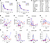 Figure 1
PK profiling of ciapavir in NHPs. Naive rhesus macaques were intravenously administered ciapavir at 0.25 mg/kg on day 0 (A) and 0.5 mg/kg on day 7 (B) to determine plasma concentrations of the molecule. PK parameters are shown for both doses in the table (C). C0, initial concentration; MRTIFN, mean residence time to infinity. Complete blood count (CBC) analysis revealed a temporary drop in lymphocyte (D) and monocyte (E) counts following drug administration as well as increases in granulocytes (F). Blood chemistry analysis showed modest and temporary increases in alanine aminotransferase (ALT) (G) and aspartate aminotransferase (AST) (H), as well as in high-sensitivity C-reactive protein (I). The second dose of ciapavir was accompanied by increases in CD69+ (J) and HLA-DR+ (K) CD4+ cells. Vertical lines indicate ciapavir administration on day 0 (0.25 mg/kg) and day 7 (0.5 mg/kg).
Tolerability assessment revealed no severe adverse effects with the administration of a single dose. Transient hematological changes, including reductions in lymphocyte and APC/monocyte counts and an increase in granulocytes, were observed postdosing but resolved within several days (Figure 1, D–F). Modest elevations in liver enzymes (alanine aminotransferase [ALT] and aspartate aminotransferase [AST]) returned to baseline levels shortly after treatment (Figure 1, G and H). High-sensitivity C-reactive protein (hsCRP) assays indicated a transient increase in this inflammatory marker 24 hours postdosing, returning to pretreatment levels by day 7 (Figure 1I). Notably, increases in CD69- and HLA-DR–positive cells were observed only after the second dose of ciapavir (Figure 1, J and K). No behavioral changes were detected in the study animals. In summary, the PK profile of ciapavir in uninfected rhesus macaques suggests effective systemic exposure and good tissue penetration. The transient changes in blood parameters and liver enzymes without severe adverse effects indicate tolerability of ciapavir but warrant careful monitoring. Since the plasma concentration of ciapavir required to effectively reverse viral latency in NHPs is not known, and both the 0.25 mg/kg and 0.5 mg/kg doses were well tolerated in the PK study, we selected the higher dose for study in SHIV-infected, ART-suppressed animals.
AAV-expressed eCD4-Ig protects against intrarectal shiv challenge. eCD4-Ig is an immunoadhesin protein that combines the properties of CD4 domains 1 and 2, which bind the viral Env protein, a sulfated coreceptor mimetic peptide, and an Ig domain. This synthetic protein represents a rationally designed broadly neutralizing agent against HIV, SIV, and SHIV (24, 26). eCD4-Ig has potential for long-term expression when administered via AAV vectors, offering a unique advantage for prophylactic as well as therapeutic applications (24, 26). Our study evaluated the expression and efficacy of 3 vectorized eCD4-Ig expression cassettes and their ability to protect against dose-escalating intrarectal (IR) SHIV challenge in 8 Indian-origin rhesus macaques (Figure 2A). Each cassette was designed to optimize eCD4-Ig expression and improve PK from our previous studies (Figure 2B). We had previously tested AAV expression cassettes that utilized the CMV promoter with a woodchuck hepatitis virus posttranscriptional regulatory element (WPRE) to express an rh-eCD4-IgG2 construct that successfully prevented SHIV-AD8 and SIVmac239 infection (24, 26). In an effort to decrease antidrug antibody (ADA) responses, we removed the WPRE and replaced it with 4 miRNA-142 target sequences in tandem to reduce expression from antigen-presenting cells (27, 28). Additionally, in the second cassette, we replaced the CMV promoter with the chicken β-actin (CBA) promoter because the CMV promoter has been associated with transcriptional silencing (29). Last, in the third cassette, we replaced rhesus IgG2 Fc with the rhesus IgG1 Fc. All cassettes included the M428L/N434S amino acid substitutions that have been described to improve FcRn binding and increase antibody half-life (30).
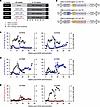 Figure 2
Optimized AAV-expressed rh-eCD4-Ig cassettes used in the NHP study. (A) Schematic of the NHP study. Four groups of 2 rhesus macaques each were used. NX, necropsy. (B) Designs of the AAV expression cassettes used to express rh-eCD4-Ig. Concentrations of rh-eCD4-Ig and ADA responses as measured by ELISA are shown for vectors using a CMV promoter expressing rh-eCD4-IgG2-LS (C), a CBA promoter expressing rh-eCD4-IgG2-LS (D), or a CMV promoter expressing rh-eCD4-IgG1-LS (E). ADA responses in panels C–E are shown as gray lines, measured by absorbance at 450 nm using a 1:10 serum dilution. Error bars in C–E represent range of duplicates per assay.
Fuchs et al. had previously demonstrated a prime/boost strategy that resulted in higher antibody expression with lower ADA in rhesus macaques using AAV8 and AAV1 vectors (31). In an effort to reduce ADA and increase eCD4-Ig expression from AAV vectors, we decided to replicate this strategy where 3 groups of 2 rhesus macaques each would receive 4 intramuscular (IM) injections of AAV8 rh-eCD4-Ig vectors on week 0 in the left and right quadriceps muscles followed by 4 IM injections of AAV1 rh-eCD4-Ig vectors in the left and right biceps and deltoid muscles at week 12. AAV8 vectors were administered at a 2.1 vector genomes (vg) per kilogram dose and were mixed with an AAV8 vector encoding TPST2 at a dose of 0.75 vg/kg to ensure the coreceptor mimetic peptide was sulfated. AAV1 vectors were administered at a 2.7 vg/kg dose and were mixed with an AAV1 vector encoding TPST2 at a dose of 0.75 vg/kg. The first group of macaques using the CMV promoter had peak rh-eCD4-IgG2-LS concentrations reach 7–15 μg/mL but ultimately ranged from <1 to 9 μg/mL before the challenge phase of the study (Figure 2C). ADA responses were observed in both animals starting at weeks 3 or 4 after AAV8 administration and lasted throughout the study. The second group of macaques using the CBA promoter had more sustained and constant expression of rh-eCD4-IgG2-LS ranging from 9 to 16 μg/mL for most of the study (Figure 2D). ADA was also observed in both animals but decreased between weeks 10 and 15 of the study. The third group of macaques using the CMV promoter to express rh-eCD4-IgG1-LS had the lowest concentrations and highest ADA of the 6 animals (Figure 2E). We observed peak concentrations after AAV1 administration ranging from 4 to 8 μg/mL, but by the end of the study, the concentrations decreased to <0.1 to 2.5 μg/mL. ADA was again observed in both animals but was markedly higher in A17039.
We next assessed the in vivo efficacy of the expressed rh-eCD4-Ig to protect against escalating, IR SHIV-CH848 challenges. A17039 was removed from the challenge phase, as it had no detectable rh-eCD4-Ig, and was used to identify anti–rh-eCD4-Ig antibodies. Two control animals became infected after the first challenge (32), and we observed delayed acquisition in the animals expressing rh-eCD4-Ig; however, statistical significance was not assessed because of the limited control sample size (Figure 3, A and B). A17015 became infected after the first challenge, A17018 after the second, A17033 after the third, and A17031 after the fourth. Notably, A17029 was protected against 6 IR challenges and only became infected after an intravenous (IV) dose that was 10-fold higher than the last IR challenge. Moreover, we observed a strong correlation between the number of challenges before infection with the concentration of expressed rh-eCD4-Ig at the start of the challenge phase (Figure 3C). Importantly, to our knowledge, this is the first NHP study to demonstrate that rh-eCD4-Ig can protect against IR SHIV challenges.
 Figure 3
AAV-expressed rh-eCD4-Ig protects from IR SHIV-CH848 challenges. (A) Kaplan-Meier curve of uninfected rhesus macaques after each SHIV-CH848 challenge. Two control macaques and 5 of the rh-eCD4-Ig–expressing macaques were IR challenged 6 times with escalating doses of SHIV-CH848. Note that the seventh challenge of A17029 was a dose 10-fold higher than the sixth challenge and administered intravenously. (B) Viral RNA measurements of each animal as determined by qPCR. (C) Correlation plot of number of challenges needed for infection compared with the amount of rh-eCD4-Ig as measured by ELISA before the start of challenges. Correlation statistics are included in the graph.
Characterization of NHP response to ART in study groups. We next tested the impact of AAV-delivered eCD4-Ig and Smac mimetics on the latent SHIV reservoir. A total of 30 rhesus macaques (Macaca mulatta; Supplemental Table 1; supplemental material available online with this article; https://doi.org/10.1172/JCI187961DS1) of Indian origin were infected IV with the 1157ipd3N4 strain (23, 33–38), a highly replicating and mucosally transmissible R5 virus that encodes an HIV-1 clade C envelope (Figure 4A). Blood draws were conducted biweekly throughout the study and twice per week during Smac mimetic dosing (Figure 4B). Seven weeks postinfection, animals were placed on ART consisting of daily tenofovir (TDF, 5.1 mg/kg), emtricitabine (FTC, 30 mg/kg), and dolutegravir (DTG, 2.5 mg/kg), administered subcutaneously for 72–73 weeks (39) (Figure 4A). Animals treated with ART only (n = 10) served as a control group (Figure 4C). Experimental groups consisting of 4 or 6 animals were used to evaluate treatment with AAV-delivered eCD4-Ig, ciapavir, or AZD5582 individually, as well as AAV-delivered eCD4-Ig in combination with ciapavir (Figure 4, D–G). Note that the treatment groups were not all studied concurrently.
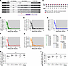 Figure 4
Study design and plasma viral load kinetics in ART-suppressed SHIV-infected NHPs. (A) Study schematic for experiments in SHIV-infected, ART-suppressed rhesus macaques. Seven weeks after infection with SHIV, animals initiated suppressive ART and were treated for at least 47 weeks before reservoir targeting with AAV-eCD4-Ig (orange arrows) and/or Smac mimetic compounds (purple arrows). ART was released after 24–25 weeks of reservoir targeting, and viral rebound was monitored for 16–25 weeks prior to end-of-study necropsy. (B) Smac mimetic dosing and sampling schedule for SHIV-infected, ART-suppressed rhesus macaques. Up to 10 doses of AZD5582 or ciapavir were administered, 1–2 weeks apart. Blue and red circles indicate sampling on the day of dosing and 2 days postdose, respectively. PVL is continuously monitored throughout the study in animals treated with ART only (negative control) (C), eCD4-Ig (D), AZD5582 (E), ciapavir (F), or ciapavir in combination with eCD4-Ig (G). Area shaded in gray represents the period of ART administration. The level of quantification (LOQ) is indicated by a black dashed line. Pre-ART peak PVL (H) and average PVL (I) indicate an equal distribution of viral loads in the animals across treatment groups. Bars indicate group median. Statistical significance was determined by pairwise comparison using the Kruskal-Wallis test with Dunn’s multiple-comparison correction.
With one exception in the control group, all animals displayed peak plasma viral loads (PVLs) between 6 × 10⁵ and 1 × 10⁸, and average PVLs between 3 × 10⁵ and 1 × 10⁷, prior to ART. At the time of ART initiation in week 7, PVLs ranged from 104 to 105 SHIV RNA copies/mL. Although difficult to discern due to ART initiation at week 7, we did observe that set point viral loads began to trend lower approximately 4 weeks postinfection in a subset of our animals, suggesting spontaneous control of SHIV. All animals were well suppressed to PVL below the LOQ (20 copies/mL) at week 60, prior to LRA treatment (Figure 4, C–G). Our study groups exhibited similar virologic parameters, without significant differences in pre-ART peak and average viral loads (Figure 4, H and I). Animals with lower average viral loads in Figure 4I are most likely to have spontaneously controlled virus replication prior to ART initiation.
AAV9 vector delivery of rh-eCD4-IgG2-LS in SHIV-infected, ART-treated rhesus macaques. Twelve macaques were administered AAV9 vectors encoding rh-eCD4-IgG2-LS and rhTPST2 at 34 weeks postinfection (Figure 4A), including 6 without subsequent ciapavir treatment (Figure 5A) and 6 with ciapavir treatment (Figure 5B). AAV9 was selected over AAV1 and AAV8 for this study, as our previous study had demonstrated AAV9 expression in rhesus macaques to be as good as AAV1 and better than AAV8 via IM administration. We elected to use rh-eCD4-IgG2-LS expressed from the CBA promoter based on our results in Figure 2. While the rhesus macaque IgG2 Fc has reduced effector function compared with the rhesus macaque IgG1 Fc, it does have the ability to kill infected cells, unlike the human IgG2 Fc. Peak rh-eCD4-IgG2-LS concentrations ranged from 10 to 40 μg/mL in all but 3 macaques (A19097, A19098, and A19102) before dropping to concentrations <1 to 5 μg/mL in all macaques for the duration of the study (Figure 5, A and B). The lower concentrations we observed before the ATI phase did not achieve therapeutic efficacy after ART was withdrawn, as viral rebound was observed in all 12 macaques (Supplemental Figure 1).
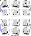 Figure 5
AAV9-delivered rh-eCD4-IgG2-LS concentrations in rhesus macaques with suppressed SHIV viremia mediated by ART. (A) Six SHIV-1157ipd3N4–infected rhesus macaques were administered AAV9 vectors encoding rh-eCD4-IgG2-LS and rhTPST2 at 34 weeks after infection while all macaques had suppressed viremia mediated by daily ART. (B) Same as A except this group of 6 rhesus macaques also received ciapavir. Rh-eCD4-IgG2-LS concentrations were measured by anti-CD4 ELISA, and concentrations are represented as the average of duplicates in blue. Error bars represent range of the duplicates. PVLs as determined by qPCR are represented in black. Area shaded in gray represents the period of ART administration.
Ciapavir induces modest increase in PVLs in SHIV-infected rhesus macaques. Latency reversal was defined as on-ART viremia where viral RNA in the plasma exceeded the LOQ (20 copies/mL). The Smac mimetics ciapavir or AZD5582 were administered weekly for 10 weeks, starting at week 62, or until adverse effects were observed. AZD5582 was administered at the previously established dose of 0.1 mg/kg (16, 18, 20), and ciapavir was administered at 0.5 mg/kg based on the PK evaluation. Study cohorts correspond to the schematic in Figure 4A and included an ART-only control group (Figure 6A) and animals dosed with AAV-delivered eCD4-Ig (Figure 6B), AZD5582 (Figure 6C), or ciapavir (Figure 6D). Animals treated with eCD4-Ig or AZD5582 largely resembled controls, lacking evidence of latency reversal during the treatment (Supplemental Figure 2). However, 3 of 4 ciapavir-treated animals (75%) responded with low but significant levels of on-ART viremia (Figure 6D and Supplemental Figure 2). Animals A18130, A18132, and A18100 showed multiple blips of viral RNA in the plasma following ciapavir administration.
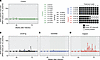 Figure 6
Treatment of SHIV-infected, ART-suppressed NHPs. Smac mimetics were administered to the animals in 10 weekly doses unless adverse effects were observed. Measurements of SHIV PVL in the control group (A) and groups treated with eCD4-Ig (B) or AZD5582 (C) did not show on-ART viremia. In the ciapavir-treated group (D), 3 animals showed blips of SHIV RNA in the plasma exceeding the LOQ. The LOQ is indicated by a black dashed line. Area shaded in gray represents the period of ART administration. LRA dosing is indicated by green vertical lines.
In addition to measuring PVL, we quantified cell-associated SHIV RNA and DNA levels in PBMCs of treated animals (Figure 7). Despite PVL increases in the ciapavir-treated group, PBMC-associated SHIV RNA levels were not significantly impacted compared with levels recorded in the other groups. Levels of PBMC-associated SHIV DNA and RNA/DNA ratios remained equally unaffected. While this indicated that ciapavir treatment alone did not impact viral reservoir size, it should be noted that the signal-to-noise ratio, particularly in the RNA/DNA proportion, may preclude detection of modest effects.
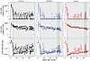 Figure 7
Cell-associated SHIV RNA and DNA levels in PBMCs of treated animals. Following ART initiation, cell-associated (CA) RNA levels remained constant in all treatment groups. CA DNA levels showed a constant decrease postinfection that converged toward a plateau consistent across groups. Area shaded in gray represents the period of ART administration. LRA dosing is indicated by green vertical lines.
Repeat dosing of ciapavir causes adverse effects in treated animals. Clinical assessment of repeat administration of ciapavir indicated tolerability through 5 doses without serious adverse events. However, following the sixth dose, 2 animals (A18130 and A18132) exhibited acute nausea and emesis, prompting cessation of treatment. One animal (A18132) also displayed serous nasal discharge and facial flushing. Adverse responses were managed with a regimen consisting of ondansetron, maropitant for nausea/emesis, diphenhydramine and dexamethasone for potential immune reaction, enrofloxacin for potential infection, and fluids for dehydration, resolving within 30 minutes. No such effects were noted in the remaining 2 animals treated with ciapavir or any animals treated with AZD5582 or AAV-delivered eCD4-Ig alone, indicating that the adverse reactions could be dose-/treatment-dependent or specific to certain individuals. CBC and lymphocyte subset analysis showed a temporary decrease in white blood cells and neutrophils postdosing of both Smac mimetics, ciapavir and AZD5582 (Supplemental Figure 3), similar to trends seen in the PK evaluation in uninfected animals (Figure 1, D–F). A sawtooth pattern was observed in several parameters, most notably in lymphocytes and monocytes, decreasing from the day of dosing to the 2-day postdose time point, and resolving by the next dose 7 days later (Supplemental Figures 3 and 4). Comprehensive metabolic panel parameters remained stable except for modest, temporary reductions in alkaline phosphatase (ALP) levels and increases in serum glucose levels, particularly in AZD5582-treated animals (Supplemental Figure 5).
Combination of AAV-delivered eCD4-Ig and ciapavir does not impact the viral reservoir size in SHIV-infected rhesus macaques. Finally, we asked whether the administration of a combined “shock” (ciapavir) and “kill” treatment (eCD4-Ig) impacts viral reservoir size following respective virus reactivation and elimination. Based on the significant induction of viremia in the ciapavir group (P = 0.0044, Supplemental Figure 2) compared with animals treated with AZD5582 at the administered doses, we selected ciapavir for evaluation in combination with eCD4-Ig to assess the effect of this shock-and-kill treatment on the viral reservoir. To reduce the likelihood of adverse effects upon repeated ciapavir administration, we altered the dosing schema for the treatment combination (Figure 8A). In weeks 1, 3, and 5, animals were treated with ciapavir on a biweekly schedule before returning to a weekly administration starting in week 6. Despite the adjusted dosing regimen, 1 of 6 animals in the treatment group (A19096) exhibited severe adverse effects after dose 6 (treatment week 8), characterized by lack of appetite/nausea and a spike in kidney toxicity markers that ultimately required euthanasia. Due to this severe adverse event, LRA dosing of all animals in the treatment group was halted and not resumed for the duration of the study. Notably, PVLs in this treatment group were reactivated during LRA treatment, with measurements repeatedly exceeding the LOQ of 20 copies/mL (Figure 8A).
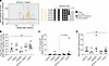 Figure 8
Impact of combined ciapavir and eCD4-Ig treatment on the viral reservoir. Following AAV delivery of eCD4-Ig, 6 animals were scheduled for biweekly (doses 1–3) administration of ciapavir, followed by weekly (doses 4–8) administration. After adverse effects were observed in A19096, dosing of all animals was halted. (A) PVL levels show repeated blips of SHIV RNA after LRA treatment. Area shaded in gray represents the period of ART administration. The LOQ is indicated by a black dashed line. (B) Post-ATI average viral loads did not significantly differ between treatment groups. (C) PVL during ATI was calculated as a percentage of average PVL during primary infection. (D) Days to rebound after ATI did not reveal significant differences between treatment groups. Bars indicate group median. Significance was determined with a Kruskal-Wallis test. Unless otherwise noted, pairwise comparisons of treatment groups were not significant (P > 0.05).
To determine the impact of the combination treatment on the viral reservoir, an analytical treatment interruption (ATI) was conducted 8 weeks after LRA administration (79–80 weeks postinfection, 72–73 weeks after ART initiation) in all study groups, including single and combination treatments (Supplemental Figure 1). Analysis of average viral loads prior to initiation of suppressive ART and reservoir-targeting treatments confirmed equal distribution between groups (Figure 4, H and I). Following LRA treatment, a post-ATI average viral load was determined for each animal by averaging measurements over the first 7 weeks of recrudescent viremia, providing a comparable window to pre-ART average viral loads (37, 40) (Figure 8B). Comparisons between pre- and post-ART PVLs did not reveal significant differences between treatment groups. Likewise, calculating post-ATI PVL as percentage of pre-ART PVL to assess rebound did not indicate a meaningful impact of treatments on the viral reservoir (Figure 8C). Time-to-rebound, calculated as days to SHIV rebound post-ATI and serving as an additional measure to detect changes in reservoir size, did not show significant differences between the study arms (Figure 8D). The data suggest that neither the individual treatments with Smac mimetics, nor AAV-delivered eCD4-Ig, nor the combination of ciapavir and eCD4-Ig mediated a measurable impact on the magnitude or kinetics of the viral reservoir.
Although our measurements of total SHIV DNA (Figure 7) and PVL kinetics post-ATI (Figure 8, B–D) failed to detect an impact of our shock-and-kill approach, these assays are often insufficient to detect partial effects. To more quantitatively assess the how eCD4-Ig and Smac mimetics may have altered SHIV reservoir size, we conducted 2 additional experiments. First, we optimized a SHIV-specific 5-target intact provirus detection assay (5T-IPDA), applying our expertise from similar assays for people living with HIV (41). Using leukapheresis products collected before and after reservoir targeting, we were able to quantify reservoir size in a total of 51 samples in our n = 30 study. In SHIV-infected, ART-suppressed controls that did not receive reservoir-targeting therapies, we observed a natural decay in reservoir size (Figure 9A) consistent with past reports in the NHP/SHIV model (42). Comparing log10 fold-changes in reservoir size between each group reinforces the notion that neither eCD4-Ig nor Smac mimetics increased or decreased viral reservoir size relative to controls (Figure 9B). Finally, we asked whether modest selective pressures mediated by our treatments affected viral diversity. Since our SHIV was not barcoded, we generated and compared full-length Env sequences as a surrogate measure of eCD4-Ig and/or Smac mimetics’ impact on virus replication following ART interruption. Plasma samples from 8 rhesus macaques representing the eCD4-Ig, eCD4-Ig + ciapavir, and untreated control groups were analyzed by deep sequencing of the full-length SHIV-1157ipd3N4 env gene after viral rebound (Supplemental Table 2). Supplemental Figure 6 shows very low amino acid change rates across the entire Env protein, including areas previously identified for resistance mutations to rh-eCD4-Ig (26, 43). Amino acid changes observed in the regions of interest were minimal and not at residues that have been shown to mediate resistance to rh-eCD4-Ig neutralization, ruling out the emergence of treatment-resistant variants as contributing to the inability to achieve significant reservoir reduction. The lack of measurable Env diversity that we observed reinforces our conclusion that our eCD4-Ig and LRA treatments failed to measurably impact recrudescent SHIV replication following ATI.
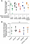 Figure 9
A SHIV-specific 5T-IPDA shows no impact of eCD4-Ig or LRAs in vivo. (A) Intact proviral SHIV DNA copies per million CD4+ T cells was measured by SHIV 5T-IPDA before (filled circles) and after (unfilled circles) treatment. Horizontal bars represent group median. (B) Log10 fold-change in intact proviral SHIV DNA copies per million CD4+ T cells calculated for each rhesus macaque with matched pre- and posttreatment 5T-IPDA measurements. Each square represents an individual animal. Missing data points indicate animals that lacked sufficient pre- or posttreatment samples for analysis. Horizontal bars denote group medians, and vertical T bars represent 95% confidence intervals for the median. No statistically significant differences in fold-change between groups were detected by Kruskal-Wallis test (P = 0.564).


Copyright © 2026 American Society for Clinical Investigation
ISSN: 0021-9738 (print), 1558-8238 (online)