
Advertisement
Research ArticleGeneticsReproductive biology
Open Access |  10.1172/JCI178862
Targeting the PRMT5/Nur77 methylation axis enhances endometrial decidualization capacity and female fertility in preclinical models
Zhiwen Cao,1,2 Xinyu Cai,1,3 Jie Mei,1,3 Na Kong,1,3 Yang Liu,1,2 Xiaoyue Shen,1,3 Min Wu,1,3 Xin Zhen,1,3 Jianxin Sun,4 Rong Li,1,3,5 Ruiwei Jiang,1,5 Haixiang Sun,1,4,5,6 and Guijun Yan1,3,5,6
1Center for Reproductive Medicine and Obstetrics and Gynecology, Nanjing Drum Tower Hospital, Affiliated Hospital of Medical School, State Key Laboratory of Pharmaceutical Biotechnology, and
2Center for Molecular Reproductive Medicine, Nanjing University, Nanjing, Jiangsu, China.
3State Key Laboratory of Reproductive Medicine and Offspring Health, Nanjing Medical University, Nanjing, Jiangsu, China.
4Department of Medicine, Center for Translational Medicine, Thomas Jefferson University, Philadelphia, Pennsylvania, USA.
5Jiangsu Human Reproductive Function Remodeling Engineering Research Center, Nanjing, Jiangsu, China.
6Nanjing Clinical Medical Center for Reproductive Medicine, Nanjing, Jiangsu, China.
Address correspondence to: Guijun Yan, Haixiang Sun, Ruiwei Jiang, or Rong Li, Zhongshanbei Road 53, Nanjing, Jiangsu 210008 China. Phone: 025.83106666; Email: yanguijun@nju.edu.cn (GY); Email: haixiang_sun@nju.edu.cn (HS); Email: rwjiang@smail.nju.edu.cn (RJ); lirongdme@163.com (RL).
Authorship note: ZC, XC, and JM contributed equally to this work.
Find articles by Cao, Z. in: PubMed | Google Scholar
1Center for Reproductive Medicine and Obstetrics and Gynecology, Nanjing Drum Tower Hospital, Affiliated Hospital of Medical School, State Key Laboratory of Pharmaceutical Biotechnology, and
2Center for Molecular Reproductive Medicine, Nanjing University, Nanjing, Jiangsu, China.
3State Key Laboratory of Reproductive Medicine and Offspring Health, Nanjing Medical University, Nanjing, Jiangsu, China.
4Department of Medicine, Center for Translational Medicine, Thomas Jefferson University, Philadelphia, Pennsylvania, USA.
5Jiangsu Human Reproductive Function Remodeling Engineering Research Center, Nanjing, Jiangsu, China.
6Nanjing Clinical Medical Center for Reproductive Medicine, Nanjing, Jiangsu, China.
Address correspondence to: Guijun Yan, Haixiang Sun, Ruiwei Jiang, or Rong Li, Zhongshanbei Road 53, Nanjing, Jiangsu 210008 China. Phone: 025.83106666; Email: yanguijun@nju.edu.cn (GY); Email: haixiang_sun@nju.edu.cn (HS); Email: rwjiang@smail.nju.edu.cn (RJ); lirongdme@163.com (RL).
Authorship note: ZC, XC, and JM contributed equally to this work.
Find articles by Cai, X. in: PubMed | Google Scholar
1Center for Reproductive Medicine and Obstetrics and Gynecology, Nanjing Drum Tower Hospital, Affiliated Hospital of Medical School, State Key Laboratory of Pharmaceutical Biotechnology, and
2Center for Molecular Reproductive Medicine, Nanjing University, Nanjing, Jiangsu, China.
3State Key Laboratory of Reproductive Medicine and Offspring Health, Nanjing Medical University, Nanjing, Jiangsu, China.
4Department of Medicine, Center for Translational Medicine, Thomas Jefferson University, Philadelphia, Pennsylvania, USA.
5Jiangsu Human Reproductive Function Remodeling Engineering Research Center, Nanjing, Jiangsu, China.
6Nanjing Clinical Medical Center for Reproductive Medicine, Nanjing, Jiangsu, China.
Address correspondence to: Guijun Yan, Haixiang Sun, Ruiwei Jiang, or Rong Li, Zhongshanbei Road 53, Nanjing, Jiangsu 210008 China. Phone: 025.83106666; Email: yanguijun@nju.edu.cn (GY); Email: haixiang_sun@nju.edu.cn (HS); Email: rwjiang@smail.nju.edu.cn (RJ); lirongdme@163.com (RL).
Authorship note: ZC, XC, and JM contributed equally to this work.
Find articles by Mei, J. in: PubMed | Google Scholar
1Center for Reproductive Medicine and Obstetrics and Gynecology, Nanjing Drum Tower Hospital, Affiliated Hospital of Medical School, State Key Laboratory of Pharmaceutical Biotechnology, and
2Center for Molecular Reproductive Medicine, Nanjing University, Nanjing, Jiangsu, China.
3State Key Laboratory of Reproductive Medicine and Offspring Health, Nanjing Medical University, Nanjing, Jiangsu, China.
4Department of Medicine, Center for Translational Medicine, Thomas Jefferson University, Philadelphia, Pennsylvania, USA.
5Jiangsu Human Reproductive Function Remodeling Engineering Research Center, Nanjing, Jiangsu, China.
6Nanjing Clinical Medical Center for Reproductive Medicine, Nanjing, Jiangsu, China.
Address correspondence to: Guijun Yan, Haixiang Sun, Ruiwei Jiang, or Rong Li, Zhongshanbei Road 53, Nanjing, Jiangsu 210008 China. Phone: 025.83106666; Email: yanguijun@nju.edu.cn (GY); Email: haixiang_sun@nju.edu.cn (HS); Email: rwjiang@smail.nju.edu.cn (RJ); lirongdme@163.com (RL).
Authorship note: ZC, XC, and JM contributed equally to this work.
Find articles by Kong, N. in: PubMed | Google Scholar
1Center for Reproductive Medicine and Obstetrics and Gynecology, Nanjing Drum Tower Hospital, Affiliated Hospital of Medical School, State Key Laboratory of Pharmaceutical Biotechnology, and
2Center for Molecular Reproductive Medicine, Nanjing University, Nanjing, Jiangsu, China.
3State Key Laboratory of Reproductive Medicine and Offspring Health, Nanjing Medical University, Nanjing, Jiangsu, China.
4Department of Medicine, Center for Translational Medicine, Thomas Jefferson University, Philadelphia, Pennsylvania, USA.
5Jiangsu Human Reproductive Function Remodeling Engineering Research Center, Nanjing, Jiangsu, China.
6Nanjing Clinical Medical Center for Reproductive Medicine, Nanjing, Jiangsu, China.
Address correspondence to: Guijun Yan, Haixiang Sun, Ruiwei Jiang, or Rong Li, Zhongshanbei Road 53, Nanjing, Jiangsu 210008 China. Phone: 025.83106666; Email: yanguijun@nju.edu.cn (GY); Email: haixiang_sun@nju.edu.cn (HS); Email: rwjiang@smail.nju.edu.cn (RJ); lirongdme@163.com (RL).
Authorship note: ZC, XC, and JM contributed equally to this work.
Find articles by Liu, Y. in: PubMed | Google Scholar
1Center for Reproductive Medicine and Obstetrics and Gynecology, Nanjing Drum Tower Hospital, Affiliated Hospital of Medical School, State Key Laboratory of Pharmaceutical Biotechnology, and
2Center for Molecular Reproductive Medicine, Nanjing University, Nanjing, Jiangsu, China.
3State Key Laboratory of Reproductive Medicine and Offspring Health, Nanjing Medical University, Nanjing, Jiangsu, China.
4Department of Medicine, Center for Translational Medicine, Thomas Jefferson University, Philadelphia, Pennsylvania, USA.
5Jiangsu Human Reproductive Function Remodeling Engineering Research Center, Nanjing, Jiangsu, China.
6Nanjing Clinical Medical Center for Reproductive Medicine, Nanjing, Jiangsu, China.
Address correspondence to: Guijun Yan, Haixiang Sun, Ruiwei Jiang, or Rong Li, Zhongshanbei Road 53, Nanjing, Jiangsu 210008 China. Phone: 025.83106666; Email: yanguijun@nju.edu.cn (GY); Email: haixiang_sun@nju.edu.cn (HS); Email: rwjiang@smail.nju.edu.cn (RJ); lirongdme@163.com (RL).
Authorship note: ZC, XC, and JM contributed equally to this work.
Find articles by Shen, X. in: PubMed | Google Scholar
1Center for Reproductive Medicine and Obstetrics and Gynecology, Nanjing Drum Tower Hospital, Affiliated Hospital of Medical School, State Key Laboratory of Pharmaceutical Biotechnology, and
2Center for Molecular Reproductive Medicine, Nanjing University, Nanjing, Jiangsu, China.
3State Key Laboratory of Reproductive Medicine and Offspring Health, Nanjing Medical University, Nanjing, Jiangsu, China.
4Department of Medicine, Center for Translational Medicine, Thomas Jefferson University, Philadelphia, Pennsylvania, USA.
5Jiangsu Human Reproductive Function Remodeling Engineering Research Center, Nanjing, Jiangsu, China.
6Nanjing Clinical Medical Center for Reproductive Medicine, Nanjing, Jiangsu, China.
Address correspondence to: Guijun Yan, Haixiang Sun, Ruiwei Jiang, or Rong Li, Zhongshanbei Road 53, Nanjing, Jiangsu 210008 China. Phone: 025.83106666; Email: yanguijun@nju.edu.cn (GY); Email: haixiang_sun@nju.edu.cn (HS); Email: rwjiang@smail.nju.edu.cn (RJ); lirongdme@163.com (RL).
Authorship note: ZC, XC, and JM contributed equally to this work.
Find articles by Wu, M. in: PubMed | Google Scholar
1Center for Reproductive Medicine and Obstetrics and Gynecology, Nanjing Drum Tower Hospital, Affiliated Hospital of Medical School, State Key Laboratory of Pharmaceutical Biotechnology, and
2Center for Molecular Reproductive Medicine, Nanjing University, Nanjing, Jiangsu, China.
3State Key Laboratory of Reproductive Medicine and Offspring Health, Nanjing Medical University, Nanjing, Jiangsu, China.
4Department of Medicine, Center for Translational Medicine, Thomas Jefferson University, Philadelphia, Pennsylvania, USA.
5Jiangsu Human Reproductive Function Remodeling Engineering Research Center, Nanjing, Jiangsu, China.
6Nanjing Clinical Medical Center for Reproductive Medicine, Nanjing, Jiangsu, China.
Address correspondence to: Guijun Yan, Haixiang Sun, Ruiwei Jiang, or Rong Li, Zhongshanbei Road 53, Nanjing, Jiangsu 210008 China. Phone: 025.83106666; Email: yanguijun@nju.edu.cn (GY); Email: haixiang_sun@nju.edu.cn (HS); Email: rwjiang@smail.nju.edu.cn (RJ); lirongdme@163.com (RL).
Authorship note: ZC, XC, and JM contributed equally to this work.
Find articles by Zhen, X. in: PubMed | Google Scholar
1Center for Reproductive Medicine and Obstetrics and Gynecology, Nanjing Drum Tower Hospital, Affiliated Hospital of Medical School, State Key Laboratory of Pharmaceutical Biotechnology, and
2Center for Molecular Reproductive Medicine, Nanjing University, Nanjing, Jiangsu, China.
3State Key Laboratory of Reproductive Medicine and Offspring Health, Nanjing Medical University, Nanjing, Jiangsu, China.
4Department of Medicine, Center for Translational Medicine, Thomas Jefferson University, Philadelphia, Pennsylvania, USA.
5Jiangsu Human Reproductive Function Remodeling Engineering Research Center, Nanjing, Jiangsu, China.
6Nanjing Clinical Medical Center for Reproductive Medicine, Nanjing, Jiangsu, China.
Address correspondence to: Guijun Yan, Haixiang Sun, Ruiwei Jiang, or Rong Li, Zhongshanbei Road 53, Nanjing, Jiangsu 210008 China. Phone: 025.83106666; Email: yanguijun@nju.edu.cn (GY); Email: haixiang_sun@nju.edu.cn (HS); Email: rwjiang@smail.nju.edu.cn (RJ); lirongdme@163.com (RL).
Authorship note: ZC, XC, and JM contributed equally to this work.
Find articles by Sun, J. in: PubMed | Google Scholar
1Center for Reproductive Medicine and Obstetrics and Gynecology, Nanjing Drum Tower Hospital, Affiliated Hospital of Medical School, State Key Laboratory of Pharmaceutical Biotechnology, and
2Center for Molecular Reproductive Medicine, Nanjing University, Nanjing, Jiangsu, China.
3State Key Laboratory of Reproductive Medicine and Offspring Health, Nanjing Medical University, Nanjing, Jiangsu, China.
4Department of Medicine, Center for Translational Medicine, Thomas Jefferson University, Philadelphia, Pennsylvania, USA.
5Jiangsu Human Reproductive Function Remodeling Engineering Research Center, Nanjing, Jiangsu, China.
6Nanjing Clinical Medical Center for Reproductive Medicine, Nanjing, Jiangsu, China.
Address correspondence to: Guijun Yan, Haixiang Sun, Ruiwei Jiang, or Rong Li, Zhongshanbei Road 53, Nanjing, Jiangsu 210008 China. Phone: 025.83106666; Email: yanguijun@nju.edu.cn (GY); Email: haixiang_sun@nju.edu.cn (HS); Email: rwjiang@smail.nju.edu.cn (RJ); lirongdme@163.com (RL).
Authorship note: ZC, XC, and JM contributed equally to this work.
Find articles by Li, R. in: PubMed | Google Scholar
1Center for Reproductive Medicine and Obstetrics and Gynecology, Nanjing Drum Tower Hospital, Affiliated Hospital of Medical School, State Key Laboratory of Pharmaceutical Biotechnology, and
2Center for Molecular Reproductive Medicine, Nanjing University, Nanjing, Jiangsu, China.
3State Key Laboratory of Reproductive Medicine and Offspring Health, Nanjing Medical University, Nanjing, Jiangsu, China.
4Department of Medicine, Center for Translational Medicine, Thomas Jefferson University, Philadelphia, Pennsylvania, USA.
5Jiangsu Human Reproductive Function Remodeling Engineering Research Center, Nanjing, Jiangsu, China.
6Nanjing Clinical Medical Center for Reproductive Medicine, Nanjing, Jiangsu, China.
Address correspondence to: Guijun Yan, Haixiang Sun, Ruiwei Jiang, or Rong Li, Zhongshanbei Road 53, Nanjing, Jiangsu 210008 China. Phone: 025.83106666; Email: yanguijun@nju.edu.cn (GY); Email: haixiang_sun@nju.edu.cn (HS); Email: rwjiang@smail.nju.edu.cn (RJ); lirongdme@163.com (RL).
Authorship note: ZC, XC, and JM contributed equally to this work.
Find articles by Jiang, R. in: PubMed | Google Scholar
1Center for Reproductive Medicine and Obstetrics and Gynecology, Nanjing Drum Tower Hospital, Affiliated Hospital of Medical School, State Key Laboratory of Pharmaceutical Biotechnology, and
2Center for Molecular Reproductive Medicine, Nanjing University, Nanjing, Jiangsu, China.
3State Key Laboratory of Reproductive Medicine and Offspring Health, Nanjing Medical University, Nanjing, Jiangsu, China.
4Department of Medicine, Center for Translational Medicine, Thomas Jefferson University, Philadelphia, Pennsylvania, USA.
5Jiangsu Human Reproductive Function Remodeling Engineering Research Center, Nanjing, Jiangsu, China.
6Nanjing Clinical Medical Center for Reproductive Medicine, Nanjing, Jiangsu, China.
Address correspondence to: Guijun Yan, Haixiang Sun, Ruiwei Jiang, or Rong Li, Zhongshanbei Road 53, Nanjing, Jiangsu 210008 China. Phone: 025.83106666; Email: yanguijun@nju.edu.cn (GY); Email: haixiang_sun@nju.edu.cn (HS); Email: rwjiang@smail.nju.edu.cn (RJ); lirongdme@163.com (RL).
Authorship note: ZC, XC, and JM contributed equally to this work.
Find articles by Sun, H. in: PubMed | Google Scholar
1Center for Reproductive Medicine and Obstetrics and Gynecology, Nanjing Drum Tower Hospital, Affiliated Hospital of Medical School, State Key Laboratory of Pharmaceutical Biotechnology, and
2Center for Molecular Reproductive Medicine, Nanjing University, Nanjing, Jiangsu, China.
3State Key Laboratory of Reproductive Medicine and Offspring Health, Nanjing Medical University, Nanjing, Jiangsu, China.
4Department of Medicine, Center for Translational Medicine, Thomas Jefferson University, Philadelphia, Pennsylvania, USA.
5Jiangsu Human Reproductive Function Remodeling Engineering Research Center, Nanjing, Jiangsu, China.
6Nanjing Clinical Medical Center for Reproductive Medicine, Nanjing, Jiangsu, China.
Address correspondence to: Guijun Yan, Haixiang Sun, Ruiwei Jiang, or Rong Li, Zhongshanbei Road 53, Nanjing, Jiangsu 210008 China. Phone: 025.83106666; Email: yanguijun@nju.edu.cn (GY); Email: haixiang_sun@nju.edu.cn (HS); Email: rwjiang@smail.nju.edu.cn (RJ); lirongdme@163.com (RL).
Authorship note: ZC, XC, and JM contributed equally to this work.
Find articles by Yan, G. in: PubMed | Google Scholar
Published July 17, 2026 - More info
J Clin Invest. 2026;136(17):e178862. https://doi.org/10.1172/JCI178862.
© 2026 Cao et al. This work is licensed under the Creative Commons Attribution 4.0 International License. To view a copy of this license, visit http://creativecommons.org/licenses/by/4.0/.
Received: January 3, 2024; Accepted: July 9, 2026


-
Results
PRMT5-mediated SDMA protein modification is compromised in RIF. Dynamic transcriptomic changes during the WOI are critical for establishing the endometrial receptivity (43), we first conducted longitudinal RNA-seq on endometrial biopsies from fertile women (FER) across the secretory phase (LH+2, LH+5, LH+7, LH+9, LH+11) (Figure 1A and Supplemental Tables 1 and 2; supplemental material available online with this article; https://doi.org/10.1172/JCI178862DS1). As previously reported (44), progesterone receptor expression was high in epithelial cells during the early secretory phase (LH+2 to LH+5), declined by the midsecretory phase (LH+7 to LH+9), and further decreased by the late secretory phase (LH+11), but persisted at the stroma throughout the stages, coinciding with the temporal mRNA changes of proliferation, differentiation, and decidualization representative genes (Supplemental Figure 1, A–C, and Supplemental Table 1). PRMT family genes exhibited distinct expression patterns: an early secretory group (PRMT6/8) that declined at WOI onset, a midsecretory transient group (PRMT3/7) peaking at LH+7, and a late-accumulating group (PRMT1/2 and type II enzymes PRMT5/9) that was maximized from LH+7 to LH+9 (Figure 1B). Accordingly, a moderate increase of ADMA and an obvious increase of MMA was observed from LH+2 to LH+7, but only SDMA showed a robust accumulation during the WOI from LH+7 to LH+9 across a broad molecular weight range (17–180 kDa) (Figure 1C), indicating a potential association with endometrial receptivity. Interestingly, the analysis of one published single-cell RNA-seq dataset of human endometrium also indicates that PRMT5 was the only SDMA catalytic enzyme that expressed much higher levels in the EnSCs at LH+7 compared with LH+2 and LH+12 (Supplemental Figure 1D), implicating its critical role in regulating SDMA during endometrial decidualization.
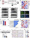 Figure 1
PRMT5-mediated arginine methylome is compromised in RIF. (A) Schematic diagram illustrating the collection of endometrium samples for transcriptome sequencing at different time points during the secretory phase of LH in normal FER, as well as Alstra-DIA methylation sequencing of endometrial samples at the LH+7 time point from both controls and patients with RIF. (B) Transcriptomic analysis reveals the expression dynamics of PRMT family expression profiles at different time points during the secretory phase. (C and D) Western blot analysis revealing expression patterns of 3 protein arginine methylation modifications (SDMA, ADMA, MMA) in human endometrium temporal dynamics from LH+2 to LH+11 days during secretory phase (C) (n = 3) and between FER and RIF patients (D) (n = 6). (E) Immunofluorescence detection of SDMA expression and localization in midsecretory-phase endometrium from FER and RIF patients. (F) The number of proteins with differentially expressed SDMA-modified peptides in RIF compared with FER endometria, using Alstra-DIA methylation sequencing and co-immunoprecipitation of anti-SDMA antibody. (G) Biological Process (Gene Ontology) enrichment analysis of differentially methylated proteins in RIF compared with FER groups. (H) Heatmap of PRMT family expression profiles in FER and RIF endometria at LH+7 (n = 6). Western blot (I) analysis of PRMT5 protein levels and IHC staining (J) and IHC quantitative analysis (K) of PRMT5 expression in endometrial epithelial and stromal cells in FER and RIF patients (n = 24). Data represent mean ± SEM. **P < 0.01; ***P < 0.001; ****P < 0.0001; Student’s t test. Black and white scale bars: 100 μm. FER, fertile controls; RIF, recurrent implantation failure; DIA, data-independent acquisition; SDMA, symmetric dimethylarginine; ADMA, asymmetric dimethylarginine; MMA, monomethylarginine; LH, luteinizing hormone; GE, glandular epithelium; LE, luminal epithelium; S, stroma.
Here, we collected endometrial biopsies from 6 patients with RIF, who did not achieve a pregnancy after at least 5 transfers of high-quality embryos, for in-depth multiomics analysis to identify the key endometrial factors implicated in the pathogenesis of RIF. We found the global SDMA abundance was markedly reduced in the LH+7 endometria from patients with RIF, in contrast with comparable ADMA and MMA signals (Figure 1D). The reduction of SDMA was more pronounced in the endometrial stroma than in the epithelium (Figure 1E and Supplemental Figure 2A). These findings suggest that the diminished endometrial SDMA modification may contribute to the pathogenesis of RIF.
Next, by integrating a PTMScan SDMA motif enrichment kit with Alstra-DIA proteomics, we obtained a high-sensitivity SDMA protein landscape in the pooled control and RIF endometrial samples mid-secretory (LH+7) (Supplemental Figure 2B and Supplemental Table 3). There were 507 upregulated and 435 downregulated SDMA-modified proteins in patients with RIF (Figure 1F). These differentially methylated proteins are prominently enriched in pathways essential for decidualization, including Actin cytoskeleton organization, Relaxin signaling, and Wnt signaling (Figure 1G). Transcriptomic analysis demonstrated multiple dysregulated decidualization pathways in these RIF endometria (Supplemental Figure 2C and Supplemental Table 4). Between the 2 primary enzymes responsible for SDMA modification, PRMT9 expression was unaltered, whereas PRMT5 was downregulated in RIF endometrium (Figure 1H), which was further confirmed by qPCR (Supplemental Figure 2D). The downregulation of PRMT5 proteins was detected in both endometrial compartments of RIF patients but was more pronounced at the stroma (Figure 1, I–K), which is consistent with the SDMA staining results (Figure 1E). Collectively, these results identify endometrial PRMT5 deficiency, particularly in the stroma of patients with RIF, which might account for the hypo-SDMA of decidual regulatory networks.
PRMT5 deficiency impairs endometrial decidualization in vivo. Consistent with human patterns, PRMT5 was present in both epithelial and stromal compartments of the pregnant mouse uterus, with higher stromal expression at gestation day 4 (D4) compared with D1, started to rise in predecidua at D5, and was robustly upregulated in decidual cells at D6 to D8, implying its potential importance in murine endometrial decidualization (Supplemental Figure 3A). To test the in vivo functions of PRMT5 on endometrial decidualization, we generated 3 transgenic mouse lines. Pgr-Cre mice have been widely used to conditionally target genes in both uterine epithelium and stroma (45, 46). But we found PgrCre/+ Prmt5fl/fl mice exhibited developmental defects, which caused severe uterine dysplasia and a complete absence of glands during adulthood (Supplemental Figure 3B). Since the developmental defects have not been observed in patients with RIF, this mouse model may not be suitable for investigating the association of PRMT5 and RIF. Ltf-iCre mainly mediates recombination at uterine epithelium (47). Unfortunately, we observed varying degrees of ovarian dysfunction in LtfiCre/+ Prmt5fl/fl mice, thereby complicating their utility to study uterine functions (Supplemental Figure 3C). In contrast, Amhr2-Cre-mediated Prmt5 knockout (Prmt5d/d) maintained normal uterine and ovarian morphology. qRT-PCR analysis of isolated uterine epithelial and stromal cells, together with immunofluorescence staining of uterine sections, revealed a variable yet pronounced reduction of PRMT5 in the uterine stroma of Prmt5d/d mice at D3.5, which was accompanied by a concurrent decrease in global SDMA levels (Figure 2, A–C, and Supplemental Figure 3D), similar to the marked reduction observed in patients with RIF (Figure 1, E and J). It has been reported that Amhr2-Cre has some penetration issues (48). But considering the essential role of PRMT5 in embryonic development (49), we were not surprised that the mice we checked only exhibited restricted deletion within the stromal compartment. Therefore, we primarily investigated the roles of stromal PRMT5 in regulating endometrial receptivity using this mouse model.
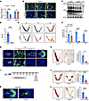 Figure 2
Stromal PRMT5 deficiency impairs endometrial decidualization and embryo development. (A) qRT-PCR analysis of PRMT5 mRNA levels in isolated primary endometrial stromal and epithelial cells from Prmt5fl/fl and Amhr2cre Prmt5fl/fl (Prmt5d/d) mice. (B and C) Immunofluorescence (B) and Western blot analysis (C) of PRMT5 protein in uteri from Prmt5fl/fl and Prmt5d/d mice. (D) Litter size in Prmt5fl/fl and Prmt5d/d mice. Gross morphology detection (E) and statistics (F) of uterine implantation sites on D5, D6, and D8 in Prmt5fl/fl and Prmt5d/d mice. (G) Immunofluorescence analysis of COX2 for embryo attachment reaction on D5 and decidual marker DTPRP for decidualization on D6 and D8. (H) Gross uterine morphology and weight ratio of stimulated to unstimulated sides in artificial decidualization model in Prmt5fl/fl and Prmt5d/d mice. (I–K) GSK591 or vehicle was administered to normal pregnant mice from D5 to D7, and uteri were collected on D8 for analysis: schematic of the treatment protocol (I), representative uterine images and quantification of implantation sites (J), and immunofluorescence analysis of the DTPRP in implantation sites (K). (L) Gross uterine morphology and weight ratio of stimulated to unstimulated sides in artificial decidualization model of GSK591-administered mice. Data represent mean ± SEM. *P < 0.05; ***P < 0.001; ****P < 0.0001; Student’s t test. White scale bars: 100 μm (B, G, and K). LE, luminal epithelium; GE, glandular epithelium; S, stroma; Em, embryo.
First of all, we found no pups were delivered by Prmt5d/d female mice (Figure 2D). To determine whether the infertility stemmed from initial implantation failure, we examined uteri at D5, when embryo implantation occurred, by intravenous Chicago Blue injection and found out the number of implantation sites was comparable between Prmt5fl/fl and Prmt5d/d mice (Figure 2, E and F). The loss of stromal PRMT5 altered neither the embryo attachment marker COX2 expression (50) nor the epithelial receptivity marker FOXO1 expression (51) on D5, indicating that the initial phase of embryo implantation remains uncompromised (Figure 2G and Supplemental Figure 3E). However, a subsequent decidualization defect was observed on D6, as evidenced by attenuated HAND2 expression (Supplemental Figure 3F). On D6 and D8, Prmt5d/d mice displayed extensive embryo resorptions (Figure 2, E and F), which could be caused by the severe decidualization defect, as indicated by markedly reduced expression of the murine decidual marker DTPRP (52) (Figure 2G). Since decidualization is tightly regulated by the ovarian hormones progesterone and estrogen (5), and Amhr2-Cre is also active in the granulosa cells of mice (53), we checked the ovaries of Prmt5d/d mice. The number of ovulated oocytes, as well as serum estradiol-17β (E2) and progesterone (P4) levels, were comparable to those of Prmt5fl/fl mice, thereby ruling out ovarian dysfunction as a confounding factor for decidualization (Supplemental Figure 3, G–I). Additionally, we employed an artificial decidualization (AD) model, which reliably recapitulates endometrial decidualization during natural pregnancy in mice while eliminating confounding interferences from ovarian and embryonic signals (54). The decidualization failure in the Prmt5d/d mice was validated in AD models, characterized by a blunted decidual weight induction and a complete absence of stromal alkaline phosphatase activity (Figure 2H and Supplemental Figure 3J). Collectively, these results demonstrate that stromal PRMT5 is indispensable for endometrial decidualization leading to successful pregnancy.
GSK591 is a well-characterized PRMT5 inhibitor that efficiently inhibits its methylation activity (55). The genetic heterogeneity of outbred ICR mice makes them suitable for medical study (56). To determine whether methylation activity is critical for endometrial decidualization, we administered GSK591 to ICR mice on D5, D6, and D7 after embryo implantation. This treatment markedly diminished stromal SDMA methylation, which resulted in the suppressed decidualization and elevated embryonic resorption at D8 (Figure 2, I–K, and Supplemental Figure 3K). Since PRMT5 is essential for embryonic and ovarian follicle development (49, 57), intraperitoneal injections of GSK591 may also adversely affect embryos and ovaries. To specifically assess the uterine decidual ability following GSK591 treatment while excluding the confounding factors from ovaries and embryos, artificial decidualization was also conducted in the ovariectomized ICR mice. GSK591 administration all through the hormone treatment period in the artificial decidualization model robustly inhibited the decidual response (Figure 2L and Supplemental Figure 3L). These findings establish that the endometrial decidualization is dependent on the enzymatic activity of PRMT5.
PRMT5 is indispensable for the decidualization of human EnSCs. The in vivo findings demonstrated the important roles of stromal PRMT5 in endometrial decidualization, probably through its methylation activity. Next, we investigated the detailed roles of stromal PRMT5 in decidualization using cultured human EnSCs. First, we characterized its expression pattern across the secretory phase, revealing that PRMT5 protein expression peaked in the midsecretory endometrium at LH+7 (Figure 3A). Reanalysis of a previously published single-cell atlas of human endometrium indicated PRMT5 was mainly expressed in the stromal fibroblasts with peak expressions at LH+7 (Supplemental Figure 4A). The decidualization of human endometrium is induced by progesterone rise and second messenger cAMP production in vivo and can be mimicked in vitro by synthetic progesterone MPA and cAMP (Pc) treatment (10). We found PRMT5 and global SDMA modification were increased after Pc treatment from 0 to 72 hours (Figure 3, B and C). Moreover, treatment with progesterone alone was sufficient to upregulate PRMT5 expression, suggesting that PRMT5 and PRMT5-mediated SDMA were downstream effectors of progesterone signaling during decidualization (Supplemental Figure 4B).
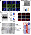 Figure 3
PRMT5 is indispensable for endometrial stromal decidualization and trophoblast invasion in vitro. (A) Immunofluorescence analysis of PRMT5 expression patterns in human endometrium across the secretory phase (LH+2 to LH+11). Western blot analysis (B) and quantification (C) of temporal changes in PRMT5 and SDMA levels in human EnSCs treated with MPA and cAMP for 0, 12, 24, 48, and 72 hours. n = 3. (D) Western blot analysis of PRMT5, SDMA, and IGFBP1 protein levels in EnSCs transfected with Ad-shRNA or Ad-shPRMT5 followed by decidualization induction (MPA+cAMP). Effects of PRMT5 knockdown on decidualization markers were assessed by RT-qPCR for IGFBP1 mRNA levels (E) and ELISA for PRL secretion (F) (n = 3). (G) Immunofluorescence staining of F-actin (red) showing morphological changes in control and PRMT5-knockdown EnSCs under decidualizing conditions. Nuclei were stained with DAPI (blue). (H) Representative images (left) and quantification (right) of trophoblast spheroid invasion into EnSC monolayers. EnSCs were treated with Ad-shRNA or Ad-shPRMT5 and induced with 8-Br-cAMP prior to coculture. The invasion area was measured at 12, 24, 48, and 72 hours. (I) Heatmap of RNA-seq data showing the expression profiles of key decidualization regulators and markers in nondecidualized (shRNA), decidualized control (shRNA_Dec), and decidualized PRMT5-knockdown (shPRMT5_Dec) groups. n = 3. Data represent mean ± SEM. *P < 0.05; **P < 0.01; ****P < 0.0001. One-way ANOVA with Tukey’s multiple comparisons test was used (C, E, and F). Two-way ANOVA with Bonferroni’s multiple comparisons test was used (H). Scale bar: 100 μm.
To directly assess the functional requirement of PRMT5, we performed adenoviral-mediated knockdown (Ad-shPRMT5) in primary EnSCs. This intervention substantially attenuated the Pc-induced upregulation of PRMT5 and SDMA (Figure 3D). Critically, PRMT5 deficiency contributed to a functional decidualization defect, evidenced by markedly reduced expressions of canonical markers (IGFBP1, PRL) and diminished PRL secretion (Figure 3, D–F, and Supplemental Figure 4C). Beyond gene expression, PRMT5 knockdown impaired the morphological transformation of stromal cells from spindle to rounded shape during decidualization (Figure 3G). Since maternal decidualization controls the invasion behavior of the fetal trophoblast (58), it is not surprising that the trophoblast invasion over decidual EnSCs was also compromised by PRMT5 knockdown in EnSCs (Figure 3H). Transcriptomic analysis revealed a broad disruption of the decidualization program by PRMT5 knockdown, characterized by the significant downregulation of key decidual regulators (e.g., BMP2, KLF12) and decidual markers (e.g., IGFBP1, LEFTY2), and dysregulated decidualization-related pathways, including extracellular matrix organization, PI3K/AKT signaling, TGF-β signaling, and ECM-receptor interaction (Figure 3I; Supplemental Figure 4, D–H; and Supplemental Table 5). Taken together, these data indicate that PRMT5 is not only upregulated in the EnSCs during the WOI but also functionally indispensable for the endometrial decidualization that facilitates trophoblast invasion.
PRMT5 methylates Nur77 and regulates its chromatin occupancy during decidualization. We already reported before that the PRMT5 inhibitor GSK591 could impair the in vitro decidualization of EnSCs, suggesting that PRMT5 regulates decidualization through its methylation activity (42). Therefore, we performed an integrated multiomics screen combining anti-PRMT5 immunoprecipitation–mass spectrometry (IP-MS) and pan-SDMA methylation proteomics in the decidual human EnSCs to identify the critical substrate of PRMT5 during decidualization (Figure 4A). By intersecting SDMA proteins downregulated in human EnSCs upon PRMT5 loss (via knockdown or inhibition) with those deficient in RIF patient endometria, we identified 110 clinically RIF-relevant, PRMT5-dependent SDMA target proteins in endometrial stroma (Figure 4B and Supplemental Table 3). Overlapping with the PRMT5 interactome pinpointed 2 core candidates: Nur77 and FAM120A (Figure 4, C and D, and Supplemental Table 6). We focused on orphan nuclear receptor Nur77, a key decidual regulator (59), whose SDMA modification was unknown. Co-immunoprecipitation confirmed a PRMT5-Nur77 complex in HEK293T cells (Figure 4E). This interaction was more obvious during decidualization in human EnSCs, probably due to elevated PRMT5 and Nur77 expressions (Figure 4F). Furthermore, PRMT5 knockdown specifically blunted SDMA modification on Nur77 (Figure 4G), thereby identifying Nur77 as a direct substrate of PRMT5.
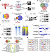 Figure 4
PRMT5 methylates Nur77 and regulates its chromatin occupancy during decidualization. (A) Schematic of multiomics strategy to identify PRMT5 downstream effectors in human EnSCs, combining anti-PRMT5 IP-MS and pan-SDMA methylation proteomics. (B) Venn diagram showing overlap between SDMA-methylated proteins downregulated upon PRMT5 deficiency (knockdown/inhibition) and those deficient in RIF patient endometria. Intersection of PRMT5-interacting proteins (IP-MS) (C) with the 110 targets from B identifies Nur77 and FAM120A as core candidates (D). n = 1 pooled one sample per group. (E) Immunoprecipitation validation of overexpressed PRMT5-Nur77 interaction in HEK293T cells. (F) Immunoprecipitation analysis of endogenous PRMT5 and Nur77 protein levels and interaction following decidualization induction in EnSCs. (G) Western blot analysis of SDMA modification of Nur77 and total Nur77 protein levels upon PRMT5 knockdown. (H) Heatmap of Nur77 CUT&Tag peak intensity and signals specifically around the transcription start site (TSS) regions in control and PRMT5-knockdown EnSCs under decidualization. (I) Representative genome browser tracks of Nur77 occupancy, monomethylation of histone H3 at lysine 4 (H3K4me1), and input at the IGFBP1 and MAOA (monoamine oxidase A, a mitochondrial enzyme) loci. (J) Functional enrichment analysis of differentially expressed genes (DEGs) associated with lost or gained Nur77 peaks upon PRMT5 knockdown. (K) Motif analysis at Nur77-bound regions gained or lost after PRMT5 knockdown, respectively. OFP, orange fluorescent protein.
We directly compared the transcriptomic changes in decidual EnSCs after PRMT5 or Nur77 knockdown. While a few genes like IGFBP1, ZNF92, and FAM20C were coregulated, the global overlap was minimal: 94.8% of PRMT5-dependent DEGs and 87.0% of Nur77-dependent DEGs were unique (Supplemental Figure 5A and Supplemental Table 7), arguing against simple functional equivalence. Considering Nur77 is a transcription factor that binds at specific chromatin loci to regulate the transcriptions of its target genes (60), we further examined the alteration of Nur77 chromatin occupancy following PRMT5 knockdown in EnSCs to elucidate the functional consequences of PRMT5 SDMA on Nur77. Strikingly, PRMT5 knockdown failed to alter the global intensity of Nur77 peaks but triggered a robust accumulation of Nur77 peaks at TSS regions (Figure 4H and Supplemental Figure 5, B and C). Specifically, elevated Nur77 occupancy upstream of the TSS coincided with reduced expression of key decidual genes (e.g., IGFBP1 and MAOA) (61), suggesting that aberrantly bound, unmethylated Nur77 may transcriptionally repress these genes (Figure 4I). Conversely, the loss of Nur77 at downstream intergenic regions was also associated with some upregulated genes (e.g., FUT2 and PLAC1) (Supplemental Figure 5D). H3K4me1 typically marks enhancers to facilitate transcription (62). We observed that the gained Nur77 peaks following PRMT5 knockdown showed a stronger overlap with H3K4me1-marked regions compared with the lost peaks, raising the possibility that unmethylated Nur77 may convert these enhancers to silencers (Figure 4I and Supplemental Figure 5, D–F). All these results demonstrate that PRMT5 depletion fundamentally alters the chromatin binding specificity of Nur77, leading to dysregulated decidual gene expression.
To globally assess the regulation of PRMT5/Nur77 axis on gene expression, we performed the integrated analysis of Nur77 CUT&Tag and PRMT5-knockdown RNA-seq data in decidual EnSCs. In total, 236 DEGs, which are coregulated by both PRMT5 knockdown and Pc treatment, associated with lost Nur77 peaks, and 443 DEGs associated with gained peaks (Figure 4J and Supplemental Table 8). Both gene sets are involved in WNT signaling, cell migration, and adhesion pathways essential for decidualization. However, coregulated DEGs associated with lost peaks were enriched in TNF and estrogen responses, which has been reported to regulate decidualization (63, 64), whereas those associated with gained peaks were enriched in phagocytosis and lipid metabolism (Figure 4J). Notably, peaks gained, but not lost, upon PRMT5 knockdown were highly enriched for the canonical Nur77 binding motif, and distinct cofactor partnerships were predicted for the lost peaks. This indicates that PRMT5 knockdown enhanced Nur77’s direct chromatin binding while altering its coordinated protein interactions at chromatin (Figure 4K). Collectively, these data demonstrate that PRMT5-mediated SDMA modification acts as a critical fidelity checkpoint, ensuring Nur77 engages with appropriate chaperones and binds at precise chromatin sites.
PRMT5-mediated SDMA of Nur77 at R346 is indispensable for decidualization. Next, we investigated the SDMA modification sites of Nur77 by PRMT5 using 2 methylation prediction tools, PRmePred and GPS-MSP, thereby identifying 4 high-scoring arginine residues, R266, R345, R346, and R348, within the Nur77 DNA-binding domain (DBD). Single point mutation of each residue indicated that only the R346K substitution effectively abolished the PRMT5-activated SDMA signal, an evolutionarily conserved site (Figure 5A). Consistently, in vitro methylation assays confirmed that PRMT5 methylates the DBD of Nur77 specifically at residue R346 (Figure 5B). Because commercial antibodies against methylated Nur77 were not available, we generated a polyclonal antibody exhibiting high affinity to symmetric dimethyl R346 (R346-me2s), with minimal crossreactivity toward the unmodified, monomethyl, or asymmetric forms (Figure 5C). The R346K mutation attenuated Nur77-mediated decidualization of EnSCs (Figure 5, D–F). Importantly, after knockdown of endogenous Nur77, PRMT5 overexpression markedly potentiated the SDMA modification on exogenous wild-type Nur77 and synergistically amplified decidual marker induction, cytoskeletal remodeling, and trophoblast invasion in EnSCs, whereas it had negligible effects on exogenous non-SDMA mutant Nur77R346K (Figure 5, G–L). Collectively, these findings support that PRMT5-mediated SDMA at R346 of Nur77 drives the decidualization program.
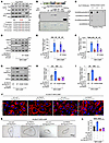 Figure 5
PRMT5-mediated SDMA of Nur77 at R346 is required for decidualization. (A) Identification of Nur77 arginine methylation sites. Top: Western blot analysis of SDMA-modified Nur77 in HEK293T cells; bottom: Evolutionary conservation of Nur77 R346 across vertebrate species. (B) In vitro methylation analysis of PRMT5 on Nur77 wild-type and various domain mutants. (C) Dot blot validation experiment of Nur77-R346me2s–specific antibody, using different antibody concentrations (111, 37, 12 ng/mL) to detect specific binding to various methylated peptides. (D) Western blot detection of intracellular Nur77 methylation status following overexpression of wild-type Nur77 and R346K mutant. (E and F) RT-qPCR analysis of decidualization markers PRL (E) and IGFBP1 (F) mRNA expression levels in Nur77 knockdown and wild-type or R346K mutant rescue experiments (n = 3). (G–L) Effects of PRMT5 overexpression on decidualization in Nur77-knockdown EnSCs rescued with wild-type or R346K mutant Nur77: Western blot analysis of PRMT5, Nur77, and Nur77-R346me2s (G); RT-qPCR analysis of PRL (H) and IGFBP1 (I) mRNA levels; immunofluorescence analysis of F-actin cytoskeletal remodeling (J); trophoblast spheroid invasion assay (K and L). Data represent mean ± SEM. **P < 0.01; ***P < 0.001; ****P < 0.0001; 1-way ANOVA with Tukey’s multiple comparisons test in (E, F, H, I, and L). Scale bar: 100 μm. NT, N-terminal domain; DBD, DNA-binding domain; LBD, ligand-binding domain.
A Nur77-derived peptide with R346K mutant (Pep-Nur77R346K) promotes decidualization. Given the critical role of the PRMT5/Nur77 methylation axis in driving decidualization, we explored whether modulating this axis could have therapeutic relevance. We designed 2 cell-permeable peptides derived from Nur77: a wild-type peptide (Pep-Nur77WT) and a methylation-deficient mutant (Pep-Nur77R346K). We found both peptides were efficiently delivered into the cytoplasm and nucleus of decidual EnSCs (Figure 6A). Surprisingly, only Pep-Nur77R346K strongly enhanced decidual marker expression (IGFBP1, PRL), while Pep-Nur77WT showed a mild suppressive effect in decidual EnSCs (Figure 6, B and C). Further analysis of the R346 flanking sequence also identified 2 serine residues, S341 and S351, that are established substrates of AKT-mediated phosphorylation, and S351 is known to regulate Nur77 activity (65) (Figure 6D). Molecular docking demonstrated that Pep-Nur77WT bound strongly to the triose-phosphate isomerase barrel region of PRMT5 and AKT LBD, thus competing with the same binding sites of Nur77 (Figure 6E and Supplemental Figure 6, A and B). Pep-Nur77R346K, however, retained binding to AKT at the canonical Nur77-binding site, but bound to PRMT5 at a non-Nur77-binding region, implying that it might compete with the AKT-mediated phosphorylation of Nur77 without interfering with the PRMT5-mediated methylation (Figure 6E and Supplemental Figure 6, C and D).
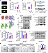 Figure 6
A Nur77-derived peptide with R346K mutant (Pep-Nur77R346K) promotes EnSC decidualization. (A) Representative fluorescence images showing the intracellular uptake of FITC-labeled wild-type (FITC-Pep-Nur77WT) and mutant (FITC-Pep-Nur77R346K) peptides in human EnSCs. (B and C) RT-qPCR analysis of decidualization markers IGFBP1 (B) and PRL (C) mRNA expression levels in EnSCs treated with control vehicle, Pep-Nur77WT, or Pep-Nur77R346K under decidualization conditions (MPA+cAMP). (D) Top: Schematic representation of the amino acid sequences of cell-permeable peptides containing the TAT transduction domain. Bottom: Domain structure of Nur77 highlighting the conserved R346 site and phosphorylation sites S341/S351. (E) Molecular docking simulation showing the interaction interfaces of AKT and PRMT5 with Pep-Nur77WT and Pep-Nur77R346K. (F) Co-immunoprecipitation analysis of endogenous Nur77 interactions with PRMT5 and AKT following treatment with Nur77-derived peptides. (G) Western blot analysis of temporal changes in Nur77 methylation (R346me2s) and phosphorylation (S341, S351) during the decidualization process (0–72 hours). (H–L) Functional rescue of PRMT5-depleted EnSCs with Pep-Nur77R346K: RT-qPCR analysis of IGFBP1 (H) and PRL (I) mRNA levels; Western blot analysis of PRMT5, Nur77-R346me2s, pNur77-S341, and pNur77-S351 status (J). (K and L) Trophoblast spheroid invasion assay with representative images (K) and quantification (L). Data represent mean ± SEM. *P < 0.05; **P < 0.01; ***P < 0.001; ****P < 0.0001; 1-way ANOVA with Tukey’s multiple comparisons test (B, C, H, I, and L). White scale bar: 50 μm.
Based on these structural insights, we hypothesized that the peptides function as molecular decoys that selectively sequester upstream enzymes. Co-immunoprecipitation confirmed that Pep-Nur77WT attenuated endogenous Nur77 interactions with both PRMT5 and AKT, reducing both R346 methylation (R346me2s) and S351 phosphorylation (Figure 6F). In contrast, Pep-Nur77R346K specifically disrupted the Nur77-AKT interaction without affecting PRMT5 binding, thereby suppressing inhibitory S351 phosphorylation while preserving R346 methylation of Nur77 (Figure 6F). Through the comparative analysis of these 2 peptides, we found out that dominant R346 methylation of Nur77, relative to its S351 phosphorylation, may play important roles in decidualization. Indeed, during in vitro decidualization, a progressive increase of Nur77-R346 SDMA coincided with the decreased Nur77 phosphorylation, particularly at Ser351 (Figure 6G). Thus, the orchestrated regulation of Nur77 methylation and phosphorylation represents a layer of posttranslational regulation mode of progesterone-driven decidualization but also emerges as a potential intervention point.
We then asked whether Pep-Nur77R346K could rescue defects caused by PRMT5 loss. We found that Pep-Nur77R346K indeed restored decidualization in PRMT5-knockdown cells and reversed the hyperphosphorylation at S351 induced by PRMT5 silencing, while leaving the methylation state at R346 undisturbed, to restore the dominance of R346 methylation of Nur77 (Figure 6, H–J). Eventually, trophoblast invasion defects over PRMT5-knockdown EnSCs were also ameliorated by Pep-Nur77R346K (Figure 6, K and L).
Pep-Nur77R346K ameliorates decidualization defects and embryo implantation failure in preclinical mouse models. Next, we assessed the therapeutic potential of functional peptide Pep-Nur77R346K in multiple preclinical models. As we described above, Prmt5d/d mice developed substantial embryonic resorption at D6 due to decidualization defects (Figure 2). Further analysis indicated Prmt5d/d mice exhibited reduced Nur77 methylation (R346me2s) and heightened Nur77 phosphorylation (S351), recapitulating the posttranslational signature observed in PRMT5-knockdown EnSCs, positioning this genetic model as a physiologically relevant platform for therapeutic validation (Supplemental Figure 7A). FITC-labeled Pep-Nur77R346K was efficiently delivered to mouse uterine cells including stromal cells through intrauterine perfusion (Supplemental Figure 7B). Two administrations of Pep-Nur77R346K before embryo implantation effectively restored the implantation sites and reversed the aberrant elevation of inhibitory phosphorylation (S351) at D6 of Prmt5d/d mice (Figure 7, A and B). The direct rescue effect of Pep-Nur77R346K on decidualization was further confirmed using an AD model of Prmt5d/d mice (Figure 7, C and D).
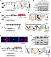 Figure 7
Pep-Nur77R346K ameliorates decidualization defects and embryo implantation failure. (A) Effects of Pep-Nur77R346K intrauterine perfusion on implantation site numbers (n = 3) on D6 in Prmt5d/d mice. (B) Western blot analysis of PRMT5, Nur77, Nur77-R346me2s, and pNur77 (S351) levels in endometrial tissues of Prmt5d/d mice treated with Pep-Nur77R346K. (C and D) Effects of Pep-Nur77R346K on artificial decidualization in Prmt5d/d mice: schematic of the experimental schedule (C); gross uterine morphology and weight ratio analysis of decidualized (oil-stimulated) to control sides (D). (E–H) Effects of Pep-Nur77R346K on GSK591-induced implantation failure: schematic of the experimental design (E); representative uterine images and quantification of implantation sites on D8 (F); immunofluorescence analysis of the decidual marker DTPRP (G); Western blot analysis of uterine PRMT5 and Nur77 modification status (H). (I and J) Effects of Pep-Nur77R346K on artificial decidualization in GSK591-treated mice, showing gross morphology and decidual weight ratio. Data represent mean ± SEM. **P < 0.01; ****P < 0.0001; Student’s t test (A and D); 1-way ANOVA with Tukey’s multiple comparisons test (F and J). Scale bar: 100 μm.
Interestingly, prophylactic treatment with Pep-Nur77R346K before implantation effectively rescued these postimplantation PRMT5 inhibitor GSK591-induced decidualization defects, restoring embryo implantation in ICR mice, probably through the downregulation of S351 phosphorylation of Nur77 (Figure 7, E–H). Besides, in the AD models, we treated the ICR mice with GSK591 all through the hormone treatment period, and 2 administrations of Pep-Nur77R346K at the estrogen and progesterone treatment period before decidual stimulation could completely reverse the impaired decidual responses (Figure 7, I and J). Together, these findings suggest that targeting the Nur77 R346/S351 modification balance with Pep-Nur77R346K may restore decidualization and improve implantation outcomes in preclinical models, warranting further evaluation.
Reduced endometrial PRMT5/Nur77 methylation is associated with RIF and pregnancy outcome. Our findings already indicated that PRMT5 deficiency, particularly within stroma, may contribute to endometrial dysfunction in patients with RIF. Immunostaining analysis revealed a pronounced global reduction in methylation in RIF endometrial samples, characterized by markedly attenuated nuclear signals of both PRMT5 and Nur77-R346me2s, along with decreased Nur77 protein levels, within both stromal and epithelial compartments in a subset of RIF patients with ≥5 failed implantations after embryo transfer cycles (Figure 8A). To elucidate the clinical relevance of the PRMT5/Nur77 methylation axis, we expanded our investigation to a larger retrospective cohort (n = 114 participants: 39 control and 75 RIF with ≥3 failed implantation after embryo transfer cycles) using semiquantitative Western blot protein analysis. Strikingly, we observed a concurrent reduction in PRMT5, Nur77, and specifically Nur77-R346me2s levels in RIF endometrial tissues (Figure 8, B–E). Moreover, correlation analysis demonstrated a strong positive association between PRMT5 protein levels and the intensity of Nur77-R346me2s modification (Figure 8F). Among these, 107 patients underwent subsequent embryo transfer (152 cycles) (Figure 8G). Spearman’s correlation analysis of 13 variables revealed significant positive correlations among PRMT5, Nur77, and Nur77-R346me2s, all of which were positively associated with clinical pregnancy outcomes (Supplemental Figure 8A and Supplemental Table 9). Logistic regression analysis demonstrated that Nur77-R346me2s levels were significantly associated with clinical pregnancy, with patients in the highest tertile exhibiting a more than 6-fold increased likelihood of pregnancy compared with the lowest tertile (OR: 6.46, 95% CI: 2.52–16.56) (Supplemental Figure 8, B and C). Receiver operating characteristic (ROC) analysis demonstrated that a clinical model incorporating parameters such as age, ovarian reserve, and endometrial thickness predicted pregnancy outcomes with an AUC of 0.679 (Figure 8H). Notably, the addition of endometrial Nur77-R346me2s alone improved predictive performance, yielding AUC of 0.828; further addition of Nur77 and/or PRMT5 did not provide substantial improvement (Figure 8H and Supplemental Figures 9 and 10). All these results demonstrated the endometrial Nur77-R346me2s levels displayed robust predictive capability for pregnancy outcomes.
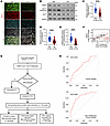 Figure 8
Reduced endometrial PRMT5 and Nur77-R346me2s are associated with RIF and pregnancy outcome. (A) Representative immunofluorescence images of PRMT5 (green), Nur77-R346me2s (red), and total Nur77 (cyan) in endometrial sections from FER and RIF patients. Nuclei were stained with DAPI (gray). (B) Representative Western blot image of PRMT5 and Nur77 methylation modification levels in endometria from FER and RIF patients. (C–E) Semiquantitative analysis of protein expression of (C) PRMT5, (D) total Nur77, and (E) Nur77-R346me2s in control (n = 39) and RIF (n = 75) endometrial tissues. (F) Correlation analysis between PRMT5 protein levels and Nur77-R346me2s intensity in human endometrial tissues. (G) Flowchart illustrating the inclusion and exclusion criteria for the clinical study participation and analysis. (H) ROC curve analysis of clinical variables alone and clinical variables plus Nur77-R346me2s. Data represent mean ± SEM. ****P < 0.0001. Student’s t test (C–E). Scale bar: 100 μm.
Beyond its predictive value, we asked whether the PRMT5/Nur77 methylation axis could be therapeutically targeted. We isolated primary EnSCs from 5 FER with normal Nur77-R346me2s expressions and from 5 patients with RIF with relatively low Nur77-R346me2s levels, then cultured these cells in vitro. Pep-Nur77R346K improved decidualization responses in RIF-derived EnSCs in a dose-dependent manner (Figure 9, A and B, and Supplemental Figure 11). Mechanistically, the peptide did not restore PRMT5 expression but selectively suppressed pathological pS351 levels without perturbing R346 methylation (Figure 9C). Consequently, it restored cytoskeletal remodeling during decidualization (Figure 9D) and enhanced trophoblast invasion over decidual cells (Figure 9, E and F). Collectively, these findings identify the loss of PRMT5-mediated R346 methylation as a molecular hallmark of RIF and highlight the therapeutic promise of Pep-Nur77R346K for restoring endometrial receptivity.
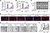 Figure 9
Pep-Nur77R346K restores decidualization-associated responses in RIF-derived EnSCs. (A and B) Effects of Pep-Nur77R346K treatment on decidualization markers PRL (A) and IGFBP1 (B) mRNA expression in EnSCs derived from FER and RIF patients. (C) Western blot analysis of PRMT5, Nur77 methylation, and phosphorylation modification levels in FER- and RIF-derived EnSCs following Pep-Nur77R346K treatment. (D) Immunofluorescence analysis of F-actin cytoskeletal remodeling and (E and F) trophoblast invasion assay (representative images left, quantification right) in RIF-derived EnSCs treated with or without Pep-Nur77R346K following decidualization induction. Data represent mean ± SEM. *P < 0.05; ***P < 0.001; ****P < 0.0001. One-way ANOVA with Tukey’s multiple comparisons test (A, B, and F). White scale bar: 100 μm (D); black scale bar: 500 μm (E).


Copyright © 2026 American Society for Clinical Investigation
ISSN: 0021-9738 (print), 1558-8238 (online)