Stem cells

Abstract
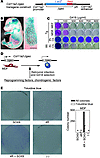
Parkinson disease (PD) involves the selective loss of midbrain dopamine (mDA) neurons and is a possible target disease for stem cell–based therapy. Human induced pluripotent stem cells (hiPSCs) are a potentially unlimited source of patient-specific cells for transplantation. However, it is critical to evaluate the safety of hiPSCs generated by different reprogramming methods. Here, we compared multiple hiPSC lines derived by virus- and protein-based reprogramming to human ES cells (hESCs). Neuronal precursor cells (NPCs) and dopamine (DA) neurons delivered from lentivirus-based hiPSCs exhibited residual expression of exogenous reprogramming genes, but those cells derived from retrovirus- and protein-based hiPSCs did not. Furthermore, NPCs derived from virus-based hiPSCs exhibited early senescence and apoptotic cell death during passaging, which was preceded by abrupt induction of p53. In contrast, NPCs derived from hESCs and protein-based hiPSCs were highly expandable without senescence. DA neurons derived from protein-based hiPSCs exhibited gene expression, physiological, and electrophysiological properties similar to those of mDA neurons. Transplantation of these cells into rats with striatal lesions, a model of PD, significantly rescued motor deficits. These data support the clinical potential of protein-based hiPSCs for personalized cell therapy of PD.
Authors
Yong-Hee Rhee, Ji-Yun Ko, Mi-Yoon Chang, Sang-Hoon Yi, Dohoon Kim, Chun-Hyung Kim, Jae-Won Shim, A-Young Jo, Byung-Woo Kim, Hyunsu Lee, Suk-Ho Lee, Wonhee Suh, Chang-Hwan Park, Hyun-Chul Koh, Yong-Sung Lee, Robert Lanza, Kwang-Soo Kim, Sang-Hun Lee

Abstract
The directed differentiation of iPS and ES cells into definitive endoderm (DE) would allow the derivation of otherwise inaccessible progenitors for endodermal tissues. However, a global comparison of the relative equivalency of DE derived from iPS and ES populations has not been performed. Recent reports of molecular differences between iPS and ES cells have raised uncertainty as to whether iPS cells could generate autologous endodermal lineages in vitro. Here, we show that both mouse iPS and parental ES cells exhibited highly similar in vitro capacity to undergo directed differentiation into DE progenitors. With few exceptions, both cell types displayed similar surges in gene expression of specific master transcriptional regulators and global transcriptomes that define the developmental milestones of DE differentiation. Microarray analysis showed considerable overlap between the genetic programs of DE derived from ES/iPS cells in vitro and authentic DE from mouse embryos in vivo. Intriguingly, iPS cells exhibited aberrant silencing of imprinted genes known to participate in endoderm differentiation, yet retained a robust ability to differentiate into DE. Our results show that, despite some molecular differences, iPS cells can be efficiently differentiated into DE precursors, reinforcing their potential for development of cell-based therapies for diseased endoderm-derived tissues.
Authors
Constantina Christodoulou, Tyler A. Longmire, Steven S. Shen, Alice Bourdon, Cesar A. Sommer, Paul Gadue, Avrum Spira, Valerie Gouon-Evans, George J. Murphy, Gustavo Mostoslavsky, Darrell N. Kotton

Abstract
Human induced pluripotent stem cells (hiPSCs) and human embryonic stem cells (hESCs) are promising candidate cell sources for regenerative medicine. However, despite the common ability of hiPSCs and hESCs to differentiate into all 3 germ layers, their functional equivalence at the single cell level remains to be demonstrated. Moreover, single cell heterogeneity amongst stem cell populations may underlie important cell fate decisions. Here, we used single cell analysis to resolve the gene expression profiles of 362 hiPSCs and hESCs for an array of 42 genes that characterize the pluripotent and differentiated states. Comparison between single hESCs and single hiPSCs revealed markedly more heterogeneity in gene expression levels in the hiPSCs, suggesting that hiPSCs occupy an alternate, less stable pluripotent state. hiPSCs also displayed slower growth kinetics and impaired directed differentiation as compared with hESCs. Our results suggest that caution should be exercised before assuming that hiPSCs occupy a pluripotent state equivalent to that of hESCs, particularly when producing differentiated cells for regenerative medicine aims.
Authors
Kazim H. Narsinh, Ning Sun, Veronica Sanchez-Freire, Andrew S. Lee, Patricia Almeida, Shijun Hu, Taha Jan, Kitchener D. Wilson, Denise Leong, Jarrett Rosenberg, Mylene Yao, Robert C. Robbins, Joseph C. Wu
Abstract
Repair of cartilage injury with hyaline cartilage continues to be a challenging clinical problem. Because of the limited number of chondrocytes in vivo, coupled with in vitro de-differentiation of chondrocytes into fibrochondrocytes, which secrete type I collagen and have an altered matrix architecture and mechanical function, there is a need for a novel cell source that produces hyaline cartilage. The generation of induced pluripotent stem (iPS) cells has provided a tool for reprogramming dermal fibroblasts to an undifferentiated state by ectopic expression of reprogramming factors. Here, we show that retroviral expression of two reprogramming factors (c-Myc and Klf4) and one chondrogenic factor (SOX9) induces polygonal chondrogenic cells directly from adult dermal fibroblast cultures. Induced cells expressed marker genes for chondrocytes but not fibroblasts, i.e., the promoters of type I collagen genes were extensively methylated. Although some induced cell lines formed tumors when subcutaneously injected into nude mice, other induced cell lines generated stable homogenous hyaline cartilage–like tissue. Further, the doxycycline-inducible induction system demonstrated that induced cells are able to respond to chondrogenic medium by expressing endogenous Sox9 and maintain chondrogenic potential after substantial reduction of transgene expression. Thus, this approach could lead to the preparation of hyaline cartilage directly from skin, without generating iPS cells.
Authors
Kunihiko Hiramatsu, Satoru Sasagawa, Hidetatsu Outani, Kanako Nakagawa, Hideki Yoshikawa, Noriyuki Tsumaki

Abstract
To be of therapeutic use, autologous stem cells derived from patients with inherited genetic disorders require genetic modification via gene repair or insertion. Here, we present proof of principle that, for diseases associated with dominant alleles (gain-of-function or haploinsufficient loss-of-function), disease allele–free ES cells can be derived from afflicted individuals without genome manipulation. This approach capitalizes on the derivation of uniparental cells, such as parthenogenetic (PG) ES cell lines from disease allele–free gametes. Diploid mammalian uniparental embryos with only maternally (oocyte-) or paternally (sperm-)derived genomes fail early in development due to the nonequivalence of parental genomes caused by genomic imprinting. However, these uniparental embryos develop to the blastocyst stage, allowing the derivation of ES cell lines. Using a mouse model for dominant beta-thalassemia, we developed disease allele–free PG ES cell lines from the oocytes of affected animals. Phenotype correction was obtained in donor-genotype recipients after transplantation of in vitro hematopoietic ES cell derivatives. This genetic correction strategy without gene targeting is potentially applicable to any dominant disease. It could also be the sole approach for larger or more complex mutations that cannot be corrected by homologous recombination.
Authors
Sigrid Eckardt, N. Adrian Leu, Ashley Yanchik, Seigo Hatada, Michael Kyba, K. John McLaughlin

Abstract
The mechanisms of BM hematopoietic stem/progenitor cell (HSPC) adhesion, engraftment, and mobilization remain incompletely identified. Here, using WT and transgenic mice, we have shown that membrane-anchored plasminogen activator, urokinase receptor (MuPAR) marks a subset of HSPCs and promotes the preservation of the size of this pool of cells in the BM. Loss or inhibition of MuPAR increased HSPC proliferation and impaired their homing, engraftment, and adhesion to the BM microenvironment. During mobilization, MuPAR was inactivated by plasmin via proteolytic cleavage. Cell-autonomous loss of the gene encoding MuPAR also impaired long-term engraftment and multilineage repopulation in primary and secondary recipient mice. These findings identify MuPAR and plasmin as regulators of the proliferation, marrow pool size, homing, engraftment, and mobilization of HSPCs and possibly also of HSCs.
Authors
Marc Tjwa, Nicolai Sidenius, Rute Moura, Sandra Jansen, Koen Theunissen, Annapaola Andolfo, Maria De Mol, Mieke Dewerchin, Lieve Moons, Francesco Blasi, Catherine Verfaillie, Peter Carmeliet

Abstract
The esophageal epithelium is a prototypical stratified squamous epithelium that exhibits an exquisite equilibrium between proliferation and differentiation. After basal cells proliferate, they migrate outward toward the luminal surface, undergo differentiation, and eventually slough due to apoptosis. The identification and characterization of stem cells responsible for the maintenance of the esophageal epithelium remains elusive. Here, we employed Hoechst dye extrusion and BrdU label–retaining assays to identify in mice a potential esophageal stem cell population that localizes to the basal cell compartment. The self-renewing capacity of this population was characterized using a clonogenic assay and a 3D organotypic culture model. The putative esophageal stem cells were also capable of epithelial reconstitution in vivo in direct esophageal epithelial injury models. In both the 3D organotypic culture and direct mucosal injury models, the putative stem cells gave rise to undifferentiated and differentiated cells. These studies therefore provide a basis for understanding the regenerative capacity and biology of the esophageal epithelium when it is faced with injurious insults.
Authors
Jiri Kalabis, Kenji Oyama, Takaomi Okawa, Hiroshi Nakagawa, Carmen Z. Michaylira, Douglas B. Stairs, Jose-Luiz Figueiredo, Umar Mahmood, J. Alan Diehl, Meenhard Herlyn, Anil K. Rustgi
Abstract
Dopamine (DA) cell replacement therapy in Parkinson disease (PD) can be achieved using human fetal mesencephalic tissue; however, limited tissue availability has hindered further developments. Embryonic stem cells provide a promising alternative, but poor survival and risk of teratoma formation have prevented their clinical application. We present here a method for generating large numbers of DA neurons based on expanding and differentiating ventral midbrain (VM) neural stem cells/progenitors in the presence of key signals necessary for VM DA neuron development. Mouse VM neurospheres (VMNs) expanded with FGF2, differentiated with sonic hedgehog and FGF8, and transfected with Wnt5a (VMN-Wnt5a) generated 10-fold more DA neurons than did conventional FGF2-treated VMNs. VMN-Wnt5a cells exhibited the transcriptional and biochemical profiles and intrinsic electrophysiological properties of midbrain DA cells. Transplantation of these cells into parkinsonian mice resulted in significant cellular and functional recovery. Importantly, no tumors were detected and only a few transplanted grafts contained sporadic nestin-expressing progenitors. Our findings show that Wnt5a improves the differentiation and functional integration of stem cell–derived DA neurons in vivo and define Wnt5a-treated neural stem cells as an efficient and safe source of DA neurons for cell replacement therapy in PD.
Authors
Clare L. Parish, Gonçalo Castelo-Branco, Nina Rawal, Jan Tonnesen, Andreas Toft Sorensen, Carmen Salto, Merab Kokaia, Olle Lindvall, Ernest Arenas
Abstract
Authors
Jeffrey J. Ross, Zhigang Hong, Ben Willenbring, Lepeng Zeng, Brett Isenberg, Eu Han Lee, Morayma Reyes, Susan A. Keirstead, E. Kenneth Weir, Robert T. Tranquillo, Catherine M. Verfaillie

Abstract
Smooth muscle formation and function are critical in development and postnatal life. Hence, studies aimed at better understanding SMC differentiation are of great importance. Here, we report that multipotent adult progenitor cells (MAPCs) isolated from rat, murine, porcine, and human bone marrow demonstrate the potential to differentiate into cells with an SMC-like phenotype and function. TGF-β1 alone or combined with PDGF-BB in serum-free medium induces a temporally correct expression of transcripts and proteins consistent with smooth muscle development. Furthermore, SMCs derived from MAPCs (MAPC-SMCs) demonstrated functional L-type calcium channels. MAPC-SMCs entrapped in fibrin vascular molds became circumferentially aligned and generated force in response to KCl, the L-type channel opener FPL64176, or the SMC agonists 5-HT and ET-1, and exhibited complete relaxation in response to the Rho-kinase inhibitor Y-27632. Cyclic distention (5% circumferential strain) for 3 weeks increased responses by 2- to 3-fold, consistent with what occurred in neonatal SMCs. These results provide evidence that MAPC-SMCs are phenotypically and functionally similar to neonatal SMCs and that the in vitro MAPC-SMC differentiation system may be an ideal model for the study of SMC development. Moreover, MAPC-SMCs may lend themselves to tissue engineering applications.
Authors
Jeffrey J. Ross, Zhigang Hong, Ben Willenbring, Lepeng Zeng, Brett Isenberg, Eu Han Lee, Morayma Reyes, Susan A. Keirstead, E. Kenneth Weir, Robert T. Tranquillo, Catherine M. Verfaillie


Copyright © 2026 American Society for Clinical Investigation
ISSN: 0021-9738 (print), 1558-8238 (online)