
Go to JCI Insight
  • About
  • Editors
  • Consulting Editors
  • For authors
  • Journal stats
  • Publication ethics
  • Publication alerts by email
  • Advertising
  • Job board
  • Contact
  • Clinical Research and Public Health
  • Current issue
  • Past issues
  • By specialty
    • COVID-19
    • Cardiology
    • Gastroenterology
    • Immunology
    • Metabolism
    • Nephrology
    • Neuroscience
    • Oncology
    • Pulmonology
    • Vascular biology
    • All ...
  • Videos
    • ASCI Milestone Awards
    • Video Abstracts
    • Conversations with Giants in Medicine
  • Reviews
    • View all reviews ...
    • The cGAS-STING pathway: DNA sensing in health and disease (Jun 2026)
    • Neurodegeneration (Mar 2026)
    • Clinical innovation and scientific progress in GLP-1 medicine (Nov 2025)
    • Pancreatic Cancer (Jul 2025)
    • Complement Biology and Therapeutics (May 2025)
    • Evolving insights into MASLD and MASH pathogenesis and treatment (Apr 2025)
    • Microbiome in Health and Disease (Feb 2025)
    • View all review series ...
  • Viewpoint
  • Collections
    • In-Press Preview
    • Clinical Research and Public Health
    • Research Letters
    • Letters to the Editor
    • Editorials
    • Commentaries
    • Editor's notes
    • Reviews
    • Viewpoints
    • 100th anniversary
    • Top read articles

  • Current issue
  • Past issues
  • Specialties
  • Reviews
  • Review series
  • ASCI Milestone Awards
  • Video Abstracts
  • Conversations with Giants in Medicine
  • In-Press Preview
  • Clinical Research and Public Health
  • Research Letters
  • Letters to the Editor
  • Editorials
  • Commentaries
  • Editor's notes
  • Reviews
  • Viewpoints
  • 100th anniversary
  • Top read articles
  • About
  • Editors
  • Consulting Editors
  • For authors
  • Journal stats
  • Publication ethics
  • Publication alerts by email
  • Advertising
  • Job board
  • Contact
Top
  • View PDF
  • Download citation information
  • Send a comment
  • Terms of use
  • Standard abbreviations
  • Need help? Email the journal
  • Top
  • Abstract
  • The mechanical and biochemical microenvironment controls fibroblast state
  • Charting the mechanosensing phosphoproteome
  • NFATC4: a phosphorylation-gated checkpoint for myofibroblast fate
  • Validation in human IPF
  • Implications and open questions
  • Author contributions
  • Conflict of interest
  • Funding support
  • Footnotes
  • References
  • Version history
  • Article usage
  • Citations to this article

Advertisement

Commentary Open Access | 10.1172/JCI209849

An NFATC4 phospho-switch links matrix stiffness to fibroblast fate

Rebecca Shelley Frabotta1 and Purushothama Rao Tata1,2,3,4,5

1Department of Cell Biology, Duke University School of Medicine, Durham, North Carolina, USA.

2Duke Regeneration Center, Duke University, Durham, North Carolina, USA.

3Division of Pulmonary, Allergy, and Critical Care Medicine, Department of Medicine, Duke University School of Medicine, Durham, North Carolina, USA.

4Center for Advanced Genomic Technologies, Duke University, Durham, North Carolina, USA.

5Duke Cancer Institute, Duke University School of Medicine, Durham, North Carolina, USA.

Address correspondence to: Purushothama Rao Tata, 303 Research Drive, Sands Building Room 451, Durham, North Carolina 27710, USA. Phone: 919.684.0624; Email: purushothamarao.tata@duke.edu.

Find articles by Frabotta, R. in: PubMed | Google Scholar

1Department of Cell Biology, Duke University School of Medicine, Durham, North Carolina, USA.

2Duke Regeneration Center, Duke University, Durham, North Carolina, USA.

3Division of Pulmonary, Allergy, and Critical Care Medicine, Department of Medicine, Duke University School of Medicine, Durham, North Carolina, USA.

4Center for Advanced Genomic Technologies, Duke University, Durham, North Carolina, USA.

5Duke Cancer Institute, Duke University School of Medicine, Durham, North Carolina, USA.

Address correspondence to: Purushothama Rao Tata, 303 Research Drive, Sands Building Room 451, Durham, North Carolina 27710, USA. Phone: 919.684.0624; Email: purushothamarao.tata@duke.edu.

Find articles by Tata, P. in: PubMed | Google Scholar |

Published September 1, 2026 - More info

Published in Volume 136, Issue 17 on September 1, 2026
J Clin Invest. 2026;136(17):e209849. https://doi.org/10.1172/JCI209849.
© 2026 Frabotta et al. This work is licensed under the Creative Commons Attribution 4.0 International License. To view a copy of this license, visit http://creativecommons.org/licenses/by/4.0/.
Published September 1, 2026 - Version history
View PDF

Related article:

Mechanosensitive phosphorylation of NFATC4 at S213/S217 drives fibroblast-to-myofibroblast transition and fibrosis
Safwen Kadri, Laura F. Mattner, Zhen Zeng, Sai Rama Sridatta Prakki, Arun Kumar Verma, Umut Cetin, Christoph H. Mayr, Meshal Ansari, Xin Wei, Sara Asgharpour, Anita A. Wasik, Nikolaus Kneidinger, Mircea-Gabriel Stoleriu, Jürgen Behr, Julien Polleux, Ali Önder Yildirim, Laurens J. De Sadeleer, Wim A. Wuyts, Gerald Burgstaller, Matthias Mann, Martin Mück-Häusl, Herbert B. Schiller
How tissue stiffness regulates identity and activity of tissue fibroblasts is unclear. Mass spectrometry based analysis of tissue stiffness dependent phosphoproteome changes reveals how primary lung fibroblasts sense the mechanical properties of their environment. Using integrative computational analysis we predicted a core mechanosensitive gene set in lung fibrosis and identified NFATC4 as a novel regulator of the stiffness dependent transition of fibroblasts to ECM secreting myofibroblasts.
Research Article Cell biology Pulmonology

Mechanosensitive phosphorylation of NFATC4 at S213/S217 drives fibroblast-to-myofibroblast transition and fibrosis

  • Text
  • PDF
Abstract

Mechanosensitive feedback between tissue stiffness and cellular contractile forces instructs cell identity. To characterize phosphorylation-mediated mechanosensing, we charted the global phosphoproteome dynamics of primary human lung fibroblasts on fibronectin-coated polydimethylsiloxane substrates of defined stiffness. We identified a key signaling threshold at 2–8 kPa, above which cells activated cytoskeletal remodeling, ECM secretion, and transition to a CTHRC1+/ACTA2+ myofibroblast state, accompanied by stiffness-dependent phosphorylation of the transcription factor NFATC4 at S213/S217. In micro-CT staged pulmonary fibrosis tissues, NFATC4 expression increased progressively, colocalizing with CTHRC1 and ACTA2 in myofibroblasts. Transcription factor regulon inference from a multicohort pulmonary fibrosis atlas confirmed elevated NFATC4 activity in disease fibroblasts, revealing a core 119-gene NFATC4-dependent fibrotic program with CTHRC1 as a top target. Phosphomimetic S213D/S217D mutants drove myofibroblast differentiation on soft substrates independently of TGFB, while phospho-dead S213A/S217A mutants blocked differentiation even on stiff matrix with TGFB, establishing the phospho-switch as both necessary and sufficient. Stiff matrix and TGFB converged on this JNK- and calcineurin-dependent switch to amplify the fibrotic response. This positions NFATC4 S213/S217 as a mechanosensitive checkpoint for CTHRC1+ myofibroblast fate and a candidate therapeutic target in multiorgan fibrosis.

Authors

Safwen Kadri, Laura F. Mattner, Zhen Zeng, Sai Rama Sridatta Prakki, Arun Kumar Verma, Umut Cetin, Christoph H. Mayr, Meshal Ansari, Xin Wei, Sara Asgharpour, Anita A. Wasik, Nikolaus Kneidinger, Mircea-Gabriel Stoleriu, Jürgen Behr, Julien Polleux, Ali Önder Yildirim, Laurens J. De Sadeleer, Wim A. Wuyts, Gerald Burgstaller, Matthias Mann, Martin Mück-Häusl, Herbert B. Schiller

×

Abstract

Fibrosis is driven by the activation of quiescent fibroblasts into contractile, matrix-secreting myofibroblasts, a transition governed jointly by biochemical signals and by the mechanical properties of the ECM. How the physical stiffness of tissue is converted into a durable transcriptional cell fate decision has remained poorly understood. In this issue of the JCI, Kadri et al. used global phosphoproteomic profiling of primary human lung fibroblasts across a defined stiffness gradient to identify phosphorylation of NFATC4 at residues S213/S217 as a mechanosensitive switch that is both necessary and sufficient for the fibroblast-to-myofibroblast transition. They validated these predictions in an independent transcriptomic dataset from patients with idiopathic pulmonary fibrosis, showing that NFATC4 expression increased with disease severity. Prior work has implicated NFATC4 activation in cardiac and hepatic fibrosis, suggesting that this single modification may serve as a convergence point for mechanical and cytokine signals across fibrotic diseases.

The mechanical and biochemical microenvironment controls fibroblast state

The healthy alveolar ECM is soft (roughly 1–5 kPa), and the parenchymal fibroblasts it harbors remain quiescent at homeostasis (1). In idiopathic pulmonary fibrosis (IPF), progressive matrix deposition and collagen cross-linking stiffen the interstitium to 10–50 kPa and give rise to contractile, matrix-secreting CTHRC1+/ACTA2+ myofibroblasts that further amplify collagen deposition and propagate fibrotic remodeling (2, 3).

TGF-β is the canonical driver of this transition, but mechanical and biochemical cues are deeply intertwined. Stiff ECM amplifies TGF-β activation, and TGF-β in turn raises contractility and matrix secretion, stiffening the matrix further in a self-reinforcing loop (3–6). Crucially, stiffness does not merely permit differentiation, it can impose it: matrix stiffening activates RhoA/ROCK-dependent actin remodeling and MKL1 nuclear translocation to drive a fibrotic gene program (7), and sustained tension can hold cells in the myofibroblast state even without TGF-β, with cells relaxing back toward quiescence as local tension falls (8). Together, these findings suggest that fibroblast activation, once mechanically established, may persist independently of soluble cues such as TGF-β. How mechanical signals are ultimately translated into a committed transcriptional cell fate decision, however, has remained poorly defined.

Charting the mechanosensing phosphoproteome

Protein phosphorylation is a central currency of mechanosensing: mechanical tension reshapes thousands of phosphorylation sites (hereafter referred to as phosphosites) at integrin-adhesion complexes, and these signals propagate inward to the nucleus to shape differentiation and gene expression (4, 9). Defining how specific phosphosites contribute to mechanosensing requires distinguishing those that are functionally decisive from the many that merely correlate with the response. This task demands both unbiased, proteome-scale discovery across a graded range of stiffness and site-specific functional validation.

In this issue, Kadri et al. applied mass spectrometry–based phosphoproteomics to map stiffness-dependent signaling in primary human lung fibroblasts plated on fibronectin-coated polydimethylsiloxane (PDMS) substrates that span a physiologically relevant range (0.5–32 kPa) (10) (Figure 1). By profiling five discrete stiffnesses rather than contrasting a single soft versus stiff condition, they captured signaling thresholds and regulatory transitions that a two-point design would miss. The resulting dataset is striking in scope: of 10,608 class I phosphosites, 1,631 were significantly regulated by stiffness, spanning essentially every major mechanotransduction compartment, including integrin-adhesion complexes, the actomyosin cytoskeleton, the nucleus, mitochondria, and the ER. A key threshold emerged at 2–8 kPa, above which cells committed to the CTHRC1+/ACTA2+ myofibroblast state; because this range overlaps the stiffness that is typically observed in early fibrotic tissue, even modest matrix remodeling may be enough to initiate activation. The dataset doubles as a standalone community resource, adding 656 previously unreported phosphosites and reporting time-resolved spreading experiments that resolve mechanosensing across time. Early attachment (10–30 minutes) was dominated by RNA processing and cytoskeletal remodeling, while later contractile phases (60–120 minutes) engaged Hippo-YAP1/TAZ, RhoA, and mesenchymal differentiation programs.
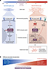
Matrix stiffness activates an NFATC4 phospho-switch gating the fibroblast-tFigure 1

Matrix stiffness activates an NFATC4 phospho-switch gating the fibroblast-to-myofibroblast transition. Kadri et al. (10) used phosphoproteomic profiling in human lung fibroblasts cultured across a range of physiological matrix stiffnesses to identify mechanosensitive phosphosites involved in fibroblast differentiation. Among the candidates, NFATC4 emerged as a key regulator based on the strong association of its downstream targets with pathological fibrosis. (A) Increasing matrix stiffness promoted JNK-dependent phosphorylation of NFATC4 at S213/S217, enabling NFATC4 nuclear localization and induction of a fibrotic gene program. This stiffness-responsive switch drove CTHRC1+/ACTA2+ myofibroblast commitment, with TGF-β/SMAD2/3 signaling reinforcing the fibrotic state. (B) Kadri et al.’s study doubles as a resource for mechanosensitive phosphosites, adding substantially to those previously reported. This panel highlights select phosphosites spanning a range of roles, including cell adhesion, cytoskeletal reorganization, cell motility, and regulation of transcription.
NFATC4: a phosphorylation-gated checkpoint for myofibroblast fate

To move from this catalog to causal candidates, Kadri et al. applied a two-step filter: they first retained phosphosites with documented regulatory function in curated databases; then, within the transcription factor category, they kept only those phosphorylated at residues with defined roles in transcriptional control (10). Six high-confidence mechanosensitive transcription factors remained: EP300, TP53, NFATC4, FOXO3, YBX1, and RB1. NFATC4 stood out: its inferred regulon in IPF fibroblasts placed CTHRC1 and COL1A1 among its top induced targets, tying it directly to the CTHRC1+/ACTA2+ state that defines pathological fibrosis. NFATC4 is a calcium- and calcineurin-regulated transcription factor controlled by phosphorylation-dependent nucleocytoplasmic shuttling, with dephosphorylation promoting nuclear accumulation and activity (11). Its phosphorylation at S213/S217 rose progressively with substrate stiffness, and both residues were previously mapped as JNK substrates (12).

Using site-specific phosphomimetic (S213D/S217D) and phospho-dead (S213A/S217A) mutants, the authors showed that phosphorylation at these two residues is both necessary and sufficient to drive fibroblasts into the CTHRC1+/ACTA2+ state. The phosphomimetic mutant forced differentiation even on soft (0.5 kPa) substrates and without TGF-β — conditions under which control fibroblasts stay quiescent — whereas the phospho-dead mutant was unable to activate myofibroblast markers even on stiff matrix in the presence of TGF-β. Critically, overexpressing WT NFATC4 at comparable levels did nothing under any condition, establishing that fate is set by the phosphorylation status of NFATC4 rather than its abundance. This mirrors the well-established logic of the NFAT family, whose activity is gated by phosphorylation-dependent localization rather than expression level (11), and it identifies posttranslational control as the decisive layer linking mechanical input to cell identity.

Pharmacological JNK inhibition phenocopied the phospho-dead mutant in healthy and IPF patient–derived fibroblasts, consistent with JNK acting upstream of the NFATC4 phospho-switch and showing that the mechanism is conserved across healthy and already activated fibrotic cells. The particular state that NFATC4 controls matters as much as the switch itself. Across independent scRNA-seq studies, CTHRC1 has emerged as the defining marker of the pathological fibroblast population that expands in fibrotic lung, heart, and liver — a transcriptionally distinct, high collagen–producing subset largely absent from healthy tissue (2, 13). By sitting upstream of CTHRC1 induction, the NFATC4 phospho-switch acts not as a regulator of generic activation but as a gatekeeper for entry into this specific, disease-driving cell state.

Validation in human IPF

Importantly, these findings were extended to human disease. Using transcription factor regulon analysis of a published multicohort scRNA-seq atlas of pulmonary fibrosis (14), the authors found that NFATC4 activity is elevated in IPF fibroblasts compared with healthy controls (10). Among the six mechanosensitive transcription factors identified by phosphoproteomics, NFATC4 showed the strongest disease association, with its regulon encompassing 119 genes upregulated in IPF fibroblasts, including key mediators of fibroblast activation, ECM remodeling, and collagen cross-linking.

Immunofluorescence analysis of micro-CT staged IPF lung tissue showed NFATC4, CTHRC1, and ACTA2 increasingly colocalized as disease severity rose. This gradient is what one would expect if NFATC4-mediated mechanosensing is engaged progressively as tissue stiffens, and it anchors the S213/S217 axis as a disease-relevant regulatory node in the human lung. The conservation of phospho-dependent regulation in fibroblasts derived from patients with IPF is particularly notable. Despite their constitutively activated state, these cells exhibited the same sensitivity to both the phosphomimetic and phospho-dead mutants. This suggests that the JNK/NFATC4 axis remains operative after pathological reprogramming, and it is not merely an initiator of disease but a potentially tractable target in established fibrosis.

Implications and open questions

The NFATC4 phospho-switch sharpens our understanding of how the physical microenvironment encodes cell identity in fibrosis. The finding that mimicking a single phosphorylation event substitutes for the stiffness cue driving differentiation on soft matrix, whereas preventing it blocks differentiation on a stiff matrix even in the presence of TGF-β, establishes this axis as a dominant, instructive regulator rather than merely a permissive one. Its relevance is unlikely to end at the lung. Work in cardiac and hepatic fibrosis already places NFATC4 downstream of calcineurin as a conserved profibrotic effector in mesenchymal cells (15, 16), and a recent spatial transcriptomic survey of interstitial lung diseases found NFATC4 activity enriched in CTHRC1hi myofibroblasts (17). The JNK/NFATC4 S213/S217 switch may thus be a broadly shared mechanosensing node across fibrotic organs and a therapeutic target with cross-organ reach.

Several key questions emerge from these findings. Although supported by pharmacological evidence and prior studies, direct phosphorylation of S213/S217 by JNK in lung fibroblasts awaits formal validation by kinase assays. Likewise, how NFATC4 integrates with other mechanosensitive transcription factors that shape the pathological program, including RUNX1, remains to be determined (18). A central translational question is whether the JNK/NFATC4 axis can be therapeutically targeted in established fibrosis rather than only at disease onset. In addition, because this regulatory switch lies upstream of CTHRC1, it is important to determine whether NFATC4 directly occupies NFAT-binding elements at the CTHRC1 locus or regulates its expression indirectly through downstream cytoskeletal programs. Distinguishing between these mechanisms will have important implications for therapeutic targeting of this pathway.

A complementary layer of this regulation lies in the noncoding genome. The transcription factors that read mechanical cues must ultimately act on cis-regulatory DNA, and those elements can themselves be mechanosensitive. Cosgrove et al. recently defined a class of stiffness-responsive enhancers, termed “mechanoenhancers,” whose chromatin accessibility and activity are tuned by ECM stiffness and that control fibrosis-associated genes including CTGF (also known as CCN2) and MYH9 (19). Strikingly, epigenetic repression of the CTGF mechanoenhancer in primary lung fibroblasts blocked roughly 96% of the TGF-β–induced increase in CTGF and suppressed activation of the fibroblast-to-myofibroblast program, indicating that stiffness is encoded not only in the phosphorylation state of transcription factors but also in the chromatin state of the enhancers they occupy. These layers are likely interdependent: the S213/S217 phospho-switch governs whether active NFATC4 reaches the nucleus, while the accessibility of NFAT-bound enhancers near CTHRC1, COL1A1, and other myofibroblast genes would dictate whether nuclear NFATC4 can productively drive their transcription. Determining whether the phospho-switch and the mechanoenhancer landscape respond to the same stiffness thresholds and whether activated NFATC4 acts through these stiffness-gated regulatory elements is an attractive avenue for unifying signaling- and chromatin-level models of mechanically encoded cell fate.

A larger question is whether the switch behaves the same way in three-dimensional and in vivo settings, where cells read more complex and dynamic mechanical inputs. The PDMS substrates used in Kadri et al.’s study impose a fixed stiffness from the outset, whereas fibrosis in patients stiffens gradually over months to years and is spatially heterogeneous once established. It is therefore unknown whether NFATC4 phosphorylation reports a cell’s current mechanical state or instead encodes a stiffness history that becomes hard to erase once the CTHRC1+ program is locked in, a distinction central to whether fibrosis can be halted or reversed. Emerging materials could settle this: dynamically tunable hydrogels and piezoelectric or magneto-responsive substrates can change stiffness in real time beneath a living cell, unlike the static gels used here. Such platforms would let NFATC4 phosphorylation and nuclear translocation be watched directly as stiffness rises and would test reversibility by softening the substrate afterward to ask whether NFATC4 dephosphorylates, exits the nucleus, and releases cells from the CTHRC1+ state or whether the switch instead locks fibroblasts into a committed fibrotic identity. Pairing such substrates with stiffness maps of patient tissue, for instance, use of atomic force microscopy coregistered with NFATC4 phosphorylation status by immunofluorescence, could further test whether the 2–8 kPa threshold defined here matches the stiffness at the leading edge of fibrotic foci.

By identifying a discrete phosphorylation event that links matrix mechanics to pathological fibroblast fate, Kadri et al. recast a complex mechanobiological process long viewed through a biophysical lens as a defined signaling mechanism with clear therapeutic potential.

Author contributions

RSF and PRT cowrote and edited the manuscript.

Conflict of interest

PRT is an inventor on patents PCT/US20/53158 and PCT/US20/53164 and received research support from Calico Life Sciences, Ono Pharmaceuticals, and United Therapeutics.

Funding support

This work is the result of NIH funding, in whole or in part, and is subject to the NIH Public Access Policy. Through acceptance of this federal funding, the NIH has been given a right to make the work publicly available in PubMed Central.

  • NIH/National Heart, Lung, and Blood Institute research awards R01HL146557, R01HL160939, R01HL153375 to PRT.
Footnotes

Copyright: © 2026, Shelley Frabotta et al. This is an open access article published under the terms of the Creative Commons Attribution 4.0 International License.

Reference information: J Clin Invest. 2026;136(17):e209849. https://doi.org/10.1172/JCI209849.

See the related article at Mechanosensitive phosphorylation of NFATC4 at S213/S217 drives fibroblast-to-myofibroblast transition and fibrosis.

References
  1. Hinz B. Mechanical aspects of lung fibrosis: a spotlight on the myofibroblast. Proc Am Thorac Soc. 2012;9(3):137–147.
    View this article via: CrossRef PubMed Google Scholar
  2. Tsukui T, et al. Collagen-producing lung cell atlas identifies multiple subsets with distinct localization and relevance to fibrosis. Nat Commun. 2020;11(1):1920.
    View this article via: CrossRef PubMed Google Scholar
  3. Liu F, et al. Feedback amplification of fibrosis through matrix stiffening and COX-2 suppression. J Cell Biol. 2010;190(4):693–706.
    View this article via: CrossRef PubMed Google Scholar
  4. Schiller HB, Fässler R. Mechanosensitivity and compositional dynamics of cell-matrix adhesions. EMBO Rep. 2013;14(6):509–519.
    View this article via: CrossRef PubMed Google Scholar
  5. Fernandez IE, Eickelberg O. The impact of TGF-β on lung fibrosis: from targeting to biomarkers. Proc Am Thorac Soc. 2012;9(3):111–116.
    View this article via: CrossRef PubMed Google Scholar
  6. Freeberg MAT, et al. Mechanical feed-forward loops contribute to idiopathic pulmonary fibrosis. Am J Pathol. 2021;191(1):18–25.
    View this article via: CrossRef PubMed Google Scholar
  7. Huang X, et al. Matrix stiffness-induced myofibroblast differentiation is mediated by intrinsic mechanotransduction. Am J Respir Cell Mol Biol. 2012;47(3):340–348.
    View this article via: CrossRef PubMed Google Scholar
  8. Kollmannsberger P, et al. Tensile forces drive a reversible fibroblast-to-myofibroblast transition during tissue growth in engineered clefts. Sci Adv. 2018;4(1):eaao4881.
    View this article via: CrossRef PubMed Google Scholar
  9. Walker CJ, et al. Nuclear mechanosensing drives chromatin remodelling in persistently activated fibroblasts. Nat Biomed Eng. 2021;5(12):1485–1499.
    View this article via: CrossRef PubMed Google Scholar
  10. Kadri S, et al. Mechanosensitive phosphorylation of NFATC4 at S213/S217 drives fibroblast-to-myofibroblast transition and fibrosis. J Clin Invest. 2026;136(17):e195121.
    View this article via: JCI CrossRef Google Scholar
  11. Okamura H, et al. Concerted dephosphorylation of the transcription factor NFAT1 induces a conformational switch that regulates transcriptional activity. Mol Cell. 2000;6(3):539–550.
    View this article via: CrossRef PubMed Google Scholar
  12. Yao K, et al. Nuclear factor of activated T3 is a negative regulator of Ras-JNK1/2-AP-1 induced cell transformation. Cancer Res. 2007;67(18):8725–8735.
    View this article via: CrossRef PubMed Google Scholar
  13. Ruiz-Villalba A, et al. Single-cell RNA sequencing analysis reveals a crucial role for CTHRC1 (collagen triple helix repeat containing 1) cardiac fibroblasts after myocardial infarction. Circulation. 2020;142(19):1831–1847.
    View this article via: CrossRef PubMed Google Scholar
  14. Mayr CH, et al. Integrative analysis of cell state changes in lung fibrosis with peripheral protein biomarkers. EMBO Mol Med. 2021;13(4):e12871.
    View this article via: CrossRef PubMed Google Scholar
  15. Herum KM, et al. Syndecan-4 signaling via NFAT regulates extracellular matrix production and cardiac myofibroblast differentiation in response to mechanical stress. J Mol Cell Cardiol. 2013;54:73–81.
    View this article via: CrossRef PubMed Google Scholar
  16. Pan X, et al. Methylation of RCAN1.4 mediated by DNMT1 and DNMT3b enhances hepatic stellate cell activation and liver fibrogenesis through calcineurin/NFAT3 signaling. Theranostics. 2019;9(15):4308–4323.
    View this article via: CrossRef PubMed Google Scholar
  17. Jaiswal A, et al. Spatial transcriptomics reveals altered communities and drivers of aberrant epithelia and pro-fibrotic fibroblasts in interstitial lung diseases. Cell Genom. 2026;6(3):101066.
    View this article via: CrossRef PubMed Google Scholar
  18. Konkimalla A, et al. Transitional cell states sculpt tissue topology during lung regeneration. Cell Stem Cell. 2023;30(11):1486–1502.
    View this article via: CrossRef PubMed Google Scholar
  19. Cosgrove BD, et al. Mechanosensitive genomic enhancers potentiate the cellular response to matrix stiffness. Science. 2025;390(6778):eadl1988.
    View this article via: CrossRef PubMed Google Scholar
Version history
  • Version 1 (September 1, 2026): Electronic publication

Article tools

  • View PDF
  • Download citation information
  • Send a comment
  • Terms of use
  • Standard abbreviations
  • Need help? Email the journal

Metrics

  • Article usage
  • Citations to this article

Go to

  • Top
  • Abstract
  • The mechanical and biochemical microenvironment controls fibroblast state
  • Charting the mechanosensing phosphoproteome
  • NFATC4: a phosphorylation-gated checkpoint for myofibroblast fate
  • Validation in human IPF
  • Implications and open questions
  • Author contributions
  • Conflict of interest
  • Funding support
  • Footnotes
  • References
  • Version history
Advertisement
Advertisement

Copyright © 2026 American Society for Clinical Investigation
ISSN: 0021-9738 (print), 1558-8238 (online)

Sign up for email alerts