
Advertisement
Commentary
Open Access |  10.1172/JCI209019
Going to the source: pancreatic lymph nodes maintain stem-like CD8+ T cells in human type 1 diabetes
Fatoumata Samassa,1 Sylvaine You,1,2 and Roberto Mallone1,2,3
1Université Paris Cité, Institut Cochin, CNRS, INSERM, Paris, France.
2Indiana Biosciences Research Institute, Indianapolis, Indiana, USA.
3Assistance Publique Hôpitaux de Paris, Service de Diabétologie et Immunologie Clinique, Cochin Hospital, Paris, France.
Address correspondence to: Roberto Mallone, INSERM U1016 Cochin Institute, 123 boulevard de Port Royal, 75014 Paris, France. Email: roberto.mallone@inserm.fr.
Find articles by
Samassa, F.
in:
PubMed
|
Google Scholar
|

1Université Paris Cité, Institut Cochin, CNRS, INSERM, Paris, France.
2Indiana Biosciences Research Institute, Indianapolis, Indiana, USA.
3Assistance Publique Hôpitaux de Paris, Service de Diabétologie et Immunologie Clinique, Cochin Hospital, Paris, France.
Address correspondence to: Roberto Mallone, INSERM U1016 Cochin Institute, 123 boulevard de Port Royal, 75014 Paris, France. Email: roberto.mallone@inserm.fr.
Find articles by You, S. in: PubMed | Google Scholar
1Université Paris Cité, Institut Cochin, CNRS, INSERM, Paris, France.
2Indiana Biosciences Research Institute, Indianapolis, Indiana, USA.
3Assistance Publique Hôpitaux de Paris, Service de Diabétologie et Immunologie Clinique, Cochin Hospital, Paris, France.
Address correspondence to: Roberto Mallone, INSERM U1016 Cochin Institute, 123 boulevard de Port Royal, 75014 Paris, France. Email: roberto.mallone@inserm.fr.
Find articles by
Mallone, R.
in:
PubMed
|
Google Scholar
|
Published September 15, 2026 - More info
J Clin Invest. 2026;136(18):e209019. https://doi.org/10.1172/JCI209019.
© 2026 Samassa et al. This work is licensed under the Creative Commons Attribution 4.0 International License. To view a copy of this license, visit http://creativecommons.org/licenses/by/4.0/.

Related article:
Abstract
Effector CD8+ T cells are key drivers of type 1 diabetes (T1D) pathogenesis, yet questions remain regarding the molecular defects leading to altered cytotoxicity, peripheral tissue phenotype, and receptor specificity. We analyzed human pancreatic lymph nodes (pLNs) using mass cytometry and single-cell RNA-seq (scRNA-seq) with combined T cell receptor (TCR) profiling. Cytometric analysis revealed enrichment of T stem cell memory–like (TSCM-like) cells (CD8+CD45RA+CD27+CD28+CCR7+CXCR3+) in T1D pLNs. scRNA-seq indicated an elevated inflammatory cytokine gene signature (IFITM3, LTB) along with regulators of terminal differentiation (BCL6, BCL3), coupled with downregulation of exhaustion-associated genes (DUSP2, NR4A2, TSC22D3) in CD8+ T cells in T1D pLNs. Immune response enrichment analysis (IREA) indicated IL-15 signaling as a significant driver of these phenotypes. Integrated TCR and transcriptomics analysis revealed a cluster of diverse naive-like CD8+ T cell clones in T1D pLNs. Comparison of pLNs and pancreatic tissue slice isolates indicated sharing of effector CD8+ T cells, with enhanced terminal effector signatures within the pancreas relative to paired pLNs. Multiplex imaging revealed differential localization of T cell factor 1 (TCF1)- and thymocyte selection-associated high mobility group box protein (TOX)-expressing T cells in the pancreas, with islet-proximal TCF1+TOX+ cells displaying a mixture of activation and exhaustion-associated phenotypes. Thus, we provide multimodal cellular profiles enriched in T1D tissues for consideration in therapeutic targeting.
Authors
Leeana D. Peters, Howard R. Seay, Justin A. Smith, Amanda L. Posgai, Reed L. Berkowitz, Clive H. Wasserfall, Mark A. Atkinson, Rhonda Bacher, Maigan A. Brusko, Todd M. Brusko
-
Knowledge gaps for future research
Several limitations of this work deserve consideration and provide inspiration for future research (Figure 1). First, as access to human PLN and pancreatic tissues remains rare, specimen numbers were modest, and so was the number of CD8+ T cells sampled, hampering a comprehensive dissection of T cell heterogeneity and antigen specificity. The inability to recover informative HLA class I dextramer signals highlights the challenge of tracking rare autoreactive T cells directly in human tissues. Although several TCR CDR3α sequences showed similarity to previously reported β cell–reactive clones, antigen specificity cannot be inferred with confidence. An intriguing aspect of the study is that the principal T1D-enriched, naive-like cell population was highly diverse and lacked evidence of clonal expansion. This observation contrasts with expectations derived from the NOD mouse model (3), in which stem-like autoreactive cells constituted an expanded reservoir of diabetogenic clones. Moreover, despite evidence of phenotypic continuity between PLN and pancreatic CD8+ T cell populations, direct clonal overlap between the two compartments was surprisingly limited. The absence of clonal expansion and the limited PLN-to-pancreas TCR sharing complicate direct extrapolation of the reservoir model proposed in NOD mice (3), where stem-like PLN CD8+ T cells were shown experimentally to sustain diabetogenic responses. Do the identified PLN populations represent the human counterpart of the murine diabetogenic reservoir or an earlier precursor state? Or do they reflect a broader T1D-associated remodeling of the CD8+ T cell compartment toward stemness? Future studies combining antigen-specific T cell tracking and paired multi-site TCR sequencing will be required to conclude whether these stem-like T cells constitute the developmental precursors of pathogenic islet-infiltrating effectors.
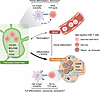 Figure 1
A proposed stemness-to-effector/exhaustion tissue axis in T1D. PLNs may serve as a reservoir for stem-like CD8+ T cells that sustain autoimmune responses in T1D. Building on the findings of Peters et al. (5) and previous observations in NOD mice, the model presented here proposes that limited β cell antigen availability in PLNs, combined with the intrinsically low affinity of autoreactive TCRs for β cell antigens, results in weak TCR signaling that favors partial differentiation and maintenance of stem-like properties. This state may be further supported by IL-15 signaling. Stem-like PLN CD8+ T cells (red) are characterized by the expression of CXCR3 and self-renewal–associated markers such as BCL2, BCL3, and BCL6, together with TCF1 and TOX. Whether circulating naive-like, islet-reactive CD8+ T cells (pink) represent a recirculating counterpart of this PLN cell population remains unknown. Upon migration to pancreatic islets, exposure to higher antigen loads and stronger TCR stimulation may promote further differentiation, acquisition of cytotoxic functions, and eventual exhaustion (brown cells). However, the developmental relationship between PLN stem-like cells and pancreatic effector/exhausted cells remains unresolved, as current evidence demonstrates phenotypic continuity but limited clonal overlap between the two compartments. The figure highlights key hypotheses and outstanding questions emerging from the study by Peters et al., including the roles of recirculation, clonal continuity, and the mechanisms governing the balance between stemness, effector differentiation, and exhaustion.
Second, the molecular signatures defining stemness remain variable across reports and disease settings. The relationships among stem-like TPEX and T stem cell memory (TSCM) states remain incompletely resolved, while the relative importance of canonical regulators such as TCF1, TOX, and other stemness-associated markers differs substantially across studies (15, 16). Furthermore, definitive demonstration that these stem-like T cells are nurtured by IL-15 signaling and directly generate pathogenic effectors in humans will require functional validation.
Third, is there a correlate of this stem-like phenotype in the blood that could provide a circulating T cell biomarker for T1D? Most studies have reported different degrees of exhaustion in circulating CD8+ T cells that correlate with disease activity (17, 18) and its therapeutic curbing (8, 19), i.e., the rates of progression from asymptomatic to clinical disease or of β cell loss after clinical onset. On the other hand, the description of different degrees of stemness is much more limited (20). B. Youngblood’s group has shown that circulating autoreactive T cells retain an epigenetically imprinted stem-like phenotype, intermediate between naive and effector/memory, that supports long-term persistence in T1D (21). Intriguingly, a predominantly naive-like (CD45RA+CCR7+) phenotype has also been described for both CD8+ T cells (1, 22) and CD4+ T cells (23) in blood, raising the possibility that this phenotype may hide intermediate stem-like states.
Fourth, what are the signals favoring the maintenance of stem-like properties? Autoreactive T cells feature low-affinity TCRs, which are likely exposed to low β cell antigen loads in PLNs (24) due to limited delivery in both soluble form (upon insulin granule exocytosis) (25, 26) and cell-bound form (ferried by antigen-presenting cells [APCs], in the frame of surface HLA molecules that also feature low binding affinity for β cell epitopes) (24). Overall, these features likely deliver very weak TCR signals, which may favor the maintenance of a partially differentiated stem-like phenotype in PLNs. This is also where the ancillary IL-15 signaling highlighted by Peters et al. may prove critical. Once partially differentiated T cells migrate to the pancreas, the higher antigen load available in the islet microenvironment may drive full differentiation into cytotoxic effector T cells and eventual exhaustion (24).
Article tools
- Download citation information
- Send a comment
- Terms of use
- Standard abbreviations
- Need help? Email the journal
Metrics
Go to
- Top
- Abstract
- Pancreatic lymph nodes: a reservoir for autoreactive T cells?
- A stem-like CD8+ T cell population in T1D PLNs
- Therapeutic implications: targeting PLN stem-like CD8+ T cells
- Knowledge gaps for future research
- Conclusions
- Conflict of interest
- Funding support
- Footnotes
- References
- Version history


Copyright © 2026 American Society for Clinical Investigation
ISSN: 0021-9738 (print), 1558-8238 (online)