
Advertisement
Research ArticleInflammationMetabolism
Open Access |  10.1172/JCI207089
Palmitic acid coordinates impaired visceral adipose ICOShi Treg-mediated immunosuppression and systemic metabolic disturbance during obesity
Weitong Su,1 Yuxiao Liu,1 Xi Yan,1 Mengyao Huang,1 Linghao Xu,2 Jing Lin,1 Xufeng Chen,1 Puyuan Hu,1 Chenlin Gao,2 Jian Wen,3,4 Hongdong Wang,5 Dong Ding,1 Zengpeng Zheng,1 Wenjing Li,1 Lianjia Li,1 Zhan Liu,1 Keyu Qian,1 Jing Gao,1 Tingting Zhang,1 Xiaobing Mao,2 Haibing Zhang,1 Wei Lu,1 Bin Li,6 Hong Li,1 Aoyuan Cui,1 Yan Bi,5 Chunxiang Zhang,7 and Yu Li1
1Shanghai Institute of Nutrition and Health, University of Chinese Academy of Sciences, Chinese Academy of Sciences, Shanghai, China.
2Department of Endocrinology and Metabolism, Metabolic Vascular Disease Key Laboratory of Sichuan Province, The Affiliated Hospital of Southwest Medical University, Luzhou, Sichuan, China.
3Department of General Surgery (Hepatobiliary Surgery), Biliary-Pancreatic Center, The Affiliated Hospital, Southwest Medical University, Luzhou, Sichuan, China.
4Metabolic Hepatobiliary and Pancreatic Diseases Key Laboratory of Luzhou City, Academician (Expert) Workstation of Sichuan Province, The Affiliated Hospital, Southwest Medical University, Luzhou, Sichuan, China.
5Department of Endocrinology, Endocrine and Metabolic Disease Medical Center, Nanjing Drum Tower Hospital, Affiliated Hospital of Medical School, Nanjing University, Nanjing, China.
6Department of Immunology and Microbiology, Shanghai Institute of Immunology, Shanghai Jiao Tong University School of Medicine, Shanghai, China.
7Metabolic Vascular Disease Key Laboratory of Sichuan Province, The Affiliated Hospital of Southwest Medical University, Key Laboratory of Medical Electrophysiology, Ministry of Education, Southwest Medical University, Luzhou, China.
Address correspondence to: Yu Li, Chinese Academy of Sciences, 320 Yue Yang Road, Life Science Research Building A1816, Shanghai 200031, China. Phone: 86.21.5492.0753; Email: liyu@sinh.ac.cn. Or to: Chunxiang Zhang, Southwest Medical University, 25 Taiping Road, Luzhou, Sichuan 646000, China. Email: zhangchx999@163.com. Or to: Yan Bi, Nanjing Drum Tower Hospital Nanjing University, 321 Zhongshan Road, Nanjing 210008, China. Phone: 86.21.136.5517.6368; Email: biyan@nju.edu.cn. Or to: Aoyuan Cui, Shanghai Institute of Nutrition and Health, Chinese Academy of Sciences, 320 Yue Yang Road, Life Science Research Building A1818, Shanghai 200031, China. Phone: 86.21.5492.0924; Email: aycui@sinh.ac.cn.
Authorship note: WS, YL, and XY contributed equally to this work.
Find articles by Su, W. in: PubMed | Google Scholar
1Shanghai Institute of Nutrition and Health, University of Chinese Academy of Sciences, Chinese Academy of Sciences, Shanghai, China.
2Department of Endocrinology and Metabolism, Metabolic Vascular Disease Key Laboratory of Sichuan Province, The Affiliated Hospital of Southwest Medical University, Luzhou, Sichuan, China.
3Department of General Surgery (Hepatobiliary Surgery), Biliary-Pancreatic Center, The Affiliated Hospital, Southwest Medical University, Luzhou, Sichuan, China.
4Metabolic Hepatobiliary and Pancreatic Diseases Key Laboratory of Luzhou City, Academician (Expert) Workstation of Sichuan Province, The Affiliated Hospital, Southwest Medical University, Luzhou, Sichuan, China.
5Department of Endocrinology, Endocrine and Metabolic Disease Medical Center, Nanjing Drum Tower Hospital, Affiliated Hospital of Medical School, Nanjing University, Nanjing, China.
6Department of Immunology and Microbiology, Shanghai Institute of Immunology, Shanghai Jiao Tong University School of Medicine, Shanghai, China.
7Metabolic Vascular Disease Key Laboratory of Sichuan Province, The Affiliated Hospital of Southwest Medical University, Key Laboratory of Medical Electrophysiology, Ministry of Education, Southwest Medical University, Luzhou, China.
Address correspondence to: Yu Li, Chinese Academy of Sciences, 320 Yue Yang Road, Life Science Research Building A1816, Shanghai 200031, China. Phone: 86.21.5492.0753; Email: liyu@sinh.ac.cn. Or to: Chunxiang Zhang, Southwest Medical University, 25 Taiping Road, Luzhou, Sichuan 646000, China. Email: zhangchx999@163.com. Or to: Yan Bi, Nanjing Drum Tower Hospital Nanjing University, 321 Zhongshan Road, Nanjing 210008, China. Phone: 86.21.136.5517.6368; Email: biyan@nju.edu.cn. Or to: Aoyuan Cui, Shanghai Institute of Nutrition and Health, Chinese Academy of Sciences, 320 Yue Yang Road, Life Science Research Building A1818, Shanghai 200031, China. Phone: 86.21.5492.0924; Email: aycui@sinh.ac.cn.
Authorship note: WS, YL, and XY contributed equally to this work.
Find articles by Liu, Y. in: PubMed | Google Scholar
1Shanghai Institute of Nutrition and Health, University of Chinese Academy of Sciences, Chinese Academy of Sciences, Shanghai, China.
2Department of Endocrinology and Metabolism, Metabolic Vascular Disease Key Laboratory of Sichuan Province, The Affiliated Hospital of Southwest Medical University, Luzhou, Sichuan, China.
3Department of General Surgery (Hepatobiliary Surgery), Biliary-Pancreatic Center, The Affiliated Hospital, Southwest Medical University, Luzhou, Sichuan, China.
4Metabolic Hepatobiliary and Pancreatic Diseases Key Laboratory of Luzhou City, Academician (Expert) Workstation of Sichuan Province, The Affiliated Hospital, Southwest Medical University, Luzhou, Sichuan, China.
5Department of Endocrinology, Endocrine and Metabolic Disease Medical Center, Nanjing Drum Tower Hospital, Affiliated Hospital of Medical School, Nanjing University, Nanjing, China.
6Department of Immunology and Microbiology, Shanghai Institute of Immunology, Shanghai Jiao Tong University School of Medicine, Shanghai, China.
7Metabolic Vascular Disease Key Laboratory of Sichuan Province, The Affiliated Hospital of Southwest Medical University, Key Laboratory of Medical Electrophysiology, Ministry of Education, Southwest Medical University, Luzhou, China.
Address correspondence to: Yu Li, Chinese Academy of Sciences, 320 Yue Yang Road, Life Science Research Building A1816, Shanghai 200031, China. Phone: 86.21.5492.0753; Email: liyu@sinh.ac.cn. Or to: Chunxiang Zhang, Southwest Medical University, 25 Taiping Road, Luzhou, Sichuan 646000, China. Email: zhangchx999@163.com. Or to: Yan Bi, Nanjing Drum Tower Hospital Nanjing University, 321 Zhongshan Road, Nanjing 210008, China. Phone: 86.21.136.5517.6368; Email: biyan@nju.edu.cn. Or to: Aoyuan Cui, Shanghai Institute of Nutrition and Health, Chinese Academy of Sciences, 320 Yue Yang Road, Life Science Research Building A1818, Shanghai 200031, China. Phone: 86.21.5492.0924; Email: aycui@sinh.ac.cn.
Authorship note: WS, YL, and XY contributed equally to this work.
Find articles by Yan, X. in: PubMed | Google Scholar
1Shanghai Institute of Nutrition and Health, University of Chinese Academy of Sciences, Chinese Academy of Sciences, Shanghai, China.
2Department of Endocrinology and Metabolism, Metabolic Vascular Disease Key Laboratory of Sichuan Province, The Affiliated Hospital of Southwest Medical University, Luzhou, Sichuan, China.
3Department of General Surgery (Hepatobiliary Surgery), Biliary-Pancreatic Center, The Affiliated Hospital, Southwest Medical University, Luzhou, Sichuan, China.
4Metabolic Hepatobiliary and Pancreatic Diseases Key Laboratory of Luzhou City, Academician (Expert) Workstation of Sichuan Province, The Affiliated Hospital, Southwest Medical University, Luzhou, Sichuan, China.
5Department of Endocrinology, Endocrine and Metabolic Disease Medical Center, Nanjing Drum Tower Hospital, Affiliated Hospital of Medical School, Nanjing University, Nanjing, China.
6Department of Immunology and Microbiology, Shanghai Institute of Immunology, Shanghai Jiao Tong University School of Medicine, Shanghai, China.
7Metabolic Vascular Disease Key Laboratory of Sichuan Province, The Affiliated Hospital of Southwest Medical University, Key Laboratory of Medical Electrophysiology, Ministry of Education, Southwest Medical University, Luzhou, China.
Address correspondence to: Yu Li, Chinese Academy of Sciences, 320 Yue Yang Road, Life Science Research Building A1816, Shanghai 200031, China. Phone: 86.21.5492.0753; Email: liyu@sinh.ac.cn. Or to: Chunxiang Zhang, Southwest Medical University, 25 Taiping Road, Luzhou, Sichuan 646000, China. Email: zhangchx999@163.com. Or to: Yan Bi, Nanjing Drum Tower Hospital Nanjing University, 321 Zhongshan Road, Nanjing 210008, China. Phone: 86.21.136.5517.6368; Email: biyan@nju.edu.cn. Or to: Aoyuan Cui, Shanghai Institute of Nutrition and Health, Chinese Academy of Sciences, 320 Yue Yang Road, Life Science Research Building A1818, Shanghai 200031, China. Phone: 86.21.5492.0924; Email: aycui@sinh.ac.cn.
Authorship note: WS, YL, and XY contributed equally to this work.
Find articles by Huang, M. in: PubMed | Google Scholar
1Shanghai Institute of Nutrition and Health, University of Chinese Academy of Sciences, Chinese Academy of Sciences, Shanghai, China.
2Department of Endocrinology and Metabolism, Metabolic Vascular Disease Key Laboratory of Sichuan Province, The Affiliated Hospital of Southwest Medical University, Luzhou, Sichuan, China.
3Department of General Surgery (Hepatobiliary Surgery), Biliary-Pancreatic Center, The Affiliated Hospital, Southwest Medical University, Luzhou, Sichuan, China.
4Metabolic Hepatobiliary and Pancreatic Diseases Key Laboratory of Luzhou City, Academician (Expert) Workstation of Sichuan Province, The Affiliated Hospital, Southwest Medical University, Luzhou, Sichuan, China.
5Department of Endocrinology, Endocrine and Metabolic Disease Medical Center, Nanjing Drum Tower Hospital, Affiliated Hospital of Medical School, Nanjing University, Nanjing, China.
6Department of Immunology and Microbiology, Shanghai Institute of Immunology, Shanghai Jiao Tong University School of Medicine, Shanghai, China.
7Metabolic Vascular Disease Key Laboratory of Sichuan Province, The Affiliated Hospital of Southwest Medical University, Key Laboratory of Medical Electrophysiology, Ministry of Education, Southwest Medical University, Luzhou, China.
Address correspondence to: Yu Li, Chinese Academy of Sciences, 320 Yue Yang Road, Life Science Research Building A1816, Shanghai 200031, China. Phone: 86.21.5492.0753; Email: liyu@sinh.ac.cn. Or to: Chunxiang Zhang, Southwest Medical University, 25 Taiping Road, Luzhou, Sichuan 646000, China. Email: zhangchx999@163.com. Or to: Yan Bi, Nanjing Drum Tower Hospital Nanjing University, 321 Zhongshan Road, Nanjing 210008, China. Phone: 86.21.136.5517.6368; Email: biyan@nju.edu.cn. Or to: Aoyuan Cui, Shanghai Institute of Nutrition and Health, Chinese Academy of Sciences, 320 Yue Yang Road, Life Science Research Building A1818, Shanghai 200031, China. Phone: 86.21.5492.0924; Email: aycui@sinh.ac.cn.
Authorship note: WS, YL, and XY contributed equally to this work.
Find articles by Xu, L. in: PubMed | Google Scholar
1Shanghai Institute of Nutrition and Health, University of Chinese Academy of Sciences, Chinese Academy of Sciences, Shanghai, China.
2Department of Endocrinology and Metabolism, Metabolic Vascular Disease Key Laboratory of Sichuan Province, The Affiliated Hospital of Southwest Medical University, Luzhou, Sichuan, China.
3Department of General Surgery (Hepatobiliary Surgery), Biliary-Pancreatic Center, The Affiliated Hospital, Southwest Medical University, Luzhou, Sichuan, China.
4Metabolic Hepatobiliary and Pancreatic Diseases Key Laboratory of Luzhou City, Academician (Expert) Workstation of Sichuan Province, The Affiliated Hospital, Southwest Medical University, Luzhou, Sichuan, China.
5Department of Endocrinology, Endocrine and Metabolic Disease Medical Center, Nanjing Drum Tower Hospital, Affiliated Hospital of Medical School, Nanjing University, Nanjing, China.
6Department of Immunology and Microbiology, Shanghai Institute of Immunology, Shanghai Jiao Tong University School of Medicine, Shanghai, China.
7Metabolic Vascular Disease Key Laboratory of Sichuan Province, The Affiliated Hospital of Southwest Medical University, Key Laboratory of Medical Electrophysiology, Ministry of Education, Southwest Medical University, Luzhou, China.
Address correspondence to: Yu Li, Chinese Academy of Sciences, 320 Yue Yang Road, Life Science Research Building A1816, Shanghai 200031, China. Phone: 86.21.5492.0753; Email: liyu@sinh.ac.cn. Or to: Chunxiang Zhang, Southwest Medical University, 25 Taiping Road, Luzhou, Sichuan 646000, China. Email: zhangchx999@163.com. Or to: Yan Bi, Nanjing Drum Tower Hospital Nanjing University, 321 Zhongshan Road, Nanjing 210008, China. Phone: 86.21.136.5517.6368; Email: biyan@nju.edu.cn. Or to: Aoyuan Cui, Shanghai Institute of Nutrition and Health, Chinese Academy of Sciences, 320 Yue Yang Road, Life Science Research Building A1818, Shanghai 200031, China. Phone: 86.21.5492.0924; Email: aycui@sinh.ac.cn.
Authorship note: WS, YL, and XY contributed equally to this work.
Find articles by Lin, J. in: PubMed | Google Scholar
1Shanghai Institute of Nutrition and Health, University of Chinese Academy of Sciences, Chinese Academy of Sciences, Shanghai, China.
2Department of Endocrinology and Metabolism, Metabolic Vascular Disease Key Laboratory of Sichuan Province, The Affiliated Hospital of Southwest Medical University, Luzhou, Sichuan, China.
3Department of General Surgery (Hepatobiliary Surgery), Biliary-Pancreatic Center, The Affiliated Hospital, Southwest Medical University, Luzhou, Sichuan, China.
4Metabolic Hepatobiliary and Pancreatic Diseases Key Laboratory of Luzhou City, Academician (Expert) Workstation of Sichuan Province, The Affiliated Hospital, Southwest Medical University, Luzhou, Sichuan, China.
5Department of Endocrinology, Endocrine and Metabolic Disease Medical Center, Nanjing Drum Tower Hospital, Affiliated Hospital of Medical School, Nanjing University, Nanjing, China.
6Department of Immunology and Microbiology, Shanghai Institute of Immunology, Shanghai Jiao Tong University School of Medicine, Shanghai, China.
7Metabolic Vascular Disease Key Laboratory of Sichuan Province, The Affiliated Hospital of Southwest Medical University, Key Laboratory of Medical Electrophysiology, Ministry of Education, Southwest Medical University, Luzhou, China.
Address correspondence to: Yu Li, Chinese Academy of Sciences, 320 Yue Yang Road, Life Science Research Building A1816, Shanghai 200031, China. Phone: 86.21.5492.0753; Email: liyu@sinh.ac.cn. Or to: Chunxiang Zhang, Southwest Medical University, 25 Taiping Road, Luzhou, Sichuan 646000, China. Email: zhangchx999@163.com. Or to: Yan Bi, Nanjing Drum Tower Hospital Nanjing University, 321 Zhongshan Road, Nanjing 210008, China. Phone: 86.21.136.5517.6368; Email: biyan@nju.edu.cn. Or to: Aoyuan Cui, Shanghai Institute of Nutrition and Health, Chinese Academy of Sciences, 320 Yue Yang Road, Life Science Research Building A1818, Shanghai 200031, China. Phone: 86.21.5492.0924; Email: aycui@sinh.ac.cn.
Authorship note: WS, YL, and XY contributed equally to this work.
Find articles by Chen, X. in: PubMed | Google Scholar
1Shanghai Institute of Nutrition and Health, University of Chinese Academy of Sciences, Chinese Academy of Sciences, Shanghai, China.
2Department of Endocrinology and Metabolism, Metabolic Vascular Disease Key Laboratory of Sichuan Province, The Affiliated Hospital of Southwest Medical University, Luzhou, Sichuan, China.
3Department of General Surgery (Hepatobiliary Surgery), Biliary-Pancreatic Center, The Affiliated Hospital, Southwest Medical University, Luzhou, Sichuan, China.
4Metabolic Hepatobiliary and Pancreatic Diseases Key Laboratory of Luzhou City, Academician (Expert) Workstation of Sichuan Province, The Affiliated Hospital, Southwest Medical University, Luzhou, Sichuan, China.
5Department of Endocrinology, Endocrine and Metabolic Disease Medical Center, Nanjing Drum Tower Hospital, Affiliated Hospital of Medical School, Nanjing University, Nanjing, China.
6Department of Immunology and Microbiology, Shanghai Institute of Immunology, Shanghai Jiao Tong University School of Medicine, Shanghai, China.
7Metabolic Vascular Disease Key Laboratory of Sichuan Province, The Affiliated Hospital of Southwest Medical University, Key Laboratory of Medical Electrophysiology, Ministry of Education, Southwest Medical University, Luzhou, China.
Address correspondence to: Yu Li, Chinese Academy of Sciences, 320 Yue Yang Road, Life Science Research Building A1816, Shanghai 200031, China. Phone: 86.21.5492.0753; Email: liyu@sinh.ac.cn. Or to: Chunxiang Zhang, Southwest Medical University, 25 Taiping Road, Luzhou, Sichuan 646000, China. Email: zhangchx999@163.com. Or to: Yan Bi, Nanjing Drum Tower Hospital Nanjing University, 321 Zhongshan Road, Nanjing 210008, China. Phone: 86.21.136.5517.6368; Email: biyan@nju.edu.cn. Or to: Aoyuan Cui, Shanghai Institute of Nutrition and Health, Chinese Academy of Sciences, 320 Yue Yang Road, Life Science Research Building A1818, Shanghai 200031, China. Phone: 86.21.5492.0924; Email: aycui@sinh.ac.cn.
Authorship note: WS, YL, and XY contributed equally to this work.
Find articles by Hu, P. in: PubMed | Google Scholar
1Shanghai Institute of Nutrition and Health, University of Chinese Academy of Sciences, Chinese Academy of Sciences, Shanghai, China.
2Department of Endocrinology and Metabolism, Metabolic Vascular Disease Key Laboratory of Sichuan Province, The Affiliated Hospital of Southwest Medical University, Luzhou, Sichuan, China.
3Department of General Surgery (Hepatobiliary Surgery), Biliary-Pancreatic Center, The Affiliated Hospital, Southwest Medical University, Luzhou, Sichuan, China.
4Metabolic Hepatobiliary and Pancreatic Diseases Key Laboratory of Luzhou City, Academician (Expert) Workstation of Sichuan Province, The Affiliated Hospital, Southwest Medical University, Luzhou, Sichuan, China.
5Department of Endocrinology, Endocrine and Metabolic Disease Medical Center, Nanjing Drum Tower Hospital, Affiliated Hospital of Medical School, Nanjing University, Nanjing, China.
6Department of Immunology and Microbiology, Shanghai Institute of Immunology, Shanghai Jiao Tong University School of Medicine, Shanghai, China.
7Metabolic Vascular Disease Key Laboratory of Sichuan Province, The Affiliated Hospital of Southwest Medical University, Key Laboratory of Medical Electrophysiology, Ministry of Education, Southwest Medical University, Luzhou, China.
Address correspondence to: Yu Li, Chinese Academy of Sciences, 320 Yue Yang Road, Life Science Research Building A1816, Shanghai 200031, China. Phone: 86.21.5492.0753; Email: liyu@sinh.ac.cn. Or to: Chunxiang Zhang, Southwest Medical University, 25 Taiping Road, Luzhou, Sichuan 646000, China. Email: zhangchx999@163.com. Or to: Yan Bi, Nanjing Drum Tower Hospital Nanjing University, 321 Zhongshan Road, Nanjing 210008, China. Phone: 86.21.136.5517.6368; Email: biyan@nju.edu.cn. Or to: Aoyuan Cui, Shanghai Institute of Nutrition and Health, Chinese Academy of Sciences, 320 Yue Yang Road, Life Science Research Building A1818, Shanghai 200031, China. Phone: 86.21.5492.0924; Email: aycui@sinh.ac.cn.
Authorship note: WS, YL, and XY contributed equally to this work.
Find articles by Gao, C. in: PubMed | Google Scholar
1Shanghai Institute of Nutrition and Health, University of Chinese Academy of Sciences, Chinese Academy of Sciences, Shanghai, China.
2Department of Endocrinology and Metabolism, Metabolic Vascular Disease Key Laboratory of Sichuan Province, The Affiliated Hospital of Southwest Medical University, Luzhou, Sichuan, China.
3Department of General Surgery (Hepatobiliary Surgery), Biliary-Pancreatic Center, The Affiliated Hospital, Southwest Medical University, Luzhou, Sichuan, China.
4Metabolic Hepatobiliary and Pancreatic Diseases Key Laboratory of Luzhou City, Academician (Expert) Workstation of Sichuan Province, The Affiliated Hospital, Southwest Medical University, Luzhou, Sichuan, China.
5Department of Endocrinology, Endocrine and Metabolic Disease Medical Center, Nanjing Drum Tower Hospital, Affiliated Hospital of Medical School, Nanjing University, Nanjing, China.
6Department of Immunology and Microbiology, Shanghai Institute of Immunology, Shanghai Jiao Tong University School of Medicine, Shanghai, China.
7Metabolic Vascular Disease Key Laboratory of Sichuan Province, The Affiliated Hospital of Southwest Medical University, Key Laboratory of Medical Electrophysiology, Ministry of Education, Southwest Medical University, Luzhou, China.
Address correspondence to: Yu Li, Chinese Academy of Sciences, 320 Yue Yang Road, Life Science Research Building A1816, Shanghai 200031, China. Phone: 86.21.5492.0753; Email: liyu@sinh.ac.cn. Or to: Chunxiang Zhang, Southwest Medical University, 25 Taiping Road, Luzhou, Sichuan 646000, China. Email: zhangchx999@163.com. Or to: Yan Bi, Nanjing Drum Tower Hospital Nanjing University, 321 Zhongshan Road, Nanjing 210008, China. Phone: 86.21.136.5517.6368; Email: biyan@nju.edu.cn. Or to: Aoyuan Cui, Shanghai Institute of Nutrition and Health, Chinese Academy of Sciences, 320 Yue Yang Road, Life Science Research Building A1818, Shanghai 200031, China. Phone: 86.21.5492.0924; Email: aycui@sinh.ac.cn.
Authorship note: WS, YL, and XY contributed equally to this work.
Find articles by Wen, J. in: PubMed | Google Scholar
1Shanghai Institute of Nutrition and Health, University of Chinese Academy of Sciences, Chinese Academy of Sciences, Shanghai, China.
2Department of Endocrinology and Metabolism, Metabolic Vascular Disease Key Laboratory of Sichuan Province, The Affiliated Hospital of Southwest Medical University, Luzhou, Sichuan, China.
3Department of General Surgery (Hepatobiliary Surgery), Biliary-Pancreatic Center, The Affiliated Hospital, Southwest Medical University, Luzhou, Sichuan, China.
4Metabolic Hepatobiliary and Pancreatic Diseases Key Laboratory of Luzhou City, Academician (Expert) Workstation of Sichuan Province, The Affiliated Hospital, Southwest Medical University, Luzhou, Sichuan, China.
5Department of Endocrinology, Endocrine and Metabolic Disease Medical Center, Nanjing Drum Tower Hospital, Affiliated Hospital of Medical School, Nanjing University, Nanjing, China.
6Department of Immunology and Microbiology, Shanghai Institute of Immunology, Shanghai Jiao Tong University School of Medicine, Shanghai, China.
7Metabolic Vascular Disease Key Laboratory of Sichuan Province, The Affiliated Hospital of Southwest Medical University, Key Laboratory of Medical Electrophysiology, Ministry of Education, Southwest Medical University, Luzhou, China.
Address correspondence to: Yu Li, Chinese Academy of Sciences, 320 Yue Yang Road, Life Science Research Building A1816, Shanghai 200031, China. Phone: 86.21.5492.0753; Email: liyu@sinh.ac.cn. Or to: Chunxiang Zhang, Southwest Medical University, 25 Taiping Road, Luzhou, Sichuan 646000, China. Email: zhangchx999@163.com. Or to: Yan Bi, Nanjing Drum Tower Hospital Nanjing University, 321 Zhongshan Road, Nanjing 210008, China. Phone: 86.21.136.5517.6368; Email: biyan@nju.edu.cn. Or to: Aoyuan Cui, Shanghai Institute of Nutrition and Health, Chinese Academy of Sciences, 320 Yue Yang Road, Life Science Research Building A1818, Shanghai 200031, China. Phone: 86.21.5492.0924; Email: aycui@sinh.ac.cn.
Authorship note: WS, YL, and XY contributed equally to this work.
Find articles by Wang, H. in: PubMed | Google Scholar
1Shanghai Institute of Nutrition and Health, University of Chinese Academy of Sciences, Chinese Academy of Sciences, Shanghai, China.
2Department of Endocrinology and Metabolism, Metabolic Vascular Disease Key Laboratory of Sichuan Province, The Affiliated Hospital of Southwest Medical University, Luzhou, Sichuan, China.
3Department of General Surgery (Hepatobiliary Surgery), Biliary-Pancreatic Center, The Affiliated Hospital, Southwest Medical University, Luzhou, Sichuan, China.
4Metabolic Hepatobiliary and Pancreatic Diseases Key Laboratory of Luzhou City, Academician (Expert) Workstation of Sichuan Province, The Affiliated Hospital, Southwest Medical University, Luzhou, Sichuan, China.
5Department of Endocrinology, Endocrine and Metabolic Disease Medical Center, Nanjing Drum Tower Hospital, Affiliated Hospital of Medical School, Nanjing University, Nanjing, China.
6Department of Immunology and Microbiology, Shanghai Institute of Immunology, Shanghai Jiao Tong University School of Medicine, Shanghai, China.
7Metabolic Vascular Disease Key Laboratory of Sichuan Province, The Affiliated Hospital of Southwest Medical University, Key Laboratory of Medical Electrophysiology, Ministry of Education, Southwest Medical University, Luzhou, China.
Address correspondence to: Yu Li, Chinese Academy of Sciences, 320 Yue Yang Road, Life Science Research Building A1816, Shanghai 200031, China. Phone: 86.21.5492.0753; Email: liyu@sinh.ac.cn. Or to: Chunxiang Zhang, Southwest Medical University, 25 Taiping Road, Luzhou, Sichuan 646000, China. Email: zhangchx999@163.com. Or to: Yan Bi, Nanjing Drum Tower Hospital Nanjing University, 321 Zhongshan Road, Nanjing 210008, China. Phone: 86.21.136.5517.6368; Email: biyan@nju.edu.cn. Or to: Aoyuan Cui, Shanghai Institute of Nutrition and Health, Chinese Academy of Sciences, 320 Yue Yang Road, Life Science Research Building A1818, Shanghai 200031, China. Phone: 86.21.5492.0924; Email: aycui@sinh.ac.cn.
Authorship note: WS, YL, and XY contributed equally to this work.
Find articles by Ding, D. in: PubMed | Google Scholar
1Shanghai Institute of Nutrition and Health, University of Chinese Academy of Sciences, Chinese Academy of Sciences, Shanghai, China.
2Department of Endocrinology and Metabolism, Metabolic Vascular Disease Key Laboratory of Sichuan Province, The Affiliated Hospital of Southwest Medical University, Luzhou, Sichuan, China.
3Department of General Surgery (Hepatobiliary Surgery), Biliary-Pancreatic Center, The Affiliated Hospital, Southwest Medical University, Luzhou, Sichuan, China.
4Metabolic Hepatobiliary and Pancreatic Diseases Key Laboratory of Luzhou City, Academician (Expert) Workstation of Sichuan Province, The Affiliated Hospital, Southwest Medical University, Luzhou, Sichuan, China.
5Department of Endocrinology, Endocrine and Metabolic Disease Medical Center, Nanjing Drum Tower Hospital, Affiliated Hospital of Medical School, Nanjing University, Nanjing, China.
6Department of Immunology and Microbiology, Shanghai Institute of Immunology, Shanghai Jiao Tong University School of Medicine, Shanghai, China.
7Metabolic Vascular Disease Key Laboratory of Sichuan Province, The Affiliated Hospital of Southwest Medical University, Key Laboratory of Medical Electrophysiology, Ministry of Education, Southwest Medical University, Luzhou, China.
Address correspondence to: Yu Li, Chinese Academy of Sciences, 320 Yue Yang Road, Life Science Research Building A1816, Shanghai 200031, China. Phone: 86.21.5492.0753; Email: liyu@sinh.ac.cn. Or to: Chunxiang Zhang, Southwest Medical University, 25 Taiping Road, Luzhou, Sichuan 646000, China. Email: zhangchx999@163.com. Or to: Yan Bi, Nanjing Drum Tower Hospital Nanjing University, 321 Zhongshan Road, Nanjing 210008, China. Phone: 86.21.136.5517.6368; Email: biyan@nju.edu.cn. Or to: Aoyuan Cui, Shanghai Institute of Nutrition and Health, Chinese Academy of Sciences, 320 Yue Yang Road, Life Science Research Building A1818, Shanghai 200031, China. Phone: 86.21.5492.0924; Email: aycui@sinh.ac.cn.
Authorship note: WS, YL, and XY contributed equally to this work.
Find articles by Zheng, Z. in: PubMed | Google Scholar
1Shanghai Institute of Nutrition and Health, University of Chinese Academy of Sciences, Chinese Academy of Sciences, Shanghai, China.
2Department of Endocrinology and Metabolism, Metabolic Vascular Disease Key Laboratory of Sichuan Province, The Affiliated Hospital of Southwest Medical University, Luzhou, Sichuan, China.
3Department of General Surgery (Hepatobiliary Surgery), Biliary-Pancreatic Center, The Affiliated Hospital, Southwest Medical University, Luzhou, Sichuan, China.
4Metabolic Hepatobiliary and Pancreatic Diseases Key Laboratory of Luzhou City, Academician (Expert) Workstation of Sichuan Province, The Affiliated Hospital, Southwest Medical University, Luzhou, Sichuan, China.
5Department of Endocrinology, Endocrine and Metabolic Disease Medical Center, Nanjing Drum Tower Hospital, Affiliated Hospital of Medical School, Nanjing University, Nanjing, China.
6Department of Immunology and Microbiology, Shanghai Institute of Immunology, Shanghai Jiao Tong University School of Medicine, Shanghai, China.
7Metabolic Vascular Disease Key Laboratory of Sichuan Province, The Affiliated Hospital of Southwest Medical University, Key Laboratory of Medical Electrophysiology, Ministry of Education, Southwest Medical University, Luzhou, China.
Address correspondence to: Yu Li, Chinese Academy of Sciences, 320 Yue Yang Road, Life Science Research Building A1816, Shanghai 200031, China. Phone: 86.21.5492.0753; Email: liyu@sinh.ac.cn. Or to: Chunxiang Zhang, Southwest Medical University, 25 Taiping Road, Luzhou, Sichuan 646000, China. Email: zhangchx999@163.com. Or to: Yan Bi, Nanjing Drum Tower Hospital Nanjing University, 321 Zhongshan Road, Nanjing 210008, China. Phone: 86.21.136.5517.6368; Email: biyan@nju.edu.cn. Or to: Aoyuan Cui, Shanghai Institute of Nutrition and Health, Chinese Academy of Sciences, 320 Yue Yang Road, Life Science Research Building A1818, Shanghai 200031, China. Phone: 86.21.5492.0924; Email: aycui@sinh.ac.cn.
Authorship note: WS, YL, and XY contributed equally to this work.
Find articles by Li, W. in: PubMed | Google Scholar
1Shanghai Institute of Nutrition and Health, University of Chinese Academy of Sciences, Chinese Academy of Sciences, Shanghai, China.
2Department of Endocrinology and Metabolism, Metabolic Vascular Disease Key Laboratory of Sichuan Province, The Affiliated Hospital of Southwest Medical University, Luzhou, Sichuan, China.
3Department of General Surgery (Hepatobiliary Surgery), Biliary-Pancreatic Center, The Affiliated Hospital, Southwest Medical University, Luzhou, Sichuan, China.
4Metabolic Hepatobiliary and Pancreatic Diseases Key Laboratory of Luzhou City, Academician (Expert) Workstation of Sichuan Province, The Affiliated Hospital, Southwest Medical University, Luzhou, Sichuan, China.
5Department of Endocrinology, Endocrine and Metabolic Disease Medical Center, Nanjing Drum Tower Hospital, Affiliated Hospital of Medical School, Nanjing University, Nanjing, China.
6Department of Immunology and Microbiology, Shanghai Institute of Immunology, Shanghai Jiao Tong University School of Medicine, Shanghai, China.
7Metabolic Vascular Disease Key Laboratory of Sichuan Province, The Affiliated Hospital of Southwest Medical University, Key Laboratory of Medical Electrophysiology, Ministry of Education, Southwest Medical University, Luzhou, China.
Address correspondence to: Yu Li, Chinese Academy of Sciences, 320 Yue Yang Road, Life Science Research Building A1816, Shanghai 200031, China. Phone: 86.21.5492.0753; Email: liyu@sinh.ac.cn. Or to: Chunxiang Zhang, Southwest Medical University, 25 Taiping Road, Luzhou, Sichuan 646000, China. Email: zhangchx999@163.com. Or to: Yan Bi, Nanjing Drum Tower Hospital Nanjing University, 321 Zhongshan Road, Nanjing 210008, China. Phone: 86.21.136.5517.6368; Email: biyan@nju.edu.cn. Or to: Aoyuan Cui, Shanghai Institute of Nutrition and Health, Chinese Academy of Sciences, 320 Yue Yang Road, Life Science Research Building A1818, Shanghai 200031, China. Phone: 86.21.5492.0924; Email: aycui@sinh.ac.cn.
Authorship note: WS, YL, and XY contributed equally to this work.
Find articles by Li, L. in: PubMed | Google Scholar
1Shanghai Institute of Nutrition and Health, University of Chinese Academy of Sciences, Chinese Academy of Sciences, Shanghai, China.
2Department of Endocrinology and Metabolism, Metabolic Vascular Disease Key Laboratory of Sichuan Province, The Affiliated Hospital of Southwest Medical University, Luzhou, Sichuan, China.
3Department of General Surgery (Hepatobiliary Surgery), Biliary-Pancreatic Center, The Affiliated Hospital, Southwest Medical University, Luzhou, Sichuan, China.
4Metabolic Hepatobiliary and Pancreatic Diseases Key Laboratory of Luzhou City, Academician (Expert) Workstation of Sichuan Province, The Affiliated Hospital, Southwest Medical University, Luzhou, Sichuan, China.
5Department of Endocrinology, Endocrine and Metabolic Disease Medical Center, Nanjing Drum Tower Hospital, Affiliated Hospital of Medical School, Nanjing University, Nanjing, China.
6Department of Immunology and Microbiology, Shanghai Institute of Immunology, Shanghai Jiao Tong University School of Medicine, Shanghai, China.
7Metabolic Vascular Disease Key Laboratory of Sichuan Province, The Affiliated Hospital of Southwest Medical University, Key Laboratory of Medical Electrophysiology, Ministry of Education, Southwest Medical University, Luzhou, China.
Address correspondence to: Yu Li, Chinese Academy of Sciences, 320 Yue Yang Road, Life Science Research Building A1816, Shanghai 200031, China. Phone: 86.21.5492.0753; Email: liyu@sinh.ac.cn. Or to: Chunxiang Zhang, Southwest Medical University, 25 Taiping Road, Luzhou, Sichuan 646000, China. Email: zhangchx999@163.com. Or to: Yan Bi, Nanjing Drum Tower Hospital Nanjing University, 321 Zhongshan Road, Nanjing 210008, China. Phone: 86.21.136.5517.6368; Email: biyan@nju.edu.cn. Or to: Aoyuan Cui, Shanghai Institute of Nutrition and Health, Chinese Academy of Sciences, 320 Yue Yang Road, Life Science Research Building A1818, Shanghai 200031, China. Phone: 86.21.5492.0924; Email: aycui@sinh.ac.cn.
Authorship note: WS, YL, and XY contributed equally to this work.
Find articles by Liu, Z. in: PubMed | Google Scholar
1Shanghai Institute of Nutrition and Health, University of Chinese Academy of Sciences, Chinese Academy of Sciences, Shanghai, China.
2Department of Endocrinology and Metabolism, Metabolic Vascular Disease Key Laboratory of Sichuan Province, The Affiliated Hospital of Southwest Medical University, Luzhou, Sichuan, China.
3Department of General Surgery (Hepatobiliary Surgery), Biliary-Pancreatic Center, The Affiliated Hospital, Southwest Medical University, Luzhou, Sichuan, China.
4Metabolic Hepatobiliary and Pancreatic Diseases Key Laboratory of Luzhou City, Academician (Expert) Workstation of Sichuan Province, The Affiliated Hospital, Southwest Medical University, Luzhou, Sichuan, China.
5Department of Endocrinology, Endocrine and Metabolic Disease Medical Center, Nanjing Drum Tower Hospital, Affiliated Hospital of Medical School, Nanjing University, Nanjing, China.
6Department of Immunology and Microbiology, Shanghai Institute of Immunology, Shanghai Jiao Tong University School of Medicine, Shanghai, China.
7Metabolic Vascular Disease Key Laboratory of Sichuan Province, The Affiliated Hospital of Southwest Medical University, Key Laboratory of Medical Electrophysiology, Ministry of Education, Southwest Medical University, Luzhou, China.
Address correspondence to: Yu Li, Chinese Academy of Sciences, 320 Yue Yang Road, Life Science Research Building A1816, Shanghai 200031, China. Phone: 86.21.5492.0753; Email: liyu@sinh.ac.cn. Or to: Chunxiang Zhang, Southwest Medical University, 25 Taiping Road, Luzhou, Sichuan 646000, China. Email: zhangchx999@163.com. Or to: Yan Bi, Nanjing Drum Tower Hospital Nanjing University, 321 Zhongshan Road, Nanjing 210008, China. Phone: 86.21.136.5517.6368; Email: biyan@nju.edu.cn. Or to: Aoyuan Cui, Shanghai Institute of Nutrition and Health, Chinese Academy of Sciences, 320 Yue Yang Road, Life Science Research Building A1818, Shanghai 200031, China. Phone: 86.21.5492.0924; Email: aycui@sinh.ac.cn.
Authorship note: WS, YL, and XY contributed equally to this work.
Find articles by Qian, K. in: PubMed | Google Scholar
1Shanghai Institute of Nutrition and Health, University of Chinese Academy of Sciences, Chinese Academy of Sciences, Shanghai, China.
2Department of Endocrinology and Metabolism, Metabolic Vascular Disease Key Laboratory of Sichuan Province, The Affiliated Hospital of Southwest Medical University, Luzhou, Sichuan, China.
3Department of General Surgery (Hepatobiliary Surgery), Biliary-Pancreatic Center, The Affiliated Hospital, Southwest Medical University, Luzhou, Sichuan, China.
4Metabolic Hepatobiliary and Pancreatic Diseases Key Laboratory of Luzhou City, Academician (Expert) Workstation of Sichuan Province, The Affiliated Hospital, Southwest Medical University, Luzhou, Sichuan, China.
5Department of Endocrinology, Endocrine and Metabolic Disease Medical Center, Nanjing Drum Tower Hospital, Affiliated Hospital of Medical School, Nanjing University, Nanjing, China.
6Department of Immunology and Microbiology, Shanghai Institute of Immunology, Shanghai Jiao Tong University School of Medicine, Shanghai, China.
7Metabolic Vascular Disease Key Laboratory of Sichuan Province, The Affiliated Hospital of Southwest Medical University, Key Laboratory of Medical Electrophysiology, Ministry of Education, Southwest Medical University, Luzhou, China.
Address correspondence to: Yu Li, Chinese Academy of Sciences, 320 Yue Yang Road, Life Science Research Building A1816, Shanghai 200031, China. Phone: 86.21.5492.0753; Email: liyu@sinh.ac.cn. Or to: Chunxiang Zhang, Southwest Medical University, 25 Taiping Road, Luzhou, Sichuan 646000, China. Email: zhangchx999@163.com. Or to: Yan Bi, Nanjing Drum Tower Hospital Nanjing University, 321 Zhongshan Road, Nanjing 210008, China. Phone: 86.21.136.5517.6368; Email: biyan@nju.edu.cn. Or to: Aoyuan Cui, Shanghai Institute of Nutrition and Health, Chinese Academy of Sciences, 320 Yue Yang Road, Life Science Research Building A1818, Shanghai 200031, China. Phone: 86.21.5492.0924; Email: aycui@sinh.ac.cn.
Authorship note: WS, YL, and XY contributed equally to this work.
Find articles by Gao, J. in: PubMed | Google Scholar
1Shanghai Institute of Nutrition and Health, University of Chinese Academy of Sciences, Chinese Academy of Sciences, Shanghai, China.
2Department of Endocrinology and Metabolism, Metabolic Vascular Disease Key Laboratory of Sichuan Province, The Affiliated Hospital of Southwest Medical University, Luzhou, Sichuan, China.
3Department of General Surgery (Hepatobiliary Surgery), Biliary-Pancreatic Center, The Affiliated Hospital, Southwest Medical University, Luzhou, Sichuan, China.
4Metabolic Hepatobiliary and Pancreatic Diseases Key Laboratory of Luzhou City, Academician (Expert) Workstation of Sichuan Province, The Affiliated Hospital, Southwest Medical University, Luzhou, Sichuan, China.
5Department of Endocrinology, Endocrine and Metabolic Disease Medical Center, Nanjing Drum Tower Hospital, Affiliated Hospital of Medical School, Nanjing University, Nanjing, China.
6Department of Immunology and Microbiology, Shanghai Institute of Immunology, Shanghai Jiao Tong University School of Medicine, Shanghai, China.
7Metabolic Vascular Disease Key Laboratory of Sichuan Province, The Affiliated Hospital of Southwest Medical University, Key Laboratory of Medical Electrophysiology, Ministry of Education, Southwest Medical University, Luzhou, China.
Address correspondence to: Yu Li, Chinese Academy of Sciences, 320 Yue Yang Road, Life Science Research Building A1816, Shanghai 200031, China. Phone: 86.21.5492.0753; Email: liyu@sinh.ac.cn. Or to: Chunxiang Zhang, Southwest Medical University, 25 Taiping Road, Luzhou, Sichuan 646000, China. Email: zhangchx999@163.com. Or to: Yan Bi, Nanjing Drum Tower Hospital Nanjing University, 321 Zhongshan Road, Nanjing 210008, China. Phone: 86.21.136.5517.6368; Email: biyan@nju.edu.cn. Or to: Aoyuan Cui, Shanghai Institute of Nutrition and Health, Chinese Academy of Sciences, 320 Yue Yang Road, Life Science Research Building A1818, Shanghai 200031, China. Phone: 86.21.5492.0924; Email: aycui@sinh.ac.cn.
Authorship note: WS, YL, and XY contributed equally to this work.
Find articles by Zhang, T. in: PubMed | Google Scholar
1Shanghai Institute of Nutrition and Health, University of Chinese Academy of Sciences, Chinese Academy of Sciences, Shanghai, China.
2Department of Endocrinology and Metabolism, Metabolic Vascular Disease Key Laboratory of Sichuan Province, The Affiliated Hospital of Southwest Medical University, Luzhou, Sichuan, China.
3Department of General Surgery (Hepatobiliary Surgery), Biliary-Pancreatic Center, The Affiliated Hospital, Southwest Medical University, Luzhou, Sichuan, China.
4Metabolic Hepatobiliary and Pancreatic Diseases Key Laboratory of Luzhou City, Academician (Expert) Workstation of Sichuan Province, The Affiliated Hospital, Southwest Medical University, Luzhou, Sichuan, China.
5Department of Endocrinology, Endocrine and Metabolic Disease Medical Center, Nanjing Drum Tower Hospital, Affiliated Hospital of Medical School, Nanjing University, Nanjing, China.
6Department of Immunology and Microbiology, Shanghai Institute of Immunology, Shanghai Jiao Tong University School of Medicine, Shanghai, China.
7Metabolic Vascular Disease Key Laboratory of Sichuan Province, The Affiliated Hospital of Southwest Medical University, Key Laboratory of Medical Electrophysiology, Ministry of Education, Southwest Medical University, Luzhou, China.
Address correspondence to: Yu Li, Chinese Academy of Sciences, 320 Yue Yang Road, Life Science Research Building A1816, Shanghai 200031, China. Phone: 86.21.5492.0753; Email: liyu@sinh.ac.cn. Or to: Chunxiang Zhang, Southwest Medical University, 25 Taiping Road, Luzhou, Sichuan 646000, China. Email: zhangchx999@163.com. Or to: Yan Bi, Nanjing Drum Tower Hospital Nanjing University, 321 Zhongshan Road, Nanjing 210008, China. Phone: 86.21.136.5517.6368; Email: biyan@nju.edu.cn. Or to: Aoyuan Cui, Shanghai Institute of Nutrition and Health, Chinese Academy of Sciences, 320 Yue Yang Road, Life Science Research Building A1818, Shanghai 200031, China. Phone: 86.21.5492.0924; Email: aycui@sinh.ac.cn.
Authorship note: WS, YL, and XY contributed equally to this work.
Find articles by
Mao, X.
in:
PubMed
|
Google Scholar
|

1Shanghai Institute of Nutrition and Health, University of Chinese Academy of Sciences, Chinese Academy of Sciences, Shanghai, China.
2Department of Endocrinology and Metabolism, Metabolic Vascular Disease Key Laboratory of Sichuan Province, The Affiliated Hospital of Southwest Medical University, Luzhou, Sichuan, China.
3Department of General Surgery (Hepatobiliary Surgery), Biliary-Pancreatic Center, The Affiliated Hospital, Southwest Medical University, Luzhou, Sichuan, China.
4Metabolic Hepatobiliary and Pancreatic Diseases Key Laboratory of Luzhou City, Academician (Expert) Workstation of Sichuan Province, The Affiliated Hospital, Southwest Medical University, Luzhou, Sichuan, China.
5Department of Endocrinology, Endocrine and Metabolic Disease Medical Center, Nanjing Drum Tower Hospital, Affiliated Hospital of Medical School, Nanjing University, Nanjing, China.
6Department of Immunology and Microbiology, Shanghai Institute of Immunology, Shanghai Jiao Tong University School of Medicine, Shanghai, China.
7Metabolic Vascular Disease Key Laboratory of Sichuan Province, The Affiliated Hospital of Southwest Medical University, Key Laboratory of Medical Electrophysiology, Ministry of Education, Southwest Medical University, Luzhou, China.
Address correspondence to: Yu Li, Chinese Academy of Sciences, 320 Yue Yang Road, Life Science Research Building A1816, Shanghai 200031, China. Phone: 86.21.5492.0753; Email: liyu@sinh.ac.cn. Or to: Chunxiang Zhang, Southwest Medical University, 25 Taiping Road, Luzhou, Sichuan 646000, China. Email: zhangchx999@163.com. Or to: Yan Bi, Nanjing Drum Tower Hospital Nanjing University, 321 Zhongshan Road, Nanjing 210008, China. Phone: 86.21.136.5517.6368; Email: biyan@nju.edu.cn. Or to: Aoyuan Cui, Shanghai Institute of Nutrition and Health, Chinese Academy of Sciences, 320 Yue Yang Road, Life Science Research Building A1818, Shanghai 200031, China. Phone: 86.21.5492.0924; Email: aycui@sinh.ac.cn.
Authorship note: WS, YL, and XY contributed equally to this work.
Find articles by Zhang, H. in: PubMed | Google Scholar
1Shanghai Institute of Nutrition and Health, University of Chinese Academy of Sciences, Chinese Academy of Sciences, Shanghai, China.
2Department of Endocrinology and Metabolism, Metabolic Vascular Disease Key Laboratory of Sichuan Province, The Affiliated Hospital of Southwest Medical University, Luzhou, Sichuan, China.
3Department of General Surgery (Hepatobiliary Surgery), Biliary-Pancreatic Center, The Affiliated Hospital, Southwest Medical University, Luzhou, Sichuan, China.
4Metabolic Hepatobiliary and Pancreatic Diseases Key Laboratory of Luzhou City, Academician (Expert) Workstation of Sichuan Province, The Affiliated Hospital, Southwest Medical University, Luzhou, Sichuan, China.
5Department of Endocrinology, Endocrine and Metabolic Disease Medical Center, Nanjing Drum Tower Hospital, Affiliated Hospital of Medical School, Nanjing University, Nanjing, China.
6Department of Immunology and Microbiology, Shanghai Institute of Immunology, Shanghai Jiao Tong University School of Medicine, Shanghai, China.
7Metabolic Vascular Disease Key Laboratory of Sichuan Province, The Affiliated Hospital of Southwest Medical University, Key Laboratory of Medical Electrophysiology, Ministry of Education, Southwest Medical University, Luzhou, China.
Address correspondence to: Yu Li, Chinese Academy of Sciences, 320 Yue Yang Road, Life Science Research Building A1816, Shanghai 200031, China. Phone: 86.21.5492.0753; Email: liyu@sinh.ac.cn. Or to: Chunxiang Zhang, Southwest Medical University, 25 Taiping Road, Luzhou, Sichuan 646000, China. Email: zhangchx999@163.com. Or to: Yan Bi, Nanjing Drum Tower Hospital Nanjing University, 321 Zhongshan Road, Nanjing 210008, China. Phone: 86.21.136.5517.6368; Email: biyan@nju.edu.cn. Or to: Aoyuan Cui, Shanghai Institute of Nutrition and Health, Chinese Academy of Sciences, 320 Yue Yang Road, Life Science Research Building A1818, Shanghai 200031, China. Phone: 86.21.5492.0924; Email: aycui@sinh.ac.cn.
Authorship note: WS, YL, and XY contributed equally to this work.
Find articles by Lu, W. in: PubMed | Google Scholar
1Shanghai Institute of Nutrition and Health, University of Chinese Academy of Sciences, Chinese Academy of Sciences, Shanghai, China.
2Department of Endocrinology and Metabolism, Metabolic Vascular Disease Key Laboratory of Sichuan Province, The Affiliated Hospital of Southwest Medical University, Luzhou, Sichuan, China.
3Department of General Surgery (Hepatobiliary Surgery), Biliary-Pancreatic Center, The Affiliated Hospital, Southwest Medical University, Luzhou, Sichuan, China.
4Metabolic Hepatobiliary and Pancreatic Diseases Key Laboratory of Luzhou City, Academician (Expert) Workstation of Sichuan Province, The Affiliated Hospital, Southwest Medical University, Luzhou, Sichuan, China.
5Department of Endocrinology, Endocrine and Metabolic Disease Medical Center, Nanjing Drum Tower Hospital, Affiliated Hospital of Medical School, Nanjing University, Nanjing, China.
6Department of Immunology and Microbiology, Shanghai Institute of Immunology, Shanghai Jiao Tong University School of Medicine, Shanghai, China.
7Metabolic Vascular Disease Key Laboratory of Sichuan Province, The Affiliated Hospital of Southwest Medical University, Key Laboratory of Medical Electrophysiology, Ministry of Education, Southwest Medical University, Luzhou, China.
Address correspondence to: Yu Li, Chinese Academy of Sciences, 320 Yue Yang Road, Life Science Research Building A1816, Shanghai 200031, China. Phone: 86.21.5492.0753; Email: liyu@sinh.ac.cn. Or to: Chunxiang Zhang, Southwest Medical University, 25 Taiping Road, Luzhou, Sichuan 646000, China. Email: zhangchx999@163.com. Or to: Yan Bi, Nanjing Drum Tower Hospital Nanjing University, 321 Zhongshan Road, Nanjing 210008, China. Phone: 86.21.136.5517.6368; Email: biyan@nju.edu.cn. Or to: Aoyuan Cui, Shanghai Institute of Nutrition and Health, Chinese Academy of Sciences, 320 Yue Yang Road, Life Science Research Building A1818, Shanghai 200031, China. Phone: 86.21.5492.0924; Email: aycui@sinh.ac.cn.
Authorship note: WS, YL, and XY contributed equally to this work.
Find articles by
Li, B.
in:
PubMed
|
Google Scholar
|
1Shanghai Institute of Nutrition and Health, University of Chinese Academy of Sciences, Chinese Academy of Sciences, Shanghai, China.
2Department of Endocrinology and Metabolism, Metabolic Vascular Disease Key Laboratory of Sichuan Province, The Affiliated Hospital of Southwest Medical University, Luzhou, Sichuan, China.
3Department of General Surgery (Hepatobiliary Surgery), Biliary-Pancreatic Center, The Affiliated Hospital, Southwest Medical University, Luzhou, Sichuan, China.
4Metabolic Hepatobiliary and Pancreatic Diseases Key Laboratory of Luzhou City, Academician (Expert) Workstation of Sichuan Province, The Affiliated Hospital, Southwest Medical University, Luzhou, Sichuan, China.
5Department of Endocrinology, Endocrine and Metabolic Disease Medical Center, Nanjing Drum Tower Hospital, Affiliated Hospital of Medical School, Nanjing University, Nanjing, China.
6Department of Immunology and Microbiology, Shanghai Institute of Immunology, Shanghai Jiao Tong University School of Medicine, Shanghai, China.
7Metabolic Vascular Disease Key Laboratory of Sichuan Province, The Affiliated Hospital of Southwest Medical University, Key Laboratory of Medical Electrophysiology, Ministry of Education, Southwest Medical University, Luzhou, China.
Address correspondence to: Yu Li, Chinese Academy of Sciences, 320 Yue Yang Road, Life Science Research Building A1816, Shanghai 200031, China. Phone: 86.21.5492.0753; Email: liyu@sinh.ac.cn. Or to: Chunxiang Zhang, Southwest Medical University, 25 Taiping Road, Luzhou, Sichuan 646000, China. Email: zhangchx999@163.com. Or to: Yan Bi, Nanjing Drum Tower Hospital Nanjing University, 321 Zhongshan Road, Nanjing 210008, China. Phone: 86.21.136.5517.6368; Email: biyan@nju.edu.cn. Or to: Aoyuan Cui, Shanghai Institute of Nutrition and Health, Chinese Academy of Sciences, 320 Yue Yang Road, Life Science Research Building A1818, Shanghai 200031, China. Phone: 86.21.5492.0924; Email: aycui@sinh.ac.cn.
Authorship note: WS, YL, and XY contributed equally to this work.
Find articles by Li, H. in: PubMed | Google Scholar
1Shanghai Institute of Nutrition and Health, University of Chinese Academy of Sciences, Chinese Academy of Sciences, Shanghai, China.
2Department of Endocrinology and Metabolism, Metabolic Vascular Disease Key Laboratory of Sichuan Province, The Affiliated Hospital of Southwest Medical University, Luzhou, Sichuan, China.
3Department of General Surgery (Hepatobiliary Surgery), Biliary-Pancreatic Center, The Affiliated Hospital, Southwest Medical University, Luzhou, Sichuan, China.
4Metabolic Hepatobiliary and Pancreatic Diseases Key Laboratory of Luzhou City, Academician (Expert) Workstation of Sichuan Province, The Affiliated Hospital, Southwest Medical University, Luzhou, Sichuan, China.
5Department of Endocrinology, Endocrine and Metabolic Disease Medical Center, Nanjing Drum Tower Hospital, Affiliated Hospital of Medical School, Nanjing University, Nanjing, China.
6Department of Immunology and Microbiology, Shanghai Institute of Immunology, Shanghai Jiao Tong University School of Medicine, Shanghai, China.
7Metabolic Vascular Disease Key Laboratory of Sichuan Province, The Affiliated Hospital of Southwest Medical University, Key Laboratory of Medical Electrophysiology, Ministry of Education, Southwest Medical University, Luzhou, China.
Address correspondence to: Yu Li, Chinese Academy of Sciences, 320 Yue Yang Road, Life Science Research Building A1816, Shanghai 200031, China. Phone: 86.21.5492.0753; Email: liyu@sinh.ac.cn. Or to: Chunxiang Zhang, Southwest Medical University, 25 Taiping Road, Luzhou, Sichuan 646000, China. Email: zhangchx999@163.com. Or to: Yan Bi, Nanjing Drum Tower Hospital Nanjing University, 321 Zhongshan Road, Nanjing 210008, China. Phone: 86.21.136.5517.6368; Email: biyan@nju.edu.cn. Or to: Aoyuan Cui, Shanghai Institute of Nutrition and Health, Chinese Academy of Sciences, 320 Yue Yang Road, Life Science Research Building A1818, Shanghai 200031, China. Phone: 86.21.5492.0924; Email: aycui@sinh.ac.cn.
Authorship note: WS, YL, and XY contributed equally to this work.
Find articles by Cui, A. in: PubMed | Google Scholar
1Shanghai Institute of Nutrition and Health, University of Chinese Academy of Sciences, Chinese Academy of Sciences, Shanghai, China.
2Department of Endocrinology and Metabolism, Metabolic Vascular Disease Key Laboratory of Sichuan Province, The Affiliated Hospital of Southwest Medical University, Luzhou, Sichuan, China.
3Department of General Surgery (Hepatobiliary Surgery), Biliary-Pancreatic Center, The Affiliated Hospital, Southwest Medical University, Luzhou, Sichuan, China.
4Metabolic Hepatobiliary and Pancreatic Diseases Key Laboratory of Luzhou City, Academician (Expert) Workstation of Sichuan Province, The Affiliated Hospital, Southwest Medical University, Luzhou, Sichuan, China.
5Department of Endocrinology, Endocrine and Metabolic Disease Medical Center, Nanjing Drum Tower Hospital, Affiliated Hospital of Medical School, Nanjing University, Nanjing, China.
6Department of Immunology and Microbiology, Shanghai Institute of Immunology, Shanghai Jiao Tong University School of Medicine, Shanghai, China.
7Metabolic Vascular Disease Key Laboratory of Sichuan Province, The Affiliated Hospital of Southwest Medical University, Key Laboratory of Medical Electrophysiology, Ministry of Education, Southwest Medical University, Luzhou, China.
Address correspondence to: Yu Li, Chinese Academy of Sciences, 320 Yue Yang Road, Life Science Research Building A1816, Shanghai 200031, China. Phone: 86.21.5492.0753; Email: liyu@sinh.ac.cn. Or to: Chunxiang Zhang, Southwest Medical University, 25 Taiping Road, Luzhou, Sichuan 646000, China. Email: zhangchx999@163.com. Or to: Yan Bi, Nanjing Drum Tower Hospital Nanjing University, 321 Zhongshan Road, Nanjing 210008, China. Phone: 86.21.136.5517.6368; Email: biyan@nju.edu.cn. Or to: Aoyuan Cui, Shanghai Institute of Nutrition and Health, Chinese Academy of Sciences, 320 Yue Yang Road, Life Science Research Building A1818, Shanghai 200031, China. Phone: 86.21.5492.0924; Email: aycui@sinh.ac.cn.
Authorship note: WS, YL, and XY contributed equally to this work.
Find articles by Bi, Y. in: PubMed | Google Scholar
1Shanghai Institute of Nutrition and Health, University of Chinese Academy of Sciences, Chinese Academy of Sciences, Shanghai, China.
2Department of Endocrinology and Metabolism, Metabolic Vascular Disease Key Laboratory of Sichuan Province, The Affiliated Hospital of Southwest Medical University, Luzhou, Sichuan, China.
3Department of General Surgery (Hepatobiliary Surgery), Biliary-Pancreatic Center, The Affiliated Hospital, Southwest Medical University, Luzhou, Sichuan, China.
4Metabolic Hepatobiliary and Pancreatic Diseases Key Laboratory of Luzhou City, Academician (Expert) Workstation of Sichuan Province, The Affiliated Hospital, Southwest Medical University, Luzhou, Sichuan, China.
5Department of Endocrinology, Endocrine and Metabolic Disease Medical Center, Nanjing Drum Tower Hospital, Affiliated Hospital of Medical School, Nanjing University, Nanjing, China.
6Department of Immunology and Microbiology, Shanghai Institute of Immunology, Shanghai Jiao Tong University School of Medicine, Shanghai, China.
7Metabolic Vascular Disease Key Laboratory of Sichuan Province, The Affiliated Hospital of Southwest Medical University, Key Laboratory of Medical Electrophysiology, Ministry of Education, Southwest Medical University, Luzhou, China.
Address correspondence to: Yu Li, Chinese Academy of Sciences, 320 Yue Yang Road, Life Science Research Building A1816, Shanghai 200031, China. Phone: 86.21.5492.0753; Email: liyu@sinh.ac.cn. Or to: Chunxiang Zhang, Southwest Medical University, 25 Taiping Road, Luzhou, Sichuan 646000, China. Email: zhangchx999@163.com. Or to: Yan Bi, Nanjing Drum Tower Hospital Nanjing University, 321 Zhongshan Road, Nanjing 210008, China. Phone: 86.21.136.5517.6368; Email: biyan@nju.edu.cn. Or to: Aoyuan Cui, Shanghai Institute of Nutrition and Health, Chinese Academy of Sciences, 320 Yue Yang Road, Life Science Research Building A1818, Shanghai 200031, China. Phone: 86.21.5492.0924; Email: aycui@sinh.ac.cn.
Authorship note: WS, YL, and XY contributed equally to this work.
Find articles by Zhang, C. in: PubMed | Google Scholar
1Shanghai Institute of Nutrition and Health, University of Chinese Academy of Sciences, Chinese Academy of Sciences, Shanghai, China.
2Department of Endocrinology and Metabolism, Metabolic Vascular Disease Key Laboratory of Sichuan Province, The Affiliated Hospital of Southwest Medical University, Luzhou, Sichuan, China.
3Department of General Surgery (Hepatobiliary Surgery), Biliary-Pancreatic Center, The Affiliated Hospital, Southwest Medical University, Luzhou, Sichuan, China.
4Metabolic Hepatobiliary and Pancreatic Diseases Key Laboratory of Luzhou City, Academician (Expert) Workstation of Sichuan Province, The Affiliated Hospital, Southwest Medical University, Luzhou, Sichuan, China.
5Department of Endocrinology, Endocrine and Metabolic Disease Medical Center, Nanjing Drum Tower Hospital, Affiliated Hospital of Medical School, Nanjing University, Nanjing, China.
6Department of Immunology and Microbiology, Shanghai Institute of Immunology, Shanghai Jiao Tong University School of Medicine, Shanghai, China.
7Metabolic Vascular Disease Key Laboratory of Sichuan Province, The Affiliated Hospital of Southwest Medical University, Key Laboratory of Medical Electrophysiology, Ministry of Education, Southwest Medical University, Luzhou, China.
Address correspondence to: Yu Li, Chinese Academy of Sciences, 320 Yue Yang Road, Life Science Research Building A1816, Shanghai 200031, China. Phone: 86.21.5492.0753; Email: liyu@sinh.ac.cn. Or to: Chunxiang Zhang, Southwest Medical University, 25 Taiping Road, Luzhou, Sichuan 646000, China. Email: zhangchx999@163.com. Or to: Yan Bi, Nanjing Drum Tower Hospital Nanjing University, 321 Zhongshan Road, Nanjing 210008, China. Phone: 86.21.136.5517.6368; Email: biyan@nju.edu.cn. Or to: Aoyuan Cui, Shanghai Institute of Nutrition and Health, Chinese Academy of Sciences, 320 Yue Yang Road, Life Science Research Building A1818, Shanghai 200031, China. Phone: 86.21.5492.0924; Email: aycui@sinh.ac.cn.
Authorship note: WS, YL, and XY contributed equally to this work.
Find articles by
Li, Y.
in:
PubMed
|
Google Scholar
|
Published July 23, 2026 - More info
J Clin Invest. 2026;136(18):e207089. https://doi.org/10.1172/JCI207089.
© 2026 Su et al. This work is licensed under the Creative Commons Attribution 4.0 International License. To view a copy of this license, visit http://creativecommons.org/licenses/by/4.0/.
Received: March 19, 2026; Accepted: July 20, 2026


-
Results
Elevated CREBZF in visceral adipose–resident T cells from humans and mice with obesity. Single-cell transcriptome data from the Human Protein Atlas (HPA) shows that CREBZF, a key regulator in metabolic homeostasis (25, 30, 31), is highly expressed in T cells compared with other immune cell types in VAT and the vasculature (Figure 1A and Supplemental Figure 1A; supplemental material available online with this article; https://doi.org/10.1172/JCI207089DS1). High levels of CREBZF expression are observed in both human and murine immune tissues, with even higher levels in T cells (Supplemental Figure 1, B–D). In VAT from individuals with obesity, CREBZF mRNA expression was positively associated with BMI and the expression of T cell markers, including the pan–T cell marker CD3, the helper T cell marker CD4, and the cytotoxic T cell marker CD8 (Figure 1B). In addition, CREBZF levels correlated with metabolic parameters of insulin resistance, specifically the homeostasis model assessment of insulin resistance (HOMA-IR) and plasma insulin levels (Supplemental Figure 1E). However, CREBZF expression in subcutaneous adipose tissue (SAT) showed a slight correlation with these T cell markers (Supplemental Figure 1F). Furthermore, we confirm that CD3+ T cells isolated from epididymal white adipose tissue (eWAT) of high-fat, high-sucrose diet–fed (HFHS diet–fed) mice expressed higher levels of CREBZF than did T cells from the spleen or chow diet–fed mice (Figure 1C). These findings indicate a previously unappreciated cell-intrinsic role for CREBZF, specifically in T cells within VAT.
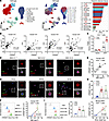 Figure 1
CREBZF is elevated in adipose tissue T cells in humans and mice with obesity. (A) CREBZF expression in different cell types (left) and clusters (middle) identified in human adipose tissue and visualized by a UMAP plot, as well as a bar chart (right). The T cell clusters are highlighted in red. (B) Pearson correlation of CREBZF mRNA levels with BMI and Tconv cell marker genes in human VAT. n = 53–55. (C) The mRNA levels of Crebzf in T cells isolated from spleens and eWAT of mice fed a chow or HFHS diet. Data are presented as the mean ± SEM. n = 6–8. Two-way ANOVA for multiple comparison. *P < 0.05, versus spleen and chow; #P < 0.05, versus eWAT and chow. (D–F) CREBZF expression in human VAT T cells positively correlated with BMI. (D) Representative immunofluorescence images of CD3+ T cells from VAT from individuals with varying BMIs. Original magnification, ×630. Scale bars: 25 µm (square side length). (E) Correlation between CREBZF intensity of CD3+ T cells and BMI. (F) Representative histograms of CREBZF in CD3+ T cells from human VAT and the correlation of CREBZF MFI with BMI. (G and H) The expression of CREBZF is increased in eWAT T cells from obese mice. (G) Representative immunofluorescence images of CREBZF in CD3+ T cells from eWAT of chow-fed, HFHS diet–fed, or ob/ob mice. Original magnification, ×630. Scale bars: 25 µm (square side length). (H) Quantification of CREBZF intensity from G. n = 6–9. *P < 0.05 versus chow diet–fed mice. (I) Representative histograms (left) and MFI (middle) of CREBZF in eWAT CD3+ T cells from chow diet–fed, HFHS diet–fed, or ob/ob mice, and the correlation (right) between CREBZF MFI and mouse body weight. Data are presented as the mean ± SEM. n = 5. One-way ANOVA for multiple comparison. *P < 0.05 versus chow diet–fed mice.
To further determine CREBZF expression in T cells from VAT in obesity, we performed immunofluorescence staining and flow cytometry. Interestingly, CREBZF expression in VAT-derived CD3+ T cells increased proportionally to BMI in obese individuals, exhibiting a significant positive correlation with BMI (Figure 1, D–F, and Supplemental Figure 1, G and H). Consistently, CREBZF levels were dramatically elevated in CD3+ T cells isolated from eWAT of both HFHS diet–fed and ob/ob mice, strongly correlating with mouse body weight (Figure 1, G–I). Taken together, these results demonstrate that CREBZF expression was elevated in CD3+ T cells from VAT and eWAT of humans and mice with obesity, suggesting that CREBZF may contribute to altered T cell function and immune homeostasis in the adipose niche during obesity.
T cell–specific Crebzf deficiency ameliorates diet-induced metabolic disturbance and epididymal adipose tissue inflammation. To assess the T cell–intrinsic function of CREBZF in adipose tissue remodeling during obesity, T cell–specific Crebzf-KO mice were generated (Supplemental Figure 2A). As expected, the mRNA and protein levels of CREBZF were markedly reduced in T cells, but not in B cells from Cd4-Cre Crebzffl/fl mice (Supplemental Figure 2, B and C). Cd4-Cre Crebzffl/fl mice exhibited no significant difference in metabolic characteristics compared with control littermates under chow diet conditions (Figure 2, A–E, and Supplemental Figure 2, D–G). Strikingly, Crebzf deficiency attenuated HFHS diet–induced metabolic disorders, including improved glucose tolerance, reduced hyperglycemia, and insulin resistance (Figure 2, A–D). Furthermore, Crebzf KO modestly reduced plasma triglyceride and total cholesterol levels (Figure 2E) and body weight, with decreased fat mass but no changes in lean mass (Supplemental Figure 2, H–L). These results demonstrate that T cell Crebzf deficiency alleviated HFHS diet–induced systemic metabolic disturbances, establishing the role of Crebzf in linking T cell–intrinsic function to obesity-associated metabolic regulation.
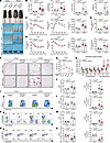 Figure 2
T cell–specific Crebzf deficiency protects against HFHS diet–induced metabolic disorders and eWAT inflammation in mice. Male Cd4-Cre Crebzffl/fl and control littermates Crebzffl/fl mice at 8 weeks were fed a chow or HFHS diet for 16 weeks. (A) Representative appearance of mice and tissues of Crebzffl/fl and Cd4-Cre Crebzffl/fl mice fed a chow or HFHS diet. (B) Body weight, fasting blood glucose levels, plasma insulin levels, and HOMA-IR. (C) GTT results and AUC. (D) ITT results and AUC. (E) Plasma triglyceride and cholesterol levels. (B–E) Data are presented as the mean ± SEM. Chow-fed group, n = 3–7; HFHS-fed group, n = 6–17. Two-way ANOVA for multiple comparison. *P < 0.05 versus Crebzffl/fl and chow diet; #P < 0.05 versus Crebzffl/fl and HFHS diet. (F) Representative H&E staining of eWAT from mice fed a chow or HFHS diet. Scale bars: 50 μm. (G) Quantification of adipocyte diameter of HFHS-fed mice. n = 6. (H) mRNA levels of proinflammatory and antiinflammatory genes in eWAT of mice fed a HFHS diet. n = 10–11. (G and H) Two-tailed, unpaired Student’s t test. *P < 0.05 versus Crebzffl/fl. (I and J) Representative flow cytometric plots (I) and quantification of frequencies and numbers (J) of CD3+ T cells in eWAT from Crebzffl/fl and Cd4-Cre Crebzffl/fl mice fed a chow or HFHS diet. (K–M) Representative flow cytometric plots (K) and quantification of the frequencies and numbers of CD4+ (L) and CD8+ (M) T cells in eWAT from mice. (N and O) Representative flow cytometric plots (N) and quantification of frequencies and numbers (O) of Tregs in eWAT from mice. Data are presented as the mean ± SEM. Chow group, n = 4–10; HFHS group, n = 6–13. Two-way ANOVA for multiple comparison. *P < 0.05 versus Crebzffl/fl and chow; #P < 0.05 versus Crebzffl/fl and HFHS.
Pathological remodeling and functional alterations of WAT mediated by T cells play crucial roles in the progression of obesity-related metabolic diseases (6, 7, 32). We analyzed the histology, cellularity, and microenvironment of eWAT to assess whether Crebzf deficiency in T cells reduces inflammation and improves metabolic parameters. Cd4-Cre Crebzffl/fl mice exhibited markedly improved obesity-associated pathology in eWAT, including reduced adipocyte size, crown-like structure (CLS) accumulation, and immune cell infiltration (Figure 2, F and G). This histologic improvement correlated with a shift in the expression of inflammatory markers in eWAT, where proinflammatory genes were downregulated and antiinflammatory genes were upregulated (Figure 2H). Flow cytometric analysis showed that T cell Crebzf deficiency had little effect on T cell populations in mice fed a chow diet, as demonstrated by similar frequencies and numbers of CD3+, CD4+, and CD8+ T cells (Figure 2, I–M). A dramatic reduction in the frequencies and total number of CD3+, CD4+, and CD8+ T cells was observed in eWAT of Cd4-Cre Crebzffl/fl mice with HFHS diet (Figure 2, I–M). Notably, eWAT Treg accumulation was not defective in Cd4-Cre Crebzffl/fl mice fed a chow diet but became impaired under obese conditions. Moreover, Crebzf deficiency in T cells specifically restored the HFHS diet–induced loss of Tregs in eWAT, significantly increasing both their percentage and absolute number (Figure 2, N and O). In contrast, the populations of Th1 and Th17 CD4+ T cells were unaltered by Crebzf deletion (Supplemental Figure 3, A–D), indicating that the attenuated inflammation in eWAT was selectively mediated by Tregs. We further confirmed that Crebzf deficiency did not disrupt Treg homeostasis in other tissues (Supplemental Figure 3, E and F). Taken together, these data establish that Crebzf deficiency remodels the T cell landscape in eWAT and that the subsequent attenuation of tissue inflammation is specifically dependent on Treg-mediated suppression.
Treg-intrinsic Crebzf is required for obesity-induced metabolic dysfunction and eWAT inflammation. To validate whether CREBZF mediates the effects on inflammation and metabolism directly through Tregs, Treg-specific deficient mice (Foxp3-Cre Crebzffl/fl) and control mice (Foxp3-Cre) were generated. Crebzf-sufficient (Foxp3-Cre) and Crebzf-deficient (Foxp3-Cre Crebzffl/fl) mice express yellow fluorescent protein (YFP), which serves as a reporter for Foxp3 expression in Tregs (33). We verified that CREBZF expression was specifically abolished in Tregs but not in CD4+ conventional T (Tconv) cells (Supplemental Figure 4, A–C).
Consistent with the findings in Cd4-Cre Crebzffl/fl mice (Figure 2), Treg-intrinsic ablation of Crebzf was sufficient to improve systemic metabolic parameters under HFHS diet conditions (Figure 3, A–E, and Supplemental Figure 4, D–L). Furthermore, Foxp3-Cre Crebzffl/fl mice displayed ameliorated eWAT pathology, characterized by reduced adipocyte hypertrophy and diminished inflammatory cell accumulation (Figure 3, F and G). Consistent with this improvement in tissue pathology, the expression of proinflammatory genes was reduced and antiinflammatory genes were elevated in eWAT of HFHS diet–fed Foxp3-Cre Crebzffl/fl mice (Figure 3H). Additionally, eWAT from Foxp3-Cre Crebzffl/fl mice showed an upregulation of gene programs for fatty acid oxidation, mitochondrial biogenesis, and lipolysis (Figure 3I). Moreover, the frequencies of CD3+, CD4+, and CD8+ T cells and total macrophages showed reduced accumulation in eWAT from Foxp3-Cre Crebzffl/fl mice fed a HFHS diet (Supplemental Figure 5, A–G). Interestingly, under HFHS diet conditions, Treg-specific Crebzf deficiency significantly reduced the proportion of the TREM2+ lipid–associated macrophage (LAM) subset in eWAT, which has emerged as a key player in obesity and in diseases involving lipid stress and inflammation (Supplemental Figure 5H). These results collectively demonstrate a broad amelioration of both inflammatory and metabolic dysfunction in eWAT upon Crebzf deficiency.
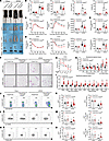 Figure 3
CREBZF in Tregs links eWAT inflammation to systemic metabolic dysfunction during obesity. Male Foxp3-Cre Crebzffl/fl and control Foxp3-Cre mice at 8 weeks were fed a chow or HFHS diet for 16 weeks. (A) Representative appearance of mice and tissues of Foxp3-Cre and Foxp3-Cre Crebzffl/fl mice fed a chow or HFHS diet. (B) Body weight, fasting blood glucose levels, plasma insulin levels, and HOMA-IR. (C) GTT results and AUC. (D) ITT results and AUC. (E) Plasma triglyceride and cholesterol levels. (B–E) Data are presented as the mean ± SEM. Chow group, n = 4–9; HFHS group, n = 6–13. Two-way ANOVA for multiple comparison. *P < 0.05 versus Foxp3-Cre and chow; #P < 0.05 versus Foxp3-Cre and HFHS. (F) Representative H&E staining of eWAT from Foxp3-Cre and Foxp3-Cre Crebzffl/fl mice fed a chow or HFHS diet. Scale bars: 50 μm. (G) Quantification of diameters of adipocytes from mice. n = 4–5. (H and I) Relative mRNA levels of inflammatory (H), lipid metabolism–, and mitochondria-related (I) genes in eWAT of mice fed a HFHS diet. (G–I) Data are presented as the mean ± SEM. n = 4–11. Two-tailed, unpaired Student’s t test. *P < 0.05 versus Foxp3-Cre. (J and K) Representative flow cytometric plots (J) and quantification of frequencies and numbers (K) of Tregs in eWAT of mice. (L and M) Representative flow cytometric plots (L) and quantification of frequencies and numbers (M) of IL-10+ Tregs in eWAT of mice. (N and O) Representative flow cytometric plots (N) and quantification of frequencies and numbers (O) of TGF-β+ Tregs in eWAT of mice. Data are presented as the mean ± SEM. Chow group, n = 3–10; HFHS group, n = 4–16. Two-way ANOVA for multiple comparison. *P < 0.05 versus Foxp3-Cre and chow; #P < 0.05 versus Foxp3-Cre and HFHS.
Given that adipose tissue Tregs suppress local inflammation and improve metabolic homeostasis via the secretion of IL-10 and TGF-β (34), we analyzed Treg abundance and function in our model. Under a chow diet, Foxp3-Cre Crebzffl/fl and Foxp3-Cre mice showed no difference in eWAT Treg abundance. In contrast, upon HFHS diet feeding, Foxp3-Cre Crebzffl/fl mice showed a significant increase in both the frequency and number of Tregs within eWAT (Figure 3, J and K). This expansion included a greater proportion of cells producing IL-10 and TGF-β (Figure 3, L–O), indicating enhanced stability and suppressive function of Crebzf-deficient Tregs in obesity. Moreover, Crebzf deficiency did not disrupt the balance in other tissues of Foxp3-Cre Crebzffl/fl mice fed a chow or HFHS diet (Supplemental Figure 5, I and J). Taken together, these results indicate that Crebzf deletion specifically enhances eWAT Treg fitness by bolstering the suppressive capacity and stability of these cells.
PA impairs Treg stability by upregulating CREBZF. VAT is enriched with a diverse array of metabolites, hormones, and inflammatory signals (35, 36). Consequently, resident immune cells undergo distinct adaptations in response to the shifting physiological and pathological landscape of obesity (37). To investigate whether obesity-associated pathological adaptations of CREBZF in T cell subsets within eWAT are driven by the nutrient overload, we screened metabolites and hormones enriched in obese VAT for their ability to regulate CREBZF. Treatment of Jurkat T cells with PA, but not oleic acid, lactic acid, insulin, or high glucose, induced a robust and dose-dependent upregulation of CREBZF at both mRNA and protein levels (Figure 4, A–C, and Supplemental Figure 6A). Moreover, this induction by PA was further confirmed in primary mouse CD3+ T cells and Tregs (Figure 4, D and E). Consistent with our observation that PA exerts an inductive effect, analysis of the Gene Expression Omnibus (GEO) public database (GSE174706) revealed that long-term high-fat diet feeding significantly induced Crebzf expression at the transcriptional level (Supplemental Figure 6B). In contrast, T cell activation did not alter CREBZF expression (Supplemental Figure 6, C–H).
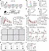 Figure 4
PA impairs Treg-mediated immunosuppression via CREBZF upregulation. (A–D) PA upregulates CREBZF expression at the mRNA and protein levels. (A) Quantitative PCR (qPCR) analysis of CREBZF expression in Jurkat T cells following treatment with different concentrations of PA, oleic acid, lactic acid, insulin, glucose, or vehicle for 24 hours. n = 4. (B and C) Representative histograms (B) and MFI (C) of CREBZF in Jurkat T cells treated with the indicated concentrations of PA or vehicle for 24 hours. n = 4. (D and E) CD3+ T cells (D) and Tregs (E) were isolated from spleens and lymph nodes of mice and treated with PA or vehicle as indicated for 24 hours. Relative Crebzf mRNA levels are shown. n = 6. (A–E) Data are presented as the mean ± SEM. One-way ANOVA for multiple comparison. *P < 0.05 versus vehicle. (F–H) Male Foxp3-Cre Crebzffl/fl and control Foxp3-Cre mice fed a chow diet were administered daily i.p. injections of PA (ethyl palmitate, 3 μM/g) or vehicle for 3 weeks. n = 3–4. (F) Schematic diagram of the animal experiment. (G) GTT results and AUC. (H) ITT results and AUC. (I) Representative H&E staining of eWAT from Foxp3-Cre and Foxp3-Cre Crebzffl/fl mice injected with PA or vehicle. Scale bars: 50 μm. (J) Relative mRNA levels of inflammatory genes in eWAT. (K and L) Representative flow cytometric plots (K) and quantification of frequencies (L) of Tregs in eWAT from Foxp3-Cre and Foxp3-Cre Crebzffl/fl mice injected with PA or vehicle. (G–L) Data are presented as the mean ± SEM. n = 3–4. Two-way ANOVA for multiple comparison. *P < 0.05 versus Foxp3-Cre and vehicle; #P < 0.05 versus Foxp3-Cre and PA.
To further evaluate the effects of PA in vivo, male Foxp3-Cre Crebzffl/fl and control Foxp3-Cre mice on a chow diet received daily i.p. injections of PA (3 μM/g) or vehicle for 3 weeks (Figure 4F). PA-treated mice displayed aggravated metabolic deficits, as evidenced by impaired glucose tolerance and insulin sensitivity (Figure 4, G and H). Three weeks of PA injection moderately increased body weight in mice fed a chow diet, and Crebzf deficiency slightly attenuated this weight gain (Supplemental Figure 6I). Concomitantly, H&E-stained histological sections of eWAT revealed aggravated pathology characterized by enhanced immune cell infiltration, increased CLSs, and tissue disruption (Figure 4I). Accordingly, the eWAT inflammatory profile was shifted, with upregulated proinflammatory and downregulated antiinflammatory gene expression (Figure 4J). Importantly, Treg-specific deletion of Crebzf significantly abrogated the PA-induced glycolipid metabolic disorder, pathological deterioration, and inflammation (Figure 4, G–J).
PA generally induces polarization of proinflammatory T cell subsets and is considered to decrease the proportion of Tregs and the stability of FOXP3 (23, 24, 38). As shown in Figure 4, K and L, PA treatment significantly reduced the frequencies of Tregs in eWAT, a loss that was reversed by Crebzf KO. Taken together, these results demonstrate that PA-dependent overactivation of CREBZF was sufficient to impair Treg stability, thereby contributing to metabolic disturbance and inflammation in eWAT during obesity progression.
Identification of 2 distinct Treg subsets in epididymal adipose tissue. The VAT Treg compartment exhibits considerable heterogeneity and contributes to the maintenance of beige fat biogenesis, insulin sensitivity, and systemic homeostasis (17–19). Therefore, the observed metabolic and inflammatory improvements upon Crebzf deletion could be attributed either to alterations in a specific VAT Treg subset or to broader reprogramming across the entire Treg compartment. To this end, we performed single-cell RNA-seq (scRNA-seq) using the 10X Genomics platform on Tregs isolated from eWAT of HFHS diet–fed mice. An unbiased integrative analysis across 2 genotypes after regressing out batch effects and other confounding factors using the Seurat R package yielded 17,702 eWAT Tregs (8,091 from control mice and 9,611 from Foxp3-Cre Crebzffl/fl mice).
Analysis based on differentially expressed genes (DEGs) reliably divided eWAT Tregs into subsets with metabolic and immune signatures, confirming the distinct features of the 2 subsets on a uniform manifold approximation and projection (UMAP) representation (Figure 5A and Supplemental Figure 7A). To further define discrete subsets of Tregs, we performed Gene Ontology (GO) pathway analysis using DEGs. Cluster 0 highlighted pathways indicating intracellular metabolism, whereas cluster 1 was enriched for pathways involved in immune response (Figure 5B). Moreover, ridge plots depicting profile scores for lipid metabolism and adaptive immune response in each cluster demonstrated that these 2 Treg subsets exhibited different functional preferences (Figure 5C). These subsets were annotated according to the most salient identified cell markers (Figure 5, D and E). Tregs from cluster 0 exhibited high expression of killer cell lectin–like receptor G1 (KLRG1), a transmembrane glycoprotein that is associated with an activated and memory phenotype, and were accordingly termed KLRG1hi Tregs. In addition, cluster 1 Tregs showed prominent expression of inducible T cell costimulator (ICOS). These were therefore termed ICOShi Tregs (Figure 5F and Supplemental Figure 7B).
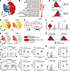 Figure 5
scRNA-seq identifies distinct KLRG1hi and ICOShi Treg subsets in epididymal adipose tissue. (A) UMAP representation of 17,702 eWAT Tregs color-coded by subset. (B) GO enrichment analysis of the DEGs between Treg clusters 0 and 1. (C) Ridge plots depicting the functional scores from the indicated Treg subtypes using AUCell. (D) UMAP visualization of signature genes of cluster 0 and cluster 1 Tregs from scRNA-seq. (E) Dot plot of top marker genes for the 2 Treg subsets. (F) Violin plot aligning Klrg1 and Icos expression at the transcriptional level for Treg subsets. (G) UMAP representations and cluster composition of Crebzf-sufficient (n = 2) and Crebzf-deficient (n = 2) Tregs. (H) Ridge plots of fatty acid oxidation in the KLRG1hi Treg cluster and IL-10 production in the ICOShi cluster, color-coded by sample condition. (I) Representative flow cytometric plots and frequencies of KLRG1hi and ICOShi Tregs in eWAT from Foxp3-Cre and Foxp3-Cre Crebzffl/fl mice fed a HFHS diet. n = 9. (J) Representative flow cytometric plots and frequencies of KLRG1hi and ICOShi Tregs in eWAT from Foxp3-Cre and Foxp3-Cre Crebzffl/fl mice treated with PA. n = 5. (I and J) Data are presented as the mean ± SEM. Two-tailed, unpaired Student’s t test. *P < 0.05 versus Foxp3-Cre. (K) Representative histograms and MFI of CREBZF in KLRG1hi (pink) and ICOShi (blue) Tregs from Foxp3-Cre mice fed a HFHS diet. n = 3–4. (L) Representative histograms and MFI of CREBZF in KLRG1hi (pink) and ICOShi (blue) Tregs from Foxp3-Cre mice treated with PA. n = 4. (K and L) Data are presented as the mean ± SEM. Two-tailed, unpaired Student’s t test. *P < 0.05 versus KLRG1hi.
We next examined the alterations in the proportions of KLRG1hi and ICOShi Tregs under chow and HFHS diet conditions. As shown in Supplemental Figure 7C, HFHS diet feeding significantly increased the frequencies of ICOShi Tregs and decreased those of KLRG1hi Tregs. Next, to determine the contribution of CREBZF to Treg identity and function, a side-by-side comparison of the scRNA-seq data from Foxp3-Cre Crebzffl/fl and Foxp3-Cre mice was conducted. Compared with control mice, loss of Crebzf increased the frequency of ICOShi Tregs, whereas the proportion of KLRG1hi Tregs was slightly decreased (Figure 5G). Furthermore, pathway analysis revealed that Crebzf deficiency specifically enhanced the IL-10 production pathway in ICOShi Tregs, whereas it did not significantly alter the fatty acid oxidation pathway in KLRG1hi Tregs (Figure 5H). Consistently, GO analysis of upregulated genes indicated that Crebzf-deficient ICOShi Tregs possessed a distinctly elevated immunosuppressive transcriptional signature and enhanced functional capacity (Supplemental Figure 7, D and E). These findings suggest that the increase in ICOShi Tregs upon Crebzf KO may represent a protective mechanism.
To further confirm the scRNA-seq findings, we performed flow cytometric analysis using the cluster-defining markers KLRG1 and ICOS. We observed that Crebzf deficiency significantly increased the frequency and number of ICOShi Tregs under both HFHS diet and palmitate treatment conditions, while it slightly elevated the number of KLRG1hi Tregs in HFHS diet–fed mice (Figure 5, I and J, and Supplemental Figure 7F). Importantly, flow cytometric and mRNA abundance analysis showed that Crebzf was specifically expressed in ICOShi Tregs, suggesting that CREBZF is a key mediator preventing ICOShi Treg accumulation in VAT during the progression of obesity (Figure 5, K and L). Notably, Crebzf deficiency did not significantly alter the frequency or number of the ICOShi Treg subset in inguinal WAT (iWAT) or spleen (Supplemental Figure 7, G–L), suggesting that the function of CREBZF is eWAT specific. Interestingly, a dietary switch from a high-fat diet to chow and caloric restriction–induced (CR-induced) weight loss also increased the proportion of Tregs in mouse eWAT, particularly ICOShi Tregs (Supplemental Figure 8), which is consistent with the changes in Treg subsets observed upon Crebzf KO, further supporting our finding that ICOShi Tregs played a protective role under obese pathological conditions. Taken together, our findings reveal 2 distinct eWAT Treg subsets with unique transcriptional and functional identities. Furthermore, we demonstrate that Crebzf specifically sustained the fitness of the ICOShi Treg subset in the obese eWAT microenvironment.
Adoptive transfer of Crebzf-deficient ICOShi Tregs attenuates the severity of HFHS diet–induced inflammation and metabolic disorders. Given the role of CREBZF in regulating ICOShi Treg immunosuppression (Figure 5, K and L), we sought to define the precise function and mechanism of CREBZF in Treg subset specialization under metabolic stress. Splenic ICOShi and KLRG1hi Tregs, either Crebzf sufficient (Crebzf+/+) or Crebzf deficient (Crebzf –/–), were sorted and transferred into Rag1–/– recipients, which were then fed a HFHS diet for 12 weeks (Figure 6A). Metabolic analysis revealed that mice receiving no Tregs developed severe metabolic dysfunction. In contrast, varying degrees of improved glycemic control and insulin sensitivity were observed in mice that received either ICOShi or KLRG1hi Tregs. Importantly, Crebzf deletion further enhanced this metabolic improvement function specifically in the ICOShi Treg subset, whereas KLRG1hi Tregs showed little change (Figure 6, B–D). Histological analysis revealed reduced eWAT weights and amelioration of obesity-associated pathology in mice receiving Crebzf –/– ICOShi Tregs, compared with other groups (Figure 6, E and F). Gene expression analysis of eWAT indicated that the transfer of Crebzf–/– ICOShi Tregs was associated with reduced expression of proinflammatory cytokine genes and increased levels of antiinflammatory markers (Figure 6G). The infiltration of macrophages and neutrophils is a hallmark of obese eWAT (4, 39). Flow cytometric analysis revealed that adoptive transfer of Crebzf –/– ICOShi Tregs into Rag1–/– mice significantly reduced the proportions of both neutrophils and macrophages in eWAT, indicating a potent attenuation of obesity-associated adipose tissue inflammation (Figure 6, H and I). These findings establish the idea that Crebzf deficiency enhances the ability of ICOShi Tregs to restrain systemic metabolic dysregulation and eWAT inflammation induced by a HFHS diet.
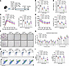 Figure 6
Crebzf-deficient ICOShi Tregs suppress eWAT inflammation. In an adoptive transfer model, Crebzf-sufficient or -deficient ICOShi or KLRG1hi Tregs were transferred into Rag1–/– recipients, followed by subsequent HFHS diet feeding for 12 weeks. n = 3–5. (A) Schematic diagram of the animal experiment. (B) Fasting blood glucose levels. (C) GTT results and AUC. (D) ITT results and AUC. (E) eWAT weights. (F) Representative H&E staining of eWAT from Rag1–/– mice transferred with Tregs followed by HFHS diet feeding. Scale bars: 50 μm. (G) Relative mRNA levels of inflammatory genes in eWAT. (H and I) Representative flow cytometric plots (H) and frequencies (I) of CD11c+Ly6G+ neutrophils and F4/80+ CD11b+ macrophages in eWAT from Rag1–/– mice transferred with Tregs followed by HFHS diet feeding. Data are presented as the mean ± SEM. n = 3–5. One-way ANOVA for multiple comparison. *P < 0.05 versus no Tregs; #P < 0.05 versus Crebzf +/+ ICOShi; &P < 0.05 versus Crebzf –/– ICOShi.
We next investigated the mechanism by which CREBZF regulates the stability and suppressive function of ICOShi Tregs. The assay for transposase accessible chromatin with high-throughput sequencing (ATAC-seq) was performed to assess its role in modulating chromatin accessibility on eWAT Tregs. Crebzf-deficient Tregs showed increased chromatin accessibility compared with Crebzf-sufficient cells (Supplemental Figure 9A). Specifically, following Crebzf deletion, chromatin accessibility increased more markedly at signature loci of ICOShi Tregs (such as Icos and Tigit) compared with those of KLRG1hi Tregs (including Klrg1 and Pparg) (Supplemental Figure 9B). The top-8 ranked motifs correspond to binding of the activator protein 1 (AP-1) family members generated based on Hypergeometric Optimization of Motif Enrichment (HOMER) (Supplemental Figure 9C).
Crebzf is established as a transcriptional coregulator that does not bind DNA directly (30). Cofactors of CREBZF were screened using our scRNA-seq dataset, and a series of transcription factors exhibited differential activity specifically in ICOShi Tregs (Supplemental Figure 9D). Co-immunoprecipitation analysis demonstrated that CREBZF directly interacted with c-JUN, the major component of the AP-1 family (40), and we detected the intracellular colocalization between c-JUN and CREBZF in Tregs (Supplemental Figure 9, E–G). AP-1 is a dimeric transcription factor composed of members from the JUN, FOS, ATF, and MAF protein families. Its activity is regulated through dimer composition, transcriptional control, posttranslational modifications, and interactions with other proteins (41). Overexpression of CREBZF attenuated the interaction between c-FOS and c-JUN, which suggested that CREBZF suppressed the AP-1 activity (Supplemental Figure 9H). In response to TCR and costimulatory signaling, the Foxp3 promoter is bound and activated by transcription factors such as nuclear factors of activated T cells (NFAT) and AP‑1 (10, 42). Given the observation that Crebzf deficiency significantly increased the expression of Foxp3 in ICOShi Tregs (Supplemental Figure 7D) and enhanced AP-1 family factor activity (Supplemental Figure 9C), we propose that CREBZF may regulate Foxp3 expression in ICOShi Tregs, and consequently their stability, by restricting AP-1 transcription factor activity. Next, we confirmed that CREBZF inhibited AP-1–driven Foxp3 luciferase activity in a dose-dependent manner (Supplemental Figure 9I). We next elucidated the causal relationship between c-JUN- and CREBZF-mediated inhibition of Foxp3 and the suppressive function of ICOShi Tregs. As expected, the suppressive effects of CREBZF on the transcription levels of Foxp3, Il10, and Tgfb were reduced by c-JUN siRNA in ICOShi Tregs (Supplemental Figure 9, J and K). Taken together, these results demonstrate that CREBZF bound to c-JUN to reduce interaction between c-JUN and c-FOS (two major components of the AP-1 family), and consequently inhibited the transcriptional activity of Foxp3, thereby impairing the stability and suppressive function of ICOShi Tregs.
Negative correlation of FOXP3 and suppressive gene expression with elevated CREBZF in VAT. Next, we examined the inhibitory effect of CREBZF on FOXP3 expression and the suppressive function of Tregs in VAT. We detected mRNA levels of CREBZF and Treg-suppressive genes (FOXP3, IL10, and TGFB) in VAT from individuals with obesity and in eWAT from mice. We observed a strong negative correlation between CREBZF and FOXP3 and inhibitory cytokine genes in both human VAT and mouse eWAT (Figure 7, A and B). This negative relationship between Crebzf and Foxp3 at the transcript level was also confirmed in a human VAT RNA-seq dataset (GSE70353) and in CD3+ T cells sorted from mouse eWAT (Figure 7, C and D).
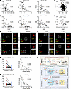 Figure 7
CREBZF inversely correlates with suppressive gene expression in Tregs from human VAT and mouse eWAT. (A–D) Pearson correlation analyses of mRNA levels in VAT. (A) Correlation of CREBZF with FOXP3, IL10, and TGFB expression in VAT from individuals with obesity. n = 55. (B) Correlation of Crebzf with Foxp3, Il10, and Tgfb in mouse eWAT. n = 45. (C) Correlation of CREBZF with FOXP3 in the GEO public dataset GSE70353. (D) Correlation of Crebzf with Foxp3 in CD3+ T cells sorted from mouse eWAT. n = 18. (E) Representative immunofluorescence images of Tregs sorted from VAT from the indicated individuals. Original magnification: x 630. Scale bars: 25 µm (square side length). (F) Representative immunofluorescence images of Tregs sorted from mouse eWAT from the indicated mice. Original magnification: x 630. Scale bars: 25 µm (square side length). (G) Quantification and correlation analysis of CREBZF and FOXP3 fluorescence intensity in Tregs sorted from human VAT. n = 15. (H) Quantification and correlation analysis of CREBZF and FOXP3 fluorescence intensity in Tregs sorted from eWAT of mice. n = 14. (I) Proposed model illustrating the mechanism by which PA mediates immunosuppression of ICOShi Tregs, thereby regulating VAT remodeling and systemic metabolic homeostasis. During the progression of obesity, PA-induced hyperactivation of CREBZF reduces the frequency and suppressive function of the ICOShi Treg subset. This occurs through CREBZF binding to c-JUN, which inhibits Foxp3 transcriptional activity. Therefore, targeting the CREBZF/c-JUN/FOXP3 axis may represent a therapeutic strategy for obesity-related metabolic diseases.
To further evaluate this observation, we isolated Tregs from human VAT and mouse eWAT and performed flow cytometry and immunofluorescence staining. We found that CREBZF expression was increased in Tregs from the VAT of individuals with a high BMI and in eWAT of HFHS diet–fed mice (Figure 7, E and F). Furthermore, CREBZF expression levels in VAT showed a significant inverse correlation with FOXP3 protein levels (Figure 7, G and H, and Supplemental Figure 10, A and B). Importantly, relative mRNA levels of CREBZF and FOXP3, IL10, and TGFB in human SAT and mouse iWAT showed no significant correlation (Supplemental Figure 10, C and D), suggesting a VAT-specific regulatory role of CREBZF. These clinical data further indicate that elevated CREBZF may deteriorate FOXP3 stability and the suppressive function of visceral adipose Tregs during obesity.


Copyright © 2026 American Society for Clinical Investigation
ISSN: 0021-9738 (print), 1558-8238 (online)