
Advertisement
Research ArticleImmunologyInfectious diseaseVirology
Open Access |  10.1172/JCI203675
Mucosal vaccination with hemagglutinin stalk provides broad protection against influenza virus subtypes in mice
Weizhong Li,1 Arunraj M. Rajendrakumar,1,2 ZiZhen Miao,1 Bibek Dhakal,1 Bogdan M. Domrachev,1 Tao Wang,1 Qiuqi Kan,1 Qiaofeng Wan,1 Hongquan Wan,3 Wenbin Tuo,2 Florian Krammer,4 Zhiping Ye,3 and Xiaoping Zhu1
1Division of Immunology, Virginia-Maryland College of Veterinary Medicine, University of Maryland, College Park, Maryland, USA.
2Animal Parasitic Diseases Laboratory, Agricultural Research Service, United States Department of Agriculture, Beltsville, Maryland, USA.
3Division of Viral Products, Center for Biologics Evaluation and Research, Food and Drug Administration, Silver Spring, Maryland, USA.
4Department of Microbiology, Icahn School of Medicine at Mount Sinai, New York, New York, USA.
Address for correspondence. Xiaoping Zhu, 8075 Greenmead Drive, University of Maryland, College Park, Maryland 20742, USA. Email: xzhu1@umd.edu.
Find articles by Li, W. in: PubMed | Google Scholar
1Division of Immunology, Virginia-Maryland College of Veterinary Medicine, University of Maryland, College Park, Maryland, USA.
2Animal Parasitic Diseases Laboratory, Agricultural Research Service, United States Department of Agriculture, Beltsville, Maryland, USA.
3Division of Viral Products, Center for Biologics Evaluation and Research, Food and Drug Administration, Silver Spring, Maryland, USA.
4Department of Microbiology, Icahn School of Medicine at Mount Sinai, New York, New York, USA.
Address for correspondence. Xiaoping Zhu, 8075 Greenmead Drive, University of Maryland, College Park, Maryland 20742, USA. Email: xzhu1@umd.edu.
Find articles by Rajendrakumar, A. in: PubMed | Google Scholar
1Division of Immunology, Virginia-Maryland College of Veterinary Medicine, University of Maryland, College Park, Maryland, USA.
2Animal Parasitic Diseases Laboratory, Agricultural Research Service, United States Department of Agriculture, Beltsville, Maryland, USA.
3Division of Viral Products, Center for Biologics Evaluation and Research, Food and Drug Administration, Silver Spring, Maryland, USA.
4Department of Microbiology, Icahn School of Medicine at Mount Sinai, New York, New York, USA.
Address for correspondence. Xiaoping Zhu, 8075 Greenmead Drive, University of Maryland, College Park, Maryland 20742, USA. Email: xzhu1@umd.edu.
Find articles by Miao, Z. in: PubMed | Google Scholar
1Division of Immunology, Virginia-Maryland College of Veterinary Medicine, University of Maryland, College Park, Maryland, USA.
2Animal Parasitic Diseases Laboratory, Agricultural Research Service, United States Department of Agriculture, Beltsville, Maryland, USA.
3Division of Viral Products, Center for Biologics Evaluation and Research, Food and Drug Administration, Silver Spring, Maryland, USA.
4Department of Microbiology, Icahn School of Medicine at Mount Sinai, New York, New York, USA.
Address for correspondence. Xiaoping Zhu, 8075 Greenmead Drive, University of Maryland, College Park, Maryland 20742, USA. Email: xzhu1@umd.edu.
Find articles by Dhakal, B. in: PubMed | Google Scholar
1Division of Immunology, Virginia-Maryland College of Veterinary Medicine, University of Maryland, College Park, Maryland, USA.
2Animal Parasitic Diseases Laboratory, Agricultural Research Service, United States Department of Agriculture, Beltsville, Maryland, USA.
3Division of Viral Products, Center for Biologics Evaluation and Research, Food and Drug Administration, Silver Spring, Maryland, USA.
4Department of Microbiology, Icahn School of Medicine at Mount Sinai, New York, New York, USA.
Address for correspondence. Xiaoping Zhu, 8075 Greenmead Drive, University of Maryland, College Park, Maryland 20742, USA. Email: xzhu1@umd.edu.
Find articles by Domrachev, B. in: PubMed | Google Scholar
1Division of Immunology, Virginia-Maryland College of Veterinary Medicine, University of Maryland, College Park, Maryland, USA.
2Animal Parasitic Diseases Laboratory, Agricultural Research Service, United States Department of Agriculture, Beltsville, Maryland, USA.
3Division of Viral Products, Center for Biologics Evaluation and Research, Food and Drug Administration, Silver Spring, Maryland, USA.
4Department of Microbiology, Icahn School of Medicine at Mount Sinai, New York, New York, USA.
Address for correspondence. Xiaoping Zhu, 8075 Greenmead Drive, University of Maryland, College Park, Maryland 20742, USA. Email: xzhu1@umd.edu.
Find articles by Wang, T. in: PubMed | Google Scholar
1Division of Immunology, Virginia-Maryland College of Veterinary Medicine, University of Maryland, College Park, Maryland, USA.
2Animal Parasitic Diseases Laboratory, Agricultural Research Service, United States Department of Agriculture, Beltsville, Maryland, USA.
3Division of Viral Products, Center for Biologics Evaluation and Research, Food and Drug Administration, Silver Spring, Maryland, USA.
4Department of Microbiology, Icahn School of Medicine at Mount Sinai, New York, New York, USA.
Address for correspondence. Xiaoping Zhu, 8075 Greenmead Drive, University of Maryland, College Park, Maryland 20742, USA. Email: xzhu1@umd.edu.
Find articles by Kan, Q. in: PubMed | Google Scholar
1Division of Immunology, Virginia-Maryland College of Veterinary Medicine, University of Maryland, College Park, Maryland, USA.
2Animal Parasitic Diseases Laboratory, Agricultural Research Service, United States Department of Agriculture, Beltsville, Maryland, USA.
3Division of Viral Products, Center for Biologics Evaluation and Research, Food and Drug Administration, Silver Spring, Maryland, USA.
4Department of Microbiology, Icahn School of Medicine at Mount Sinai, New York, New York, USA.
Address for correspondence. Xiaoping Zhu, 8075 Greenmead Drive, University of Maryland, College Park, Maryland 20742, USA. Email: xzhu1@umd.edu.
Find articles by Wan, Q. in: PubMed | Google Scholar
1Division of Immunology, Virginia-Maryland College of Veterinary Medicine, University of Maryland, College Park, Maryland, USA.
2Animal Parasitic Diseases Laboratory, Agricultural Research Service, United States Department of Agriculture, Beltsville, Maryland, USA.
3Division of Viral Products, Center for Biologics Evaluation and Research, Food and Drug Administration, Silver Spring, Maryland, USA.
4Department of Microbiology, Icahn School of Medicine at Mount Sinai, New York, New York, USA.
Address for correspondence. Xiaoping Zhu, 8075 Greenmead Drive, University of Maryland, College Park, Maryland 20742, USA. Email: xzhu1@umd.edu.
Find articles by Wan, H. in: PubMed | Google Scholar
1Division of Immunology, Virginia-Maryland College of Veterinary Medicine, University of Maryland, College Park, Maryland, USA.
2Animal Parasitic Diseases Laboratory, Agricultural Research Service, United States Department of Agriculture, Beltsville, Maryland, USA.
3Division of Viral Products, Center for Biologics Evaluation and Research, Food and Drug Administration, Silver Spring, Maryland, USA.
4Department of Microbiology, Icahn School of Medicine at Mount Sinai, New York, New York, USA.
Address for correspondence. Xiaoping Zhu, 8075 Greenmead Drive, University of Maryland, College Park, Maryland 20742, USA. Email: xzhu1@umd.edu.
Find articles by Tuo, W. in: PubMed | Google Scholar
1Division of Immunology, Virginia-Maryland College of Veterinary Medicine, University of Maryland, College Park, Maryland, USA.
2Animal Parasitic Diseases Laboratory, Agricultural Research Service, United States Department of Agriculture, Beltsville, Maryland, USA.
3Division of Viral Products, Center for Biologics Evaluation and Research, Food and Drug Administration, Silver Spring, Maryland, USA.
4Department of Microbiology, Icahn School of Medicine at Mount Sinai, New York, New York, USA.
Address for correspondence. Xiaoping Zhu, 8075 Greenmead Drive, University of Maryland, College Park, Maryland 20742, USA. Email: xzhu1@umd.edu.
Find articles by
Krammer, F.
in:
PubMed
|
Google Scholar
|

1Division of Immunology, Virginia-Maryland College of Veterinary Medicine, University of Maryland, College Park, Maryland, USA.
2Animal Parasitic Diseases Laboratory, Agricultural Research Service, United States Department of Agriculture, Beltsville, Maryland, USA.
3Division of Viral Products, Center for Biologics Evaluation and Research, Food and Drug Administration, Silver Spring, Maryland, USA.
4Department of Microbiology, Icahn School of Medicine at Mount Sinai, New York, New York, USA.
Address for correspondence. Xiaoping Zhu, 8075 Greenmead Drive, University of Maryland, College Park, Maryland 20742, USA. Email: xzhu1@umd.edu.
Find articles by Ye, Z. in: PubMed | Google Scholar
1Division of Immunology, Virginia-Maryland College of Veterinary Medicine, University of Maryland, College Park, Maryland, USA.
2Animal Parasitic Diseases Laboratory, Agricultural Research Service, United States Department of Agriculture, Beltsville, Maryland, USA.
3Division of Viral Products, Center for Biologics Evaluation and Research, Food and Drug Administration, Silver Spring, Maryland, USA.
4Department of Microbiology, Icahn School of Medicine at Mount Sinai, New York, New York, USA.
Address for correspondence. Xiaoping Zhu, 8075 Greenmead Drive, University of Maryland, College Park, Maryland 20742, USA. Email: xzhu1@umd.edu.
Find articles by Zhu, X. in: PubMed | Google Scholar
Published November 16, 2026 - More info
J Clin Invest. 2026;136(22):e203675. https://doi.org/10.1172/JCI203675.
© 2026 Li et al. This work is licensed under the Creative Commons Attribution 4.0 International License. To view a copy of this license, visit http://creativecommons.org/licenses/by/4.0/.
Received: December 17, 2025; Accepted: September 22, 2026


-
Results
Expression and characterization of HA Stalk-Fc fusion proteins. The HA Stalk is a promising target for eliciting broadly neutralizing or cross-reactive Abs (23). However, the immunogenicity of HA Stalk is believed to be masked by the highly immunogenic HA head (23, 24). To induce broad and high levels of Stalk-reactive Abs, several studies have designed and expressed stable headless HA Stalk domains using structure-based rational approaches (18, 19, 25–28), which include the HA2 domain and partial regions of HA1 that contribute to Stalk folding.
To generate a stabilized, trimeric HA Stalk protein, the HA (GenBank AAP34324.1) of influenza virus A/New Caledonia/20/1999 (H1N1) was used as a backbone. Several changes were made to remove the head and stabilize the Stalk as a prefusion conformation. First, 33–310 aa (mature HA protein) were deleted and replaced with a GSG linker. Second, the SIQS sequence before the HA1-HA2 cleavage site (R326/G327) was substituted with the QRET sequence. Third, 386–418 aa were replaced with the GSGGSG linker. Fourth, to stabilize the HA Stalk conformation under low-pH conditions, several mutations (K377M, Y420D, N421L, and E429L) were introduced. To promote trimer formation, the bacteriophage T4 foldon domain (PDB ID 1RFO) was added to the C-terminus of the Stalk. The HA Stalk was expressed as a His-tagged protein (Stalk, Figure 1A).
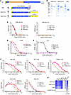 Figure 1
Expression and characterization of prefusion HA Stalk and Stalk-Fc proteins. (A) Schematic of influenza A virus HA protein, including signal peptide (SP), cleavage site (CS), transmembrane domain (TM), and cytoplasmic tail (CT). The graphic shows the fusion of the HA Stalk with the T4 fibritin foldon domain (Fd) and His tag to produce a soluble, prefusion-stabilized HA Stalk protein. Specific mutations, indicated by asterisks, were introduced in the HA Stalk to stabilize the prefusion form. Additionally, a Stalk-Fc/wt protein was generated by fusing Stalk with foldon and mouse Fcγ2a Fc. Mutations were created in the hinge region by replacing 3 cysteines with serine residues and in the Fc region by modifying Glu318, Lys320, and Lys322 to alanine residues to eliminate the C1q binding site. The Stalk-Fc/mut construct was also generated by incorporating HQ/AD (310/311) and HN/AQ (433/434) mutations (indicated by triangles) to disrupt FcRn binding. To ensure proper domain folding, G/S linkers are included. (B) The Stalk-Fc and Stalk proteins were purified from stable CHO cell lines. Both soluble Stalk-Fc and Stalk proteins were isolated using affinity columns and subsequently analyzed by SDS-PAGE under reducing conditions, followed by Coomassie blue staining. (C–E) The binding of Stalk-Fc to mouse FcRn/β2m, FcγRI, and C1q was measured by ELISA to assess the specific interactions. Stalk protein served as a negative control and mouse IgG as a positive control. (F) The interactions of Stalk-Fc or Stalk alone with HA Stalk–specific and conformation-dependent mAbs KB2, 6F12, CR6261, and FI6V3 were examined by ELISA. Stalk-specific polyclonal Ab (pAb) was used as a positive control, and a trimeric SARS-CoV-2 Spike (S) protein as a negative control. (G) A heatmap displaying the EC50 values derived from the results in F and Supplemental Figure 3.
To target FcRn, we fused the modified HA Stalk and foldon domain with the Fc portion of mouse IgG2a, creating Stalk-Fc. Mouse IgG2a Fc, which is like human IgG1 Fc, was chosen due to its high affinity for activating FcγRI (29). Since IgG Fc usually forms a disulfide-bonded dimer, we made a monomeric Fc by replacing cysteine residues at positions 224, 227, and 229 with serine in the hinge region to remove the disulfide bonds (Stalk-Fc/wt, Figure 1A). We also generated a mutant Fc (Stalk-Fc/mut, Figure 1A) that cannot bind FcRn by making HQ/AD mutations at positions 310/311and HN/AQ mutations at positions 433/434 (30). In both the wild-type (wt) and mutant (mut) Fc, the C1q-binding motif was removed to eliminate C1q binding (31). As a result, we fused either the monomeric IgG Fc/wt or Fc/mut with HA Stalk-foldon (Supplemental Table 1; supplemental material available online with this article; https://doi.org/10.1172/JCI203675DS1). The soluble Stalk-Fc or Stalk proteins were secreted from the transfected CHO cells and purified using affinity columns (Figure 1B). The Stalk-Fc/wt, Stalk-Fc/mut, and Stalk proteins were verified by Western blots using Abs against mouse IgG, HA, or His tag (Supplemental Figure 1). Importantly, Stalk-Fc/wt and Stalk-Fc/mut were expressed as monomers under both nonreducing (Supplemental Figure 1, left) and reducing conditions (Supplemental Figure 1, middle), confirming that the removal of disulfide bonds prevented Fc dimerization. The trimeric structure of the Stalk-Fc/wt or Spike was predicted by AlphaFold 3, showing the independent folding of the HA Stalk and Fc domains.
FcRn binds IgG in a pH-dependent manner (11). To test whether the Stalk-Fc/wt protein binds to FcRn, FcγRI, or C1q, we performed an ELISA. The results showed that, like mouse IgG, Stalk-Fc/wt bound to FcRn at pH 6.0 but not at pH 7.4 (Figure 1C). In contrast, both Stalk-Fc/mut and the standalone Stalk protein failed to bind FcRn at pH 6.0. Stalk-Fc/wt and Stalk-Fc/mut bound to the high-affinity receptor FcγRI (CD64) (Figure 1D). Surface plasmon resonance assay further confirmed that the affinity constant (KD) for Stalk-Fc binding to immobilized FcRn or FcγRI was 8.73 × 10⁻⁸ M (Supplemental Figure 2, left) or 1.67 × 10⁻⁷ M (Supplemental Figure 2, right), respectively. Unlike IgG, Stalk-Fc/wt, Stalk-Fc/mut, and Stalk proteins did not react with C1q, confirming the successful removal of the C1q binding site in the Fc portion of Stalk-Fc (Figure 1E). Taken together, these results indicate that the Stalk-Fc/wt protein maintains the structural integrity necessary for interaction with FcRn or FcγRI, but not with C1q protein, enabling Stalk-Fc to be nonlytic.
HA Stalk fused to an Fc fragment more effectively mimics the native HA Stalk structure by maintaining conformation-dependent B cell epitopes (32). We tested a panel of HA Stalk–specific mAbs known for their broad binding to conformational epitopes in the HA Stalk (33–36). Both Stalk-Fc fusion and Stalk were recognized by the mAbs KB2, CR6261, FI6v3, and 6F12 and a polyclonal Ab against Stalk in ELISAs (Figure 1F). However, the CR8020 mAb showed low-affinity binding (Supplemental Figure 3); this is expected because CR8020 mainly interacts with the HA Stalk from group 2 influenza viruses (37). SARS-CoV-2 Spike protein (38) was used as a negative control, demonstrating specific Ab binding. Results of Ab binding are summarized in a heatmap and presented as EC50 (log ng/mL) (Figure 1G).
The stability of HA Stalk-Fc was tested under various conditions, including repeated freeze-thaw cycles (5 or 10 times), heating at 55°C for 30 min or 1 h, incubating at 37°C for 1 or 2 days, and storage at room temperature for 4 or 6 days (Supplemental Figure 4A). The Stalk-Fc protein exhibited significant stability, as demonstrated by a Coomassie blue–stained SDS-PAGE gel. A minor band at 37 kDa was present on the SDS-PAGE gel; however, this band was undetectable by either anti-HA or anti-mouse IgG Abs in a Western blot (Supplemental Figure 1). Additionally, in comparison with the untreated protein, the treated Stalk-Fc protein under the above conditions maintained the ability to bind to FcRn; FcγRI; anti-mouse IgG2a Ab; mAbs KB2, CR6261, FI6v3, and 6F12; and a polyclonal Ab against the Stalk in ELISA (Supplemental Figure 4, B–I). Overall, these findings indicate that both the Fc and Stalk portions of the Stalk-Fc fusion protein retain their ability to interact with FcRn and FcγRI under rigorous conditions while preserving their conformational B cell epitopes.
FcRn-dependent delivery of Stalk-Fc/wt antigen across the epithelial barrier. To determine whether Stalk-Fc/wt, but not Stalk-Fc/mut, fusion protein is transported across the epithelial barrier by FcRn, we used an MDCKFcRn cell line to study the transport of Stalk-Fc fusion proteins. MDCK cells expressing rat FcRn and rat β2m have been shown to specifically transport murine IgG in vitro (39). When Stalk-Fc/wt was applied to the apical reservoir (Figure 2A), it was transported to the basolateral reservoir and detected by ELISA (Figure 2B). In contrast, Stalk-Fc/mut or Stalk alone were poorly transported across the MDCKrFcRn+rb2m monolayer (Figure 2B), indicating that Stalk-Fc/wt was transported specifically by FcRn.
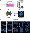 Figure 2
FcRn-mediated transport of the HA Stalk-Fc/wt protein across polarized epithelial cells. (A) In vitro transcytosis of the Stalk-Fc/wt across MDCK cell monolayers expressing FcRn and β2m. MDCKFcRn/β2m cells were grown on the apical side of Transwell inserts to become fully polarized. Stalk-Fc (50 μg/mL) or Stalk (27.5 μg/mL) proteins were added to the apical side and incubated at 37°C for 90 min. An ELISA was conducted to measure the amount of protein transported to the basolateral side. The data presented are the mean ± SEM from 9 individual wells per protein across 2 independent assays. Statistical analysis was conducted using Welch’s 1-way ANOVA (with Dunnett’s T3 multiple-comparison test). ****P < 0.0001. (B) Schematic showing the process of making a nose cryosection for the imaging analysis of in vivo transcytosis. (C) In vivo transcytosis of Stalk-Fc/wt proteins across the epithelial cells lining the nasal cavities of mice. Stalk-Fc or Stalk proteins were conjugated with the fluorescent dye AF647. A total of 10 μg of Stalk-Fc/wt, Stalk-Fc/mut, or 5.5 μg of Stalk proteins was i.n. inoculated into C57BL/6 or FcRn KO mice (n = 3) in a volume of 20 μL. 7 h later, nasal tissue samples were fixed in 10% formalin, decalcified, embedded in OCT compound, and sectioned using a cryostat. Protein distributions in the nasal epithelium (arrows) were visualized using confocal microscopy after DAPI staining. Representative images display a ventral view of the mouse noses. Scale bars: 60 μm (top), 10 μm (bottom).
To further examine whether Stalk-Fc is transported across the nasal epithelial cells in vivo following i.n. administration, 10 μg of the AF647-labeled Stalk-Fc/wt, Stalk-Fc/mut, or 5.5 μg (equal molar number) of the Stalk protein was i.n. administered to mice; after 7 h, the nose was harvested, decalcified, and subjected to cryosection at the middle part of the nasal passages. The coronal planes were visualized under a confocal microscope (Figure 2B). As shown in Figure 2C, Stalk-Fc/wt was readily detected as punctate staining in the nasal epithelial tissue (arrows) of WT mice but not in that of FcRn-KO animals. Additionally, Stalk-Fc/mut or Stalk protein alone remained mainly on the apical surface of nasal epithelial cells, which was barely transported. Based on these results, we conclude that the Stalk-Fc/wt fusion protein was efficiently transported across the polarized epithelial cells lining the airway in an FcRn-dependent manner.
FcRn-mediated nasal delivery significantly enhances Stalk-specific systemic and local immune responses. To determine whether FcRn-dependent transport enhances the immunogenicity of the HA Stalk antigen, mice were i.n. immunized with 10 μg of Stalk-Fc, 5.5 μg Stalk (in equal molar amount), or PBS (Supplemental Table 2), followed by boosting with the same dose twice at 3-week intervals (Figure 3A). Using WT mice immunized with Stalk-Fc/mut and FcRn-KO mice immunized with Stalk-Fc/wt, we demonstrated the necessity of FcRn binding for enhancing immune responses. Using Stalk alone allowed us to assess the FcRn-independent effects in vivo. The inclusion of these control groups also removed potential concerns regarding mucosal leakage. A mucosal adjuvant, CpG (10 �g/mouse) or c-di-GMP (5 �g), was used to overcome potential mucosal tolerance (40, 41). Previous studies demonstrated that neither CpG nor c-di-GMP compromises the structural integrity of the respiratory epithelial barrier (41, 42).
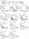 Figure 3
FcRn-mediated i.n. immunization significantly enhances HA Stalk–specific Ab immune responses. (A) 10 μg of Stalk-Fc, 5.5 μg Stalk (equivalent molar), or PBS in combination with 10 μg of CpG or 5 μg of c-di-GMP in 20 μL was i.n. administered into 7-week-old C57BL/6 or FcRn KO mice (n = 10). Mice were boosted twice at a 3-week interval. Sampling was done at the indicated time points. (B) The levels of Stalk-specific IgG were measured by ELISA. Symbols represent individual mice, and the horizontal lines indicate the mean ± SEM. (C) The broad bindings of the sera from the Stalk-Fc/wt– or PBS-immunized mice were examined by interacting with the HA1, HA2, HA5, HA9, and HA13 proteins from different influenza subtypes in ELISA. (D) The competitive Stalk binding of the sera from the mice immunized with Stalk-Fc/wt or PBS with the Stalk-specific mAbs KB2, 6F12, and FI6V3 was measured by ELISA. (E–G) Cytokine-secreting cells in the spleen. The spot-forming cells (SFCs) secreting IFN-γ (E), TNF-α (F), and IL-4 (G) per 1 × 10⁶ splenocytes from the immunized mice were quantified by ELISpot. The representative images (5 × 10⁵ cells /well) are shown below the plot. Data are presented as box-and-whisker plots with minimum, maximum, median, and interquartile ranges. (H and I) The levels of Stalk-specific IgA and IgG Abs in nasal wash (H) or BAL (I) were measured by ELISA; data are presented as mean ± SEM. Statistical analyses were conducted using ordinary 1-way ANOVA with Dunnett’s multiple-comparison tests for G and I; Welch’s 1-way ANOVA with Dunnett’s T3 multiple comparisons for B, E, F, and H (left); or Kruskal-Wallis test with Dunn’s multiple comparisons for H (right). *P < 0.05, **P < 0.01, ***P < 0.001, ****P < 0.000. ND: not detected, which was excluded from statistical analysis.
Two weeks after the final boost (Figure 3A), significantly higher titers of Stalk-specific IgG in the sera were detected in the Stalk-Fc/wt–immunized WT mice compared with the Stalk-Fc/wt–immunized FcRn-KO mice or Stalk-, Stalk-Fc/mut–, and PBS-treated groups (Figure 3B). To show the breadth and cross-reactive potency of the Ab response induced by the Stalk-Fc/wt protein, we performed an ELISA by coating various HA proteins. Our results showed that the serum Abs from the Stalk-Fc/wt–immunized mice interacted with various HA proteins (HA1, HA2, HA5, HA9, and HA13) from group 1 influenza virus subtypes more extensively than the sera from PBS control mice (Figure 3C). In a competitive ELISA, the serum Abs from the Stalk-Fc/wt–immunized mice were found to significantly diminish the binding of the HA Stalk–specific mAbs, such as KB2, 6F12, and F16v3, by competitively binding to HA Stalk (Figure 3D). KB2 and 6F12 are effective against the infection of a wide range of group 1 influenza strains (34), and F16v3 effectively neutralizes both groups 1 and 2 influenza A viruses (43). Hence, the broad reactivity with different HAs and recognition of conserved neutralizing epitopes by the serum Abs indicate the potent efficacy of FcRn-mediated nasal delivery of the Stalk-Fc against various influenza A virus subtypes. Additionally, Stalk-Fc/wt proteins stimulated robust T cell immune responses in the spleens of WT mice, as demonstrated by IFN-γ–, TNF-α–, or IL-4–producing (Figure 3, E–G, respectively) cell responses, as shown by an enzyme-linked immunospot (ELISpot) assay, compared with other control groups after the boost.
Influenza virus initiates its infection in the airways (3). Therefore, a key objective of FcRn-mediated mucosal delivery of HA Stalk is to elicit stronger local immune responses at these specific anatomical sites. To this end, we measured the levels of Stalk-specific Abs in mucosal secretions following i.n. immunizations. Nasal wash and bronchoalveolar lavage (BAL) fluids were collected 2 weeks after the final boost and analyzed for Stalk-specific IgG and IgA using ELISA. The results showed significantly increased levels of Stalk-specific IgA and IgG in nasal washes (Figure 3H) and BAL (Figure 3I) from mice immunized with Stalk-Fc/wt compared with other control groups. During the initial assessment, it was noted that Stalk alone induced a barely detectable IgG and IgA immune response (Figure 3, B, H, and I) following i.n. administration. To verify its immunogenicity, we i.m. administered it along with aluminum adjuvant in mice. After boosting, Stalk alone induced a similar level of serum IgG compared with that of the Stalk-Fc/wt (Supplemental Figure 5). Overall, our study indicates that targeting HA Stalk to FcRn via an i.n. immunization strategy significantly enhances the levels of serum IgG Abs and mucosal IgA and IgG Abs.
FcRn-mediated nasal delivery significantly induces TRM CD4+ and CD8+ T cells in the lung. Memory CD4+ and CD8+ T cells play a crucial role in protecting against the influenza virus (44, 45). A key subset of these cells, known as TRM cells, are noncirculating and remain in the nasal and lung tissues, providing a rapid response upon reexposure to the influenza virus (7–9). In our study, we evaluated whether FcRn-mediated nasal delivery of HA Stalk-Fc/wt could induce the formation of TRM T cells in the lungs (Figure 4A). To distinguish circulating T cells from lung-resident T cells, PE-CD45 Ab was intravenously injected 5 min before the euthanasia of mice, and the cells were obtained immediately for analysis. The gating strategy for TRM T cells is depicted in Supplemental Figure 6. We found a significant number of CD4+CD69+CD103+/– (Figure 4B) and CD8+CD69+CD103+ TRM cells (Figure 4C) in the lungs of mice that were i.n. immunized with Stalk-Fc/wt. Most importantly, we observed a significant increase in the proportion of both monofunctional and polyfunctional CD4+ TRM T cells producing IFN-γ, TNF-α, and IL-2 upon peptide pool stimulation (Figure 4D). This remarkable increase is proportional to the total number of CD4+ T cells detected in the lungs (Supplemental Figure 7A). We also noted that the functional CD8+ TRM T cells in the lungs of Stalk-Fc/wt–immunized mice exhibited similar responsiveness to HA peptides, as assessed by the percentages of monofunctional TRM cells producing IFN-γ, IL-2, and TNF-α, as well as cells producing both IFN-γ and TNF-α (Figure 4E). However, the total number of functional CD8+ TRM T cells detected in the lungs was relatively low, which is consistent with the fact that the total numbers of CD8+ T cells did not differ significantly across different groups (Supplemental Figure 7B). We did not include an i.m. control group because we did not observe significant increases in CD4+ or CD8+ TRM T cells in the lungs after i.m. immunization with HA-Fc (22) or Spike-Fc (38).
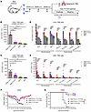 Figure 4
TRM CD4+ and CD8+ T cell responses in the lungs. (A) Schematic depicting procedures for immunization and collection of TRM cells (top) and influenza virus challenge in mice with or without FTY720 treatment (bottom). For the FTY720 treatment, Stalk-Fc/wt– or PBS-immunized mice received daily i.p. injection with 20 �g FTY720 in 100 �L 0.9% saline 2 days before and throughout the challenge period. The immunized mice administered with 0.9% saline served as controls. (B and C) TRM cells in the lungs (n = 10/group). The number of CD4+ TRM (CD69+CD103+/–) (B) or CD8+ TRM (CD69+CD103+) (C) T cells from the lungs of each mouse were quantified and plotted as mean ± SEM. The data were combined from 2 independent experiments. (D and E) The TRM cells were functionally evaluated by measuring the production of IFN-γ, TNF-α, and IL-2 cytokines after HA peptide stimulation. The number of CD4+ (D) or CD8+ (E) TRM-secreting single or multiple cytokines in the lung is presented as mean ± SEM. (F and G) The immunized mice treated with or without FTY720 were i.n. infected with rH5N1 virus (5 × 10² TCID50), and body weight changes were monitored daily for 14 days (F). Protection is shown as a Kaplan-Meier survival curve (G). Statistical analyses were conducted using an ordinary 1-way ANOVA with Dunnett’s multiple-comparison test for TNF-α (D); Welch’s 1-way ANOVA with Dunnett’s T3 multiple-comparison test for CD4+ TRM (B), CD8+ TRM (C), and IL-2 cytokine (D); or Kruskal-Wallis test followed by Dunn’s multiple comparisons (D and E). *P < 0.05, **P < 0.01, ***P < 0.001, ****P < 0.0001.
To further evaluate protection mediated by TRM T cells, which is independent of the protection conferred by circulating T cells, we treated mice with FTY720 prior to and during the infection (Figure 4A). This treatment sequesters circulating T cells within secondary lymphoid tissues, while TRM T cells remain in the lungs (7). After challenging mice with the rH5N1 virus, we found that most of the Stalk-Fc/wt–immunized mice treated with (90%) or without FTY720 (80%) were protected from virus infection, as indicated by minimal weight loss in both groups (Figure 4, F and G). In contrast, the PBS control mice treated with FTY720 experienced significant weight loss of up to 25% within 7–9 days following the viral challenge, ultimately resulting in 100% mortality (Figure 4, F and G). These results demonstrate that i.n. immunization with Stalk-Fc/wt protects mice from the morbidity and mortality associated with H5N1 infection, a protective efficacy independent of circulating lymphocytes. Therefore, our findings suggest that FcRn-mediated nasal delivery of Stalk-Fc can effectively induce protective TRM T cells in the lungs. Further investigation is warranted to determine the protective capacity of adoptively transferred TRM T cells.
FcRn-mediated nasal immunization with Stalk-Fc provides increased protection against lethal challenges of various influenza virus subtypes. To evaluate the effectiveness and breadth of the humoral and cellular immune responses generated by FcRn-mediated i.n. vaccination with Stalk-Fc/wt, we challenged immunized mice with a lethal dose (5 MLD50) of rH5N1, H1N1 (PR8), and rH6N2 viruses 3 weeks after the final boost (Figure 5A). All or most mice in the control groups, including Stalk-Fc/wt/KO, Stalk-Fc/mut, Stalk, and PBS, experienced significant weight loss, up to 25%, within 6–9 days following the viral challenges (Figure 5, B–D) and succumbed to death with a low survival rate ranging from 0% to 27.3% (Figure 5, E–G). In contrast, most WT mice immunized with Stalk-Fc/wt antigen did not exhibit severe body weight loss or other symptoms (Figure 5, B–D). Consequently, the Stalk-Fc/wt–immunized WT mice demonstrated substantial protective survival rates against rH5N1 (90%; Figure 5E), H1N1 (91%; Figure 5F), and rH6N2 (80%; Figure 5G) viruses. We also tested whether Stalk-Fc provides cross-protection against infection by group 2 H3N2 virus. Similar to the PBS control mice, the majority of Stalk-Fc/wt–immunized mice (4 out of 5) succumbed to H3N2 infection (Supplemental Figure 8).
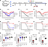 Figure 5
The i.n. immunization by HA Stalk-Fc/wt induces broad protection against the challenge of different subtypes of influenza A virus. (A) Schematic of the immunization and challenge procedures. The 7-week-old C57BL/6 or FcRn KO mice were i.n. immunized with 10 μg of Stalk-Fc or 5.5 mg of Stalk protein. 10 μg CpG or 5 μg c-di-GMP was used as an adjuvant. Mice were boosted twice at a 3-week interval. 3 weeks after the final boost, the mice were i.n. challenged with 50 mL of a lethal dosage of mouse-adapted influenza virus rH5N1, H1N1, or rH6N2 or a field strain of H9N2. (B–D) The body weights were measured daily for 14 days in each experiment, and the data represent the mean ± SEM (n = 10–12 per group). Mice were euthanized when 25% body weight loss or a humane endpoint was reached. (E–G) The survival rates are plotted by Kaplan-Meier curves (n = 10–12 per group). (H–K) rH5N1 or H9N2 virus replication in the nasal turbinate (H and J) and lungs (I and K) at 4 days post infection was also evaluated. The virus titers in the samples (n = 10–11 per group) were determined by cytopathic effect (CPE) in MDCK cells and shown as TCID50. Data are presented as box-and-whisker plots with minimum, maximum, median, and interquartile ranges. Statistical analyses were conducted using a log-rank (Mantel-Cox) test (E–G), Kruskal-Wallis test followed by Dunn’s multiple comparisons (H and I), unpaired 2-tailed t test (J), or an unpaired 2-tailed t test with Welch’s correction (K). *P < 0.05, **P < 0.01, ***P < 0.001, ****P < 0.0001.
We further evaluated virus replication in the nasal turbinate (Figure 5, H and J) and lungs (Figure 5, I and K) 4 days after the rH5N1 and H9N2 challenge. The results demonstrated a 1.5 to 2.5 log reduction of the live virus levels in the nasal turbinate or lungs of the Stalk-Fc/wt–immunized mice compared with the other control groups, which corresponds with the immune response results (Figure 3). Taken together, our data clearly indicate that FcRn-mediated nasal delivery of Stalk-Fc is essential for broad protection against infections from different influenza virus subtypes or strains.
Given that most people have received seasonal influenza vaccines or have been exposed to influenza virus infections, we further investigated whether i.n. immunization with Stalk-Fc could enhance preexisting immunity and provide additional protection. To establish preexisting immunity, we either i.m. immunized mice (n = 10) with HA1 and HA3 proteins, along with CpG and Alum adjuvants, or i.n. infected mice (n = 10) with a sublethal dose of the PR8 virus (Supplemental Figure 9A). The establishment of preexisting immunity was confirmed by detecting HA- or Stalk-specific IgG Abs in the sera of HA-immunized or PR8-infected mice (Supplemental Figure 9B). One month later, all mice with preexisting immunity were i.n. boosted with either 10 �g of Stalk-Fc (n = 5) or PBS (n = 5). Mice that received the Stalk-Fc boost developed significantly higher levels of HA-specific (Supplemental Figure 9C) or Stalk-specific (Supplemental Figure 9D) IgG compared with those of the PBS control mice. To further assess whether the i.n. boost with Stalk-Fc could reduce viral replication, we challenged the mice with a lethal dose of the rH5N1 virus. The results showed that the single Stalk-Fc boost significantly reduced rH5N1 virus replication in the nasal turbinate (Supplemental Figure 9E) and lungs (Supplemental Figure 9F) at 4 days post infection compared with the PBS control mice. In conclusion, an i.n. boost with Stalk-Fc protein can significantly enhance the preexisting immune response and lower viral replication in mice.
FcRn-mediated nasal delivery of Stalk-Fc induces strong memory immune responses. A universal influenza mucosal vaccine is expected to induce a long-lasting immune memory response (46). We determined whether the nasal delivery of Stalk-Fc/wt via the FcRn pathway could induce a durable memory immune response lasting at least 10 months after the final boost (Figure 6A). As shown in Figure 6B, the highest levels of Stalk-specific serum IgG were detected in WT mice immunized with Stalk-Fc/wt compared with those of the control groups. Additionally, we observed significantly higher levels of Stalk-specific IgA and IgG in the nasal washes (Figure 6C) and BAL fluid (Figure 6D) from the Stalk-Fc/wt–immunized mice. Interestingly, we were still able to detect remarkably high levels of Stalk-specific IgG in the sera (Figure 6E), IgA in nasal washes (Supplemental Figure 10A), and IgA and IgG in BAL fluid (Supplemental Figure 10B) from the Stalk-Fc/wt–immunized mice 16 months after the final boost, but not from other control groups. Using ELISpot, we found a substantially high number of Stalk-specific Ab-secreting cells in the bone marrow of mice immunized with Stalk-Fc/wt antigen after 10-month (Figure 6F) or 16-month (Supplemental Figure 10, C and D) immunizations. The presence of long-lived plasma cells in the bone marrow niche likely contributes to the sustained high levels of Stalk-specific IgG in the blood (47). These results reveal that Stalk-specific B cell memory is sustained at a high level for at least 10 months after immunization.
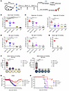 Figure 6
FcRn-mediated i.n. immunization with HA Stalk-Fc enhances memory immune responses. (A) Schematic of the experimental design to evaluate the memory immune responses. Mice were i.n. immunized at the indicated time and euthanized for sampling sera, nasal washes, BAL, and bone marrow. Some mice were subjected to rH5N1 virus challenge. (B–E) The duration of Stalk-specific Ab responses was assessed 10 or 16 months after the final boost. Stalk-specific IgG Ab in mouse sera (B and E), nasal washes (C, right), and BAL (D, right) or IgA in nasal washes (C, left) and BAL (D, left) were quantified by ELISA and exhibited as mean ± SEM (n = 9–10 for 10 month samples, and n = 4–5 for 16 month samples). (F) Long-lived Stalk-specific Ab-secreting cells (ASCs) in BM were isolated after 10 months of final boost. The 2.5 × 10⁵ cells/well from the Stalk-Fc/wt group and 1 × 10⁶ cells/well from other groups were placed on Stalk-coated plates and quantified by ELISpot analysis of IgA-secreting (left) or IgG-secreting (right) plasma cells. Boxes illustrate median values as well as 25th and 75th percentile values (n = 5 per group). Representative spot images are shown. (G) Body weight changes following rH5N1 virus challenge. 6 months after the final boost, groups of 5 mice were i.n. challenged with rH5N1 virus (5 × 10² TCID50) and weighed daily for 14 days. (H) Protection is shown as a Kaplan-Meier survival curve. Statistical analyses were conducted using Welch’s 1-way ANOVA with Dunnett’s T3 multiple comparisons (B), Kruskal-Wallis test followed by Dunn’s multiple comparisons (C and D [left]), ordinary 1-way ANOVA with Dunnett’s multiple comparisons (D [right] and F), unpaired 2-tailed t test (E), or log-rank (Mantel-Cox) test (H). *P < 0.05, **P < 0.01, ***P < 0.001, ****P < 0.0001. ND: not detected, which was excluded from statistical analysis.
To evaluate whether these memory immune responses offer protection, we challenged the mice with a lethal dosage of rH5N1 virus 6 months after the boost. Most mice in the control groups experienced a severe body weight drop within 4–9 days after the challenge and either succumbed to the infection or were euthanized after losing 25% of their initial body weight (Figure 6G). In contrast, mice immunized with Stalk-Fc/wt antigen showed significantly reduced disease severity, with a survival rate of 80% (Figure 6H). Overall, the mucosal delivery of Stalk-Fc via the FcRn pathway resulted in an effective memory immune response and protected against the lethal challenge of influenza viruses.
Stalk-specific Ab-mediated neutralizing activity and inhibition of membrane fusion. To explore the mechanisms of protection against lethal influenza virus infection in mice i.n. immunized with the Stalk-Fc antigen, we tested the immune sera using pseudotype microneutralization (pMN) assay. Typically, the pMN assay is more sensitive than the conventional MN assay (43, 48), especially for measuring HA Stalk–specific Abs. The pH5N1 virus, a pseudotyped lentiviral particle expressing HA5 and NA1 surface proteins of the H5N1 virus and harboring a luciferase reporter construct, was used. As shown in Figure 7A, the pooled Stalk-Fc/wt–immunized mouse sera exhibited powerful neutralization activity against pH5N1 virus (pMN titer = 960), while the sera from other control groups exhibited much less neutralization ability (pMN titer = 100 or <20), demonstrating that the HA Stalk-Fc/wt can induce Stalk-specific neutralizing Abs.
 Figure 7
Effects of immune serum–mediated neutralizing activity and the inhibition of the membrane fusion. (A) The microneutralization titer of the pooled mouse sera against the pseudotyped H5N1 reporter virus is expressed as the average ID50 from 2 independent tests. (B–F) Sera samples were evaluated for their ability to inhibit the pH-dependent cell–cell fusion in influenza A virus–infected cells. Vero cells were infected with different influenza viruses for 12 h: H1N1 (B, MOI = 0.2), rH5N1 (C, MOI = 0.04), rH6N2 (D, MOI = 0.4), rH9N2 (E, MOI = 0.4), or H3N2 (F, MOI = 0.4). Following infection, the cells were treated with TPCK-trypsin (5 �g/mL) for 7 min. Pooled sera from the Stalk-Fc/wt–immunized mice or PBS-control mice were diluted and added to cells for 1.5 h incubation. Cell–cell fusion was triggered by exposing the cells to pH 5.0 PBS for 10 min, whereas pH 7.2 PBS served as a control. After an additional 3 h of culture at 37°C in fresh media, the cells were stained with a nucleoprotein (NP)-specific Ab and DAPI. Membrane fusion and polykaryon formation were observed using fluorescence microscopy. Scale bars: 127 μm. (G) The fusion index of the virus-infected cells treated with sera from Stalk-Fc/wt–immunized or PBS control mice was calculated from 5 random fields under microscope. Fusion index (%) = total number of nuclei in the infected cells forming polykaryon/total number of nuclei in all infected cells.
We next tested whether the serum Abs utilized the inhibitory mechanisms typically observed for Stalk-binding mAbs (43, 49). The HA0 precursor of influenza A virus HA is found on the surfaces of infected cells. When exposed to trypsin treatment and the low pH of the environment, HA0 undergoes a conformational change from its native form to a fusion-active form, leading to the formation of polykaryons (50). Indeed, Vero cells infected with H1N1 at pH 5.0 displayed polykaryon formation, whereas no polykaryons were visualized at pH 7.2 (Figure 7B, left). We further examined the effects of immune sera from Stalk-Fc–immunized mice on the fusion process of Vero cells infected with different influenza strains. Notably, the immune sera significantly inhibited polykaryon formation in Vero cells infected with H1N1 (Figure 7B, right, and Supplemental Figure 11). In contrast, sera from PBS control mice did not prevent this fusion. Additionally, the immune sera effectively blocked polykaryon formation in Vero cells infected with rH5N1, rH6N2, and rH9N2 (Figure 7, C–E) compared with the sera from PBS control mice. We expected that the sera from Stalk-Fc–immunized mice would not inhibit polykaryon formation in Vero cells infected with H3N2 virus (Figure 7F), as H3N2 belongs to group 2 influenza A virus. In a quantitative assay (Figure 7G), we calculated the fusion indexes of each group; the indexes represent the ratio of polykaryon-forming cells to the total number of cells counted. The values were significantly lower in groups treated with Stalk antisera specific to group 1 H1N1, rH5N1, rH6N2, and rH9N2 when compared with the same groups treated with sera from PBS control mice. The fusion indexes did not differ in the group 2 H3N2 virus group when cells were treated with sera from PBS control mice. Overall, these results indicate that immune sera from Stalk-Fc–immunized mice block viral fusion with endosomal membranes induced by low pH, a crucial step in the viral replication cycle.
Stalk-specific Ab Fc-mediated effector mechanisms. Beyond direct virus neutralization, influenza virus HA Stalk–specific Abs have been shown to possess potent Fc-mediated effector functions, which are important in ameliorating disease severity by eliminating virus-infected cells (51–54). Several HA Stalk–based vaccines have been shown to stimulate Ab-dependent cellular cytotoxicity (ADCC) and Ab-dependent cellular phagocytosis (ADCP) in both animal models and humans (55, 56). Meanwhile, some HA Stalk–specific Abs promote the lysis of virus-infected cells via complement-dependent cytotoxicity (CDC) (57) that correlates with protection against influenza illness after seasonal influenza vaccination in pregnant women (54). Hence, we evaluated the ADCC, ADCP, and CDC activities of HA Stalk–specific Abs against influenza virus–infected cells. We first measured ADCC activity using a high-throughput mouse FcγRIV activated reporter assay. The H1N1 influenza virus–infected MDCK cells were subjected to treatment with antisera from different immunization groups, followed by incubation with Jurkat reporter cells expressing mouse FcγRIV. The reporter gene can only be activated by the sera from Stalk-Fc/wt vaccinated WT mice, but not those from other groups (Figure 8A, left). The elevated reporter signal by Stalk-Fc/wt sera was also remarkable in MDCK cells infected with other group 1 viruses, including rH5N1, rH6N2, and rH9N2, but was not detectable in H3N2 (group 2) virus-infected cells (Figure 8A, right). These results suggest that the cross-reactive Abs in immune sera predominantly target HA from group 1 but not group 2 influenza A virus. We further examined the functional CDC activity of Stalk Ab by incubating virus-infected MDCK cells with the diluted mouse sera and guinea pig complement. The subsequent cell death was measured using a cell viability detection kit and the percentage of cytotoxicity caused by the Stalk-specific Ab (Figure 8B). The combination of Stalk-Fc/wt–stimulated mouse sera, but not the PBS-immunized sera, with exogenous complement resulted in significantly higher cytotoxicity in target cells infected with H1N1, rH5N1, rH6N2, and rH9N2 strains. However, the Ab-specific cell lysis was minimal in H3N2 strain–infected cells.
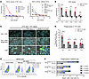 Figure 8
Evaluation of ADCC, CDC, and ADCP activities of immune sera. (A) ADCC assay. MDCK cells that were infected with the indicated influenza viruses were incubated with serially diluted immune sera. Jurkat reporter cells were added for measuring luciferase activity. Each symbol represents the mean value from duplicate wells. Data are shown as the ADCC fold change between wells treated with or without serum. IAV, influenza A virus. (B) CDC assay. Influenza virus–infected MDCK cells were incubated with the pooled immune or control sera in the presence of 10% complement to determine the lysis. Each symbol represents the values of duplicate samples from 2 independent experiments. (C) ADCP assay. HA1-coated fluorescence beads that were incubated with immune sera were added to J774 cells for phagocytosis. The cell nucleus was stained with DAPI. Fluo, fluorescence. Scale bars = 127 μm. (D) Quantitative analysis of phagocytosis in J774 was performed using beads coated with the indicated HA proteins. For each sample, images were captured from 5 random fields. The areas of beads and nuclei in each field were calculated with ImageJ (NIH), and the resulting ratio represents the phagocytosis score. (E and F) Phagocytosis analysis in BMDMs. Cells containing HA-coated beads were identified by flow cytometry and are shown in dot plots from a representative result (E). A stacked column graph (F) illustrates the percentage of cells internalizing different numbers of HA-coated beads (≥1, 2, 3, or 4 beads) within the total cell populations. The fractions within each column indicate the mean values from 2 experiments. Statistical analyses were conducted using an unpaired 2-tailed t test for the H1N1, rH5N1, rH6N2, and rH9N2 viruses or an unpaired 2-tailed t test with Welch’s correction for H3N2 virus in B, or an unpaired 2-tailed t test for HA1, HA5, and HA4 or a Mann-Whitney test for HA9 in D. *P < 0.05, **P < 0.01, ***P < 0.001.
Phagocytic macrophages (58) and neutrophils (59) have been shown to recognize the Fc portion of Abs via their FcRns, then engulf and destroy the Ab-coated target cells or virus particles, a process known as ADCP. To assess the ADCP function of Stalk Ab in immune sera, we employed a microsphere-based approach as previously described (60). The yellow/green fluorescent beads coated with various influenza HA proteins were used as a surrogate of virus-infected target cells. Beads were incubated with mouse sera and loaded on J774 or BMDM cells. The internalization of beads was visualized using a fluorescence microscope (Figure 8, C and D, and Supplemental Figure 12) or monitored using flow cytometry (Figure 8, E and F, and Supplemental Figure 13). In the presence of serum from Stalk-Fc/wt–immunized mice, a significantly high number of beads coated with HA1, HA5, or HA9 proteins were internalized by both cells (Figure 8C and Supplemental Figure 12, A and B, and Supplemental Figure 14), indicating the enhanced engulfment by Stalk Ab is broad for group 1 HA proteins. As expected, this enhancement was not observed in the PBS serum–treated samples. Furthermore, immune sera containing Stalk Abs showed negligible ADCP activity against HA4-coated beads, which can be attributed to the high diversity of group 2 HA4 protein from the group 1 HA protein (Figure 8D and Supplemental Figure 12C). Therefore, i.n. administration of HA Stalk-Fc/wt resulted in a significant increase in HA Stalk–specific ADCP activity within the immune sera.
Taken together, our results demonstrate that serum Abs elicited by FcRn-mediated nasal delivery of Stalk-Fc/wt protein directly protect through the Fc-mediated effector function, which effectively eliminates the virus-infected cells via multiple mechanisms, including ADCC, ADCP, or CDC. This may partially explain the protection against the heterologous or heterosubtypic infection from group 1 influenza virus strains in mice (Figures 4 and 5).


Copyright © 2026 American Society for Clinical Investigation
ISSN: 0021-9738 (print), 1558-8238 (online)