
Advertisement
Research ArticleCardiologyVascular biology
Open Access |  10.1172/JCI203310
Endocardial transcription factor HAND2 orchestrates hypoxic and TGF-β signaling to modulate coronary artery formation
Huijuan Wang,1 Haosheng Zhang,2 Leiyin Zheng,1 Peihan Zhang,1 Yuqian Wang,1 Sijia Ding,1 Wenping Liu,1 Yuanming Cheng,2,3 Zhongzhou Yang,3,4 and Wen Luo1,3
1Institute of Modern Biology (IMB), Department of Vascular Surgery, Cardiovascular Medical Center, and
2IMB, Department of Hematology, Nanjing Drum Tower Hospital, The Affiliated Hospital of Nanjing University Medical School, Nanjing University, Nanjing, China.
3State Key Laboratory of Pharmaceutical Biotechnology, Nanjing University, Nanjing, China.
4Jiangsu Key Laboratory of Molecular Medicine, Nanjing University Medical School, Nanjing University, Nanjing, China.
Address correspondence to: Wen Luo, Nanjing University, 163 Xianlin Road, Qixia District, Nanjing, Jiangsu Province, 210023, China. Email: wenluo@nju.edu.cn. Or to: Zhongzhou Yang, Nanjing University, 22 Hankou Road, Gulou District, Nanjing, Jiangsu Province, 210093, China. Email: zhongzhouyang@nju.edu.cn.
Authorship note: HW and HZ contributed equally to this work.
Find articles by Wang, H. in: PubMed | Google Scholar
1Institute of Modern Biology (IMB), Department of Vascular Surgery, Cardiovascular Medical Center, and
2IMB, Department of Hematology, Nanjing Drum Tower Hospital, The Affiliated Hospital of Nanjing University Medical School, Nanjing University, Nanjing, China.
3State Key Laboratory of Pharmaceutical Biotechnology, Nanjing University, Nanjing, China.
4Jiangsu Key Laboratory of Molecular Medicine, Nanjing University Medical School, Nanjing University, Nanjing, China.
Address correspondence to: Wen Luo, Nanjing University, 163 Xianlin Road, Qixia District, Nanjing, Jiangsu Province, 210023, China. Email: wenluo@nju.edu.cn. Or to: Zhongzhou Yang, Nanjing University, 22 Hankou Road, Gulou District, Nanjing, Jiangsu Province, 210093, China. Email: zhongzhouyang@nju.edu.cn.
Authorship note: HW and HZ contributed equally to this work.
Find articles by Zhang, H. in: PubMed | Google Scholar
1Institute of Modern Biology (IMB), Department of Vascular Surgery, Cardiovascular Medical Center, and
2IMB, Department of Hematology, Nanjing Drum Tower Hospital, The Affiliated Hospital of Nanjing University Medical School, Nanjing University, Nanjing, China.
3State Key Laboratory of Pharmaceutical Biotechnology, Nanjing University, Nanjing, China.
4Jiangsu Key Laboratory of Molecular Medicine, Nanjing University Medical School, Nanjing University, Nanjing, China.
Address correspondence to: Wen Luo, Nanjing University, 163 Xianlin Road, Qixia District, Nanjing, Jiangsu Province, 210023, China. Email: wenluo@nju.edu.cn. Or to: Zhongzhou Yang, Nanjing University, 22 Hankou Road, Gulou District, Nanjing, Jiangsu Province, 210093, China. Email: zhongzhouyang@nju.edu.cn.
Authorship note: HW and HZ contributed equally to this work.
Find articles by Zheng, L. in: PubMed | Google Scholar
1Institute of Modern Biology (IMB), Department of Vascular Surgery, Cardiovascular Medical Center, and
2IMB, Department of Hematology, Nanjing Drum Tower Hospital, The Affiliated Hospital of Nanjing University Medical School, Nanjing University, Nanjing, China.
3State Key Laboratory of Pharmaceutical Biotechnology, Nanjing University, Nanjing, China.
4Jiangsu Key Laboratory of Molecular Medicine, Nanjing University Medical School, Nanjing University, Nanjing, China.
Address correspondence to: Wen Luo, Nanjing University, 163 Xianlin Road, Qixia District, Nanjing, Jiangsu Province, 210023, China. Email: wenluo@nju.edu.cn. Or to: Zhongzhou Yang, Nanjing University, 22 Hankou Road, Gulou District, Nanjing, Jiangsu Province, 210093, China. Email: zhongzhouyang@nju.edu.cn.
Authorship note: HW and HZ contributed equally to this work.
Find articles by Zhang, P. in: PubMed | Google Scholar
1Institute of Modern Biology (IMB), Department of Vascular Surgery, Cardiovascular Medical Center, and
2IMB, Department of Hematology, Nanjing Drum Tower Hospital, The Affiliated Hospital of Nanjing University Medical School, Nanjing University, Nanjing, China.
3State Key Laboratory of Pharmaceutical Biotechnology, Nanjing University, Nanjing, China.
4Jiangsu Key Laboratory of Molecular Medicine, Nanjing University Medical School, Nanjing University, Nanjing, China.
Address correspondence to: Wen Luo, Nanjing University, 163 Xianlin Road, Qixia District, Nanjing, Jiangsu Province, 210023, China. Email: wenluo@nju.edu.cn. Or to: Zhongzhou Yang, Nanjing University, 22 Hankou Road, Gulou District, Nanjing, Jiangsu Province, 210093, China. Email: zhongzhouyang@nju.edu.cn.
Authorship note: HW and HZ contributed equally to this work.
Find articles by Wang, Y. in: PubMed | Google Scholar
1Institute of Modern Biology (IMB), Department of Vascular Surgery, Cardiovascular Medical Center, and
2IMB, Department of Hematology, Nanjing Drum Tower Hospital, The Affiliated Hospital of Nanjing University Medical School, Nanjing University, Nanjing, China.
3State Key Laboratory of Pharmaceutical Biotechnology, Nanjing University, Nanjing, China.
4Jiangsu Key Laboratory of Molecular Medicine, Nanjing University Medical School, Nanjing University, Nanjing, China.
Address correspondence to: Wen Luo, Nanjing University, 163 Xianlin Road, Qixia District, Nanjing, Jiangsu Province, 210023, China. Email: wenluo@nju.edu.cn. Or to: Zhongzhou Yang, Nanjing University, 22 Hankou Road, Gulou District, Nanjing, Jiangsu Province, 210093, China. Email: zhongzhouyang@nju.edu.cn.
Authorship note: HW and HZ contributed equally to this work.
Find articles by Ding, S. in: PubMed | Google Scholar
1Institute of Modern Biology (IMB), Department of Vascular Surgery, Cardiovascular Medical Center, and
2IMB, Department of Hematology, Nanjing Drum Tower Hospital, The Affiliated Hospital of Nanjing University Medical School, Nanjing University, Nanjing, China.
3State Key Laboratory of Pharmaceutical Biotechnology, Nanjing University, Nanjing, China.
4Jiangsu Key Laboratory of Molecular Medicine, Nanjing University Medical School, Nanjing University, Nanjing, China.
Address correspondence to: Wen Luo, Nanjing University, 163 Xianlin Road, Qixia District, Nanjing, Jiangsu Province, 210023, China. Email: wenluo@nju.edu.cn. Or to: Zhongzhou Yang, Nanjing University, 22 Hankou Road, Gulou District, Nanjing, Jiangsu Province, 210093, China. Email: zhongzhouyang@nju.edu.cn.
Authorship note: HW and HZ contributed equally to this work.
Find articles by Liu, W. in: PubMed | Google Scholar
1Institute of Modern Biology (IMB), Department of Vascular Surgery, Cardiovascular Medical Center, and
2IMB, Department of Hematology, Nanjing Drum Tower Hospital, The Affiliated Hospital of Nanjing University Medical School, Nanjing University, Nanjing, China.
3State Key Laboratory of Pharmaceutical Biotechnology, Nanjing University, Nanjing, China.
4Jiangsu Key Laboratory of Molecular Medicine, Nanjing University Medical School, Nanjing University, Nanjing, China.
Address correspondence to: Wen Luo, Nanjing University, 163 Xianlin Road, Qixia District, Nanjing, Jiangsu Province, 210023, China. Email: wenluo@nju.edu.cn. Or to: Zhongzhou Yang, Nanjing University, 22 Hankou Road, Gulou District, Nanjing, Jiangsu Province, 210093, China. Email: zhongzhouyang@nju.edu.cn.
Authorship note: HW and HZ contributed equally to this work.
Find articles by Cheng, Y. in: PubMed | Google Scholar
1Institute of Modern Biology (IMB), Department of Vascular Surgery, Cardiovascular Medical Center, and
2IMB, Department of Hematology, Nanjing Drum Tower Hospital, The Affiliated Hospital of Nanjing University Medical School, Nanjing University, Nanjing, China.
3State Key Laboratory of Pharmaceutical Biotechnology, Nanjing University, Nanjing, China.
4Jiangsu Key Laboratory of Molecular Medicine, Nanjing University Medical School, Nanjing University, Nanjing, China.
Address correspondence to: Wen Luo, Nanjing University, 163 Xianlin Road, Qixia District, Nanjing, Jiangsu Province, 210023, China. Email: wenluo@nju.edu.cn. Or to: Zhongzhou Yang, Nanjing University, 22 Hankou Road, Gulou District, Nanjing, Jiangsu Province, 210093, China. Email: zhongzhouyang@nju.edu.cn.
Authorship note: HW and HZ contributed equally to this work.
Find articles by Yang, Z. in: PubMed | Google Scholar
1Institute of Modern Biology (IMB), Department of Vascular Surgery, Cardiovascular Medical Center, and
2IMB, Department of Hematology, Nanjing Drum Tower Hospital, The Affiliated Hospital of Nanjing University Medical School, Nanjing University, Nanjing, China.
3State Key Laboratory of Pharmaceutical Biotechnology, Nanjing University, Nanjing, China.
4Jiangsu Key Laboratory of Molecular Medicine, Nanjing University Medical School, Nanjing University, Nanjing, China.
Address correspondence to: Wen Luo, Nanjing University, 163 Xianlin Road, Qixia District, Nanjing, Jiangsu Province, 210023, China. Email: wenluo@nju.edu.cn. Or to: Zhongzhou Yang, Nanjing University, 22 Hankou Road, Gulou District, Nanjing, Jiangsu Province, 210093, China. Email: zhongzhouyang@nju.edu.cn.
Authorship note: HW and HZ contributed equally to this work.
Find articles by Luo, W. in: PubMed | Google Scholar
Published August 6, 2026 - More info
J Clin Invest. 2026;136(19):e203310. https://doi.org/10.1172/JCI203310.
© 2026 Wang et al. This work is licensed under the Creative Commons Attribution 4.0 International License. To view a copy of this license, visit http://creativecommons.org/licenses/by/4.0/.
Received: December 8, 2025; Accepted: July 28, 2026


-
Results
Gene signature analysis of distinct cardiac endothelial populations and identification of the core endocardial TFs. The two distinct cellular origins of coronary endothelium, the Endo and the SV, make distinct contributions to cardiac coronary formation. While SV derivatives give rise to the dorsal myocardium, the endocardial derivatives contribute to the ventral portions of the heart and ventricular septum (12, 26). Nonetheless, the key regulators or transcriptional regulatory networks to control the fate conversion of SV and endocardial cells remain unclear.
To identify the critical regulators for coronary vascular development, we performed analysis of the RNA-seq data of isolated coronary ECs and endocardial cells from mouse heart at E14.5 (the key stage for arterial differentiation) (27). Gene Ontology (GO) term enrichment and Kyoto Encyclopedia of Genes and Genomes (KEGG) pathway analysis displayed upregulation and enrichment of the gene signatures involved in metabolism (e.g., glycolysis, mTOR, lysosome, AMPK), angiogenesis (ECM, TGF-β, ErbB, VEGF), and cell cycle as well as Wnt signaling pathway, in endocardial population, whereas those implicated in inflammatory (e.g., NF-κB, TNF, JAK-STAT, cytokine, PPAR, cAMP), Apelin, FoxO, MAPK, HIF, and NOTCH signaling pathways were enriched in coronary EC population. These findings suggested that SV- and Endo-derived cells incorporate and establish coronary vasculature in response to diverse signaling stimulations, while also modulating cardiac septation, structural morphogenesis, and contractility (Figure 1, A and B). Owing to the high correlation between the GRN and signaling activation, we spatially analyzed a series of key cardiac TFs in the SV and Endo based on the RNA-seq data. Interestingly, these core TFs in the endothelium were more enriched in the Endo than in the coronary endothelium (Figure 1C), suggesting the high plasticity and multidifferentiation potential of endocardial cells during cardiovascular development. We further identified Hand2 and Gata4 as the top TFs exclusively expressed in endocardial cells of the endothelium (Figure 1D). Immunostaining revealed the specific colocalization of HAND2 with ERG (an endothelial marker) in the Endo of the developing heart from E12.5 to E17.5 (Figure 1E). These findings suggest the unique role of HAND2 and GATA4 in regulating endocardial cell fate conversion and coronary artery development.
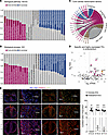 Figure 1
Transcriptome analysis of coronary endothelium identified endocardial enriched transcription factor. (A) Kyoto Encyclopedia of Genes and Genomes (KEGG) analysis of SV-enriched and Endo-enriched ECs from E14.5 hearts revealed different signaling pathways. (B) Gene Ontology (GO) enrichment analysis of endocardium and coronary ECs showing different functional categories. (C) Circular plot illustrating the expression patterns of core cardiac transcription factors across distinct endothelial cell populations. (D) Scatter plot showing the expression level [x axis, log2(FPKM)] and tissue specificity (y axis, log2 ratio) of transcription factors in the endocardium. (E) Immunofluorescence staining of HAND2 and ERG showing specific expression of HAND2 in endocardium from E12.5 to E17.5. Original magnification ×5 (upper), ×6.3 (middle), ×6.8 (lower). Boxed regions are shown at higher magnification to the right. n = 3 hearts per group. Data represent mean ± SEM. Statistical significance was assessed using 2-tailed paired t tests with Benjamini-Krieger-Yekutieli FDR correction. **P < 0.01, ***P < 0.0001. Endoc., endocardium; Myoc., myocardium. Scale bars: 200 μm.
Endothelial Hand2 deletion impairs coronary vessel development. Previous study has shown that endocardial deletion of Hand2 using Nfatc1-Cre results in embryonic lethality by E14.0. These mice displayed tricuspid atresia, multiple ventricular septa, and hypervascularized myocardium (24), which prevented investigating the details of coronary vessel development and remodeling at late developmental stage. To overcome this obstacle and study the role of Hand2 in coronary development, we intercrossed Hand2-floxed mice with the inducible EC-specific Cdh5-CreERT2 mice to generate Hand2 EC-specific knockout alleles (Hand2-EKO). Tamoxifen was administered at E11.5–12.5 when the coronary vascular plexus began to emerge on the surface of the heart, and embryos were collected at E15.5 (Figure 2A). We first verified Cre recombination and Hand2 deletion efficiency using an antibody against HAND2 (Figure 2B) and observed the absence of HAND2+ERG+ double-positive cells in the Endo of Hand2-EKO mice (Figure 2C), a finding that was further verified in Hand2-EKO; iSuRe-Cre mice (iSuRe-Cre, inducible dual reporter-Cre) (28) (Supplemental Figure 1, A and B; supplemental material available online with this article; https://doi.org/10.1172/JCI203310DS1). The Hand2 deletion mice were embryonic lethal at approximately E16.0, exhibiting edema and hemorrhage (Figure 2D). Histological analysis with H&E staining, and immunofluorescence (IF) staining for EMCN, showed marked thinning of the ventricular myocardial wall with excessive trabeculae in the Hand2-EKO hearts. This hypertrabeculation/noncompaction phenotype indicates that endocardial Hand2 is required for myocardial development and meanwhile suggests of participation in the crosstalk between ECs and cardiomyocytes (Figure 2, E–G).
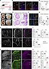 Figure 2
Hand2 is required for normal cardiovascular development. (A) Experimental strategy for Hand2 deletion in cardiac endothelium. (B) Costaining of HAND2 and ERG of E15.5 WT and Hand2-EKO sections showing Hand2 deletion efficiency. The yellow arrows indicate ERG+HAND2+ ECs in the endocardium in controls; the white arrows represent ERG+HAND2– ECs in the endocardium in Hand2-EKO mice, which demonstrate the gene deletion efficiency. (C) Quantification of HAND2+ERG+ cells in compact myocardium ECs (CM ECs) and trabecular myocardium ECs (TM ECs). n = 4 hearts per group. (D) Bright-field images of E15.5 gross embryos and hearts showing defective embryogenesis. (E) H&E staining showing marked thinning of ventricular myocardial wall and hypertrabeculation in Hand2-EKO hearts at E15.5. n = 3 hearts per group. (F and G) Immunostaining of EMCN showing decreased myocardial thickness in E15.5 Hand2-EKO hearts and (G) quantifications. n = 5 hearts per group. (H and I) Whole-mount staining of E15.5 coronary veins showing decreased density, diameter of veins, and ERG+ cell numbers with (I) quantification analysis. n = 4 hearts per group. (J and K) Confocal images showing absence of normal coronary artery formation and extension in E15.5 Hand2-EKO hearts and (K) quantifications. n = 4 hearts per group. Data represent mean ± SEM. Statistical significance was assessed using multiple unpaired 2-tailed t tests with Benjamini-Krieger-Yekutieli FDR correction for (C) and (G) and unpaired 2-tailed Student’s t tests for (I and K). ***P < 0.0001. Scale bars: 200 μm (B, E, F, H and J); 500 μm (D).
To test the function of endothelial HAND2 in coronary vessel development, we performed whole-mount IF staining of the coronary veins and arteries in control and Hand2-EKO mice. In control embryos, the coronary vasculature spread on the dorsal side of the hearts to establish a mature venous network (Figure 2H). However, in Hand2-EKO embryos, coronary venous plexus sprouting and branching were stunted, and vessel density and diameter of large veins were profoundly reduced, together with a significant decrease in the percentage of ERG+ cells in the vessels (Figure 2, H and I). Moreover, IF staining of prearterial markers, DLL4 and UNC5B, demonstrated substantially enhanced numbers of arterial capillary, in parallel with reduction of CX40, JAG1, and SM22, the matured arterial markers. Consistently, the large coronary arteries demonstrated a massive decrease in lumen size and defective remodeling and expansion in Hand2-EKO mice (Figure 2, J and K, and Supplemental Figure 1, C–E).
Taken together, these results indicate a crucial role of the endothelial HAND2 in coronary vessel formation and arterialization.
Endothelial Hand2 deletion disrupts cell proliferation and ventricle compaction. Abnormal coronary angiogenesis causes defective ventricular chamber development (22, 29–31). The above results suggest that endothelial Hand2 deletion leads to the ventricular noncompaction phenotype, we therefore examined myocardial trabeculation and compaction. We first employed a 5-ethynyl-2′-deoxyuridine (EdU) incorporation assay to measure cardiomyocyte proliferation in compact and trabecular layers (Supplemental Figure 1F). Quantification of NKX2.5+ and EdU+ double-positive cells revealed that Hand2-EKO mice had decreased cardiomyocyte proliferation in the compact layer but inversely enhanced proliferation in the trabecular layer (Supplemental Figure 1, G and H). Moreover, IF staining for α-actinin, a microfilament protein that mediates cardiac sarcomeric protein interaction, exhibited highly organized sarcomeric structure in the trabecular layer compared with the compact layer (myocardial wall) in controls (Supplemental Figure 1I). However, this type of well-organized sarcomeric structure was observed in intermediate myocardium of Hand2-EKO hearts (Supplemental Figure 1I). These noncompaction features were further supported by qPCR assay and IF staining that showed downregulation of compact myocardial marker genes (Mycn, Hey2 and Tbx20) but enhancement of trabecular myocardial marker genes (Bmp10, Gja5 and Nppb) (Supplemental Figure 1, J and K). Furthermore, we detected increased cell proliferation of ECs in the trabecular layer but decreased cell proliferation of ECs in the compact layer (Supplemental Figure 1, L and M).
Both the Endo and the coronary vasculature affect myocardial compaction (22, 29). To specifically understand endocardial HAND2 regulation on myocardial development, we delayed induction of Hand2 deletion at E13.5–14.5, when ECs passed the periods of sprouting and early specification, and harvested embryos at E17.5 (Supplemental Figure 2A). Histological analysis revealed thinning of the compact layer and thickening of the trabecular layer in Hand2 mutants, which was further visualized by IF staining for EMCN (Supplemental Figure 2, B and C). In addition, we detected increased cell proliferation of cardiomyocytes and ECs in the trabecular layer (Supplemental Figure 2, D–G). IF staining for BMP10 and CX40, two well-known trabecular markers, revealed abnormally extended expression into compacted myocardium (Supplemental Figure 2H). Strikingly, endothelial Hand2 deletion reduced the expression of several noncompaction-associated genes, including Ptbp1, Casz1, Jarid2, Prdm16, Myh7, and Nrg1, whose deletion/mutation would cause ventricular noncompaction phenotype (21, 32, 33) (Supplemental Figure 2I). Although Hand2-EKO hearts showed changes in ventricular patterning, coronary artery remodeling, and maturation were not affected (Supplemental Figure 2J).
Collectively, these results indicate that cardiac endothelial Hand2 influences ventricular chamber development through both endocardial and coronary vascular regulation on myocardial development.
Endocardial Hand2 deficiency impaired coronary arterial formation. To more precisely explore the function of endocardial HAND2 in coronary vascular and cardiac chamber development, we crossed Bmx-CreERT2 mice, in which Cre is specifically expressed in the Endo and arterial cells, with Hand2-floxed mice to generate Bmx-CreERT2; Hand2 fl/fl (Hand2-EKOBmx) mice. Deletion of Hand2 was induced at E10.5–11.5 by administration of 4-hydroxy-tamoxifen (4-OH), and the embryos were harvested at E15.5 (Figure 3A). Cre expression and Hand2 deletion efficiency were examined by IF imaging of TdTomato+ and staining for ERG+HAND2+ in the Endo (Figure 3, B and C) and were also tested using isolated endocardial ECs for Hand2 expression through qPCR assay (Figure 3D). Histological analysis of the hearts displayed poorly developed hearts, including thinned myocardial wall but thickened trabecular layer, a phenotype of ventricular noncompaction in Hand2-EKOBmx mice (Figure 3, E–H). Furthermore, we detected less coronary artery extension and remodeling with normal coronary veins in Hand2-EKOBmx mice (Figure 3, I–L).
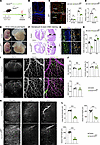 Figure 3
Endocardial deletion of Hand2 caused defective cardiovascular development. (A and B) Experimental strategy of Hand2 ablation in endocardium with Bmx-creERT2 lineage and Cre expression pattern in iSuRe-Cre; Bmx-creERT2 mice. (C) Confocal images showing deletion of Hand2 in endocardium of E15.5 Hand2-EKOBmx hearts with quantification analysis. n = 4 hearts per group. (D) qPCR analysis of Hand2 mRNA level in Hand2-EKOBmx heart ECs. n = 4 samples per group. (E) Bright-field images of E15.5 gross embryos and hearts showing abnormal embryonic and cardiac morphologies. (F) H&E staining showing decreased myocardial wall thickness in Hand2-EKOBmx hearts at E15.5. n = 3 hearts per group. (G and H) Representative images (G) and quantitative analysis (H) of EMCN+ endocardium indicate hypertrabeculation in Hand2-EKOBmx hearts at E15.5. n = 5 hearts per group. (I and J) Whole-mount staining of EMCN and ERG in coronary veins showing normal venous plexuses formation with quantification analysis. n = 4 hearts per group. (K and L) Whole-mount staining of arterial markers showing abnormal coronary artery development and remodeling in Hand2 mutants, with decreased diameter and length of arteries (K), and quantification analysis (L). n = 6 hearts per group. Data represent mean ± SEM. Statistical significance was assessed using multiple unpaired 2-tailed t tests with Benjamini-Krieger-Yekutieli FDR correction for (C and H) and unpaired 2-tailed Student’s t tests for (D, J, and L). **P < 0.01, ***P < 0.001. Scale bars: 200 μm (B, F, G, I and K); 100 μm (C); 500 μm (E).
Next, we generated Hand2-EKOPdgfb mice, which selectively inactivated Hand2 in SV-derived coronary ECs. Deletion of Hand2 was induced at E11.5–12.5 by administration of tamoxifen, and the embryos were collected at E15.5 or E17.5 (Supplemental Figure 3A). As expected, no obvious phenotypes were observed in the Hand2-EKOPdgfb mice (Supplemental Figure 3, B–G).
Overall, these results show that endocardial HAND2 plays a predominant role in cardiac arterialization and chamber development.
Spatiotemporal gene expression changes in Hand2-EKO mice impairs endocardial angiogenesis. To understand the regulatory mechanisms of HAND2 in the Endo, we performed bulk and single-cell transcriptomics analysis of the ECs in Hand2-EKO mice. Since the Cdh5-creERT2 line shows more robust Hand2 recombination efficiency than the Bmx-creERT2 line (Figure 3D and Supplemental Figure 7A), we assumed that Hand2-EKO (namely Hand2-EKOCdh5) is an ideal model for studying the role of Hand2 in Endo. We isolated CD31+TdTomato+ cardiac ECs from E14.5 Cdh5-CreERT2; iSuRe-Cre controls and Hand2-EKO; iSuRe-Cre mutants after tamoxifen induction at E11.5–12.5 (Figure 4A). We detected significant downregulation of both arterial marker genes (Slc45a4, Gja4, Gja5, Cxcl12, Hey1) and venous marker genes (Tbx20, Ptgs1, Wnt2, Aplnr, Ephb4), as well as Endo-enriched marker genes (Adgrg6, Blvrb, Irx5, Tmem108, Npr3, Nrk, Igf2r, Ece1, Plvap) in Hand2-EKO mice (Figure 4, B and C). In contrast, the SV-enriched marker genes (Pdgfb, Fabp4, Kitl, Cd36, Jag2, Pcdh17, Dhah1) were profoundly enhanced in these mice (Figure 4C). IF staining further demonstrated that reduced expression of endocardial-specific marker PLVAP and ectopic expression of SV-enriched protein FABP4 in the Endo of Hand2 mutant mice (Figure 4D). RNA-seq data revealed a distinct gene expression profile, with 1,586 genes downregulated and 1,496 genes upregulated (q < 0.05, |log2FC|> 0.25) (Supplemental Figure 4A). GO terms and KEGG pathway analysis of differentially expressed genes showed that several gene signatures involved in hypoxia and metabolism (p53 and HIF-1 pathway and glycolysis) were upregulated, whereas those signatures implicated in angiogenesis (Endo-MT, MAPK, and TGF-β pathways) were downregulated (Supplemental Figure 4, B and C).
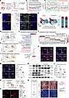 Figure 4
Bulk and single-cell RNA-Seq analysis in Hand2-EKO hearts revealed decreased coronary angiogenesis. (A) Schematic illustration of strategies for tamoxifen injection and EC preparation for bulk and single-cell RNA-seq. (B) qPCR analysis of arterial marker genes and venous marker genes. n = 4 samples per group. (C) qPCR analysis of SV-enriched and Endo-enriched genes. n = 4 samples per group. (D) Confocal images showing decreased expression of endocardial marker PLVAP and ectopic increased SV marker FABP4 in Hand2-EKO endocardium. n = 3 hearts per group. (E) UMAP representation of the ECs of control and Hand2-EKO hearts. Bar graph showing each cluster proportion in the ECs of control and Hand2-EKO hearts. (F) Dot plot showing the top 6 marker genes of subclusters. (G) The expression of Hand2 in the ECs of control and Hand2-EKO hearts. (H and I) Tip cell (H) and Endo-MT (I) module scores in Endo-Cap cell cluster. (J) Enriched GO terms of Endo-Cap differentially expressed genes. (K–M) Immunofluorescence staining demonstrates decreased Endo-MT process in Hand2 mutants. n = 3 hearts per group. (N) Western blotting analysis of Endo-MT associated markers. n = 3 samples per group. (O) Schematic showing Matrigel coronary angiogenesis assays using E12.0 left ventricular explant of Hand2-EKOBmx and control mice. (P) Representative images and quantitative analysis of EC migration. n = 8 hearts per group. (Q) Whole-mount immunostaining of E15.5 hearts for tdTomato, PECAM, and DLL4 showing migration of endocardial cells, labeled by induction at E10.5, from the endocardial layer into the myocardial layer. n = 3 hearts per group. Data represent mean ± SEM. Statistical significance was assessed using an unpaired 2-tailed Student’s t test (B, C, L, M and P). **P < 0.01, ***P < 0.001. Scale bars: 100 μm (D, K, M, P and Q). Endo-Cap, endocardial-derived capillaries; Pre-Vein, prevenous cells.
In addition, we performed single-cell RNA-seq (scRNA-seq) analysis on isolated CD31+TdTomato+ ECs from control and Hand2-EKO mice. These double-positive ECs were subsequently classified to be endocardial cells, capillaries derived from the Endo (Endo-Cap), prearterial cells, arterial cells, and prevenous cells, with well-accepted specific markers (Figure 4, E and F). Module scores were calculated and compared with the reported scRNA-seq datasets that served as quality controls (Supplemental Figure 5, A–C). Moreover, a systematic comparative bioinformatic evaluation validated the lineage trajectory rather than the cell-state transitions that were altered in Hand2-EKO mice (Supplemental Figure 5, D–H). UMAP analysis confirmed decreased Hand2 expression in Endo (Figure 4G). Cell cluster analysis uncovered augmented cell numbers of Endo-Cap clusters in Hand2-EKO mice compared with controls, suggesting of high potency to differentiate into coronary arteries (Figure 4E). A compensatory increase in cell numbers of prearterial cell and prevenous cell clusters from SV-derived ECs in Hand2-EKO mice was also identified (Figure 4E). Interestingly, the Endo-Cap cluster exhibited high expression of tip cell–enriched markers but reduced Endo-MT-associated gene expression (Figure 4, H and I). GO enrichment analysis further revealed that TGF-β and hypoxia pathways were involved in HAND2-mediated cell fate conversion (Figure 4J). Furthermore, scRNA-seq analysis also revealed ectopic expression of SV-enriched genes in Hand2-deficient Endo and reduction of Endo- and arterial-enriched genes (Supplemental Figure 4, G–J), which is consistent with the bulk RNA-seq results. These findings indicate a reciprocal compensation between SV and Endo-derived vessels during coronary development.
In Hand2-EKO hearts, we found that expression of cell adhesion proteins E-cadherin and ZO-1 was enhanced, while that of vimentin, a well-known mesenchymal marker, was downregulated (Figure 4, K–M). Western blotting and qPCR analysis disclosed disrupted Endo-MT in Hand2-EKO hearts and isolated ECs (Figure 4N and Supplemental Figure 4D). For instance, SNAIL-1, a downstream target of HAND2 involved in cardiac cushion mesenchyme formation (34), and SLUG, another well-known Endo-MT marker (35), were reduced in the Endo of Hand2-EKO mice (Figure 4N), suggesting a conserved mechanism for Endo-MT regulation. Moreover, we detected that several genes encoding ECM proteins (36), including Col1a1, Col3a1, Col6a1, Postn, Olfml2a, Tgfbi, and Ecm2, were downregulated in Hand2-EKO cells (Supplemental Figure 4E).
To further determine whether endocardial Hand2 regulates coronary angiogenesis through Endo-MT, we performed a ventricular explant assay. The ventricles from control (Bmx-creERT2 line only) and Hand2-EKOBmx mice coupled with the iSuRe-Cre reporter allele at E12.0 were dissected for in vitro ventricular explant culture (Figure 4O). Compared with controls, Hand2-EKOBmx explants showed a significant decrease in vessel sprouting and branching (Figure 4P). We next performed an in vivo lineage-tracing experiment to assess Hand2-mediated migration of endocardial ECs into myocardium. 1.5 mg 4-OH tamoxifen was given to Bmx-CreERT2; iSuRe-Cre reporter mice and Hand2-EKOBmx; iSuRe-Cre reporter mice for monitoring cell migration. Normally, endocardial ECs could ingress and integrate into the coronary artery of the myocardium, but this migration failed in Hand2 mutants, indicating that HAND2 positively controls EC migration in the Endo (Figure 4Q).
Taken together, these results demonstrate that endocardial HAND2 is required for Endo-MT and regulation of coronary angiogenesis in the developing heart.
Induction of hypoxia and tip cell signatures in Hand2-EKO mice. Gene set enrichment analysis of our RNA-seq data and the data from E9.0–E9.25 Hand2-knockout atrioventricular canals revealed an overlapping coverage of hypoxia- and glycolysis-associated genes in Hand2 mutants (34) (Supplemental Figure 6, A and B). Subsequently, we examined the expression of key TFs HIF1α and HIF2α in Hand2-EKO mice. IF staining showed that the expression of both HIF1α and HIF2α, as well as their downstream targets, was increased upon loss of Hand2 in Endo (Figure 5, A and B), suggesting that HAND2 acts upstream to suppress HIF pathway. Accordingly, glycolysis genes were upregulated at both mRNA and protein levels in Hand2-EKO mice (Figure 5, C and D). Upon exposure to hypoxia, the expression of p53 and its downstream target genes in regulating cell cycle were enhanced in Hand2-EKO mice (Supplemental Figure 4F). Strikingly, functional augmentation of the hypoxia pathway through endocardial deletion of Vhl, a negative modulator of HIF pathway, recapitulates cardiac and coronary vascular defects observed in Hand2-EKO mice (Supplemental Figure 6, C–E).
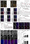 Figure 5
Hand2 deficiency in endothelium elevated hypoxia signaling and enhanced tip cell activity. (A) Representative images and quantitative analysis of hypoxia response factors HIF1α and HIF2α in E15.5 Hand2-EKO hearts. n = 3 hearts per group. (B) qPCR analysis of hypoxia-associated genes from ECs of E15.5 Hand2-EKO hearts. n = 3 samples per group. (C) Confocal images showing upregulated downstream targets, including GLUT1, ENO1, HK2, LDHA, PDK1, and TPI in Hand2 mutants. n = 3 hearts per group. (D) qPCR analysis of glycolysis-associated genes from ECs of E15.5 Hand2-EKO hearts. n = 3 samples per group. (E) Schematic illustration of strategies of EMCN+ endocardial cells sorting for qPCR analysis. (F) qPCR analysis of tip cell–enriched genes from EMCN+ ECs of E14.5 hearts. n = 3 samples per group. (G) Immunostaining of tip cell markers, such as ANGPT2, vWF, and CAV1, showing increased expression in endocardium. n = 3 hearts per group. (H) Whole-mount staining showing the partial rescue in coronary arteries after suppressing Hif pathway in Hand2-EKO hearts with quantification analysis. n = 6 hearts per group. The width of myocardial region and quantification analysis of JAG1+ arterial vessels of each group. n = 3 hearts per group. Data represent mean ± SEM. Statistical significance was assessed using an unpaired 2-tailed Student’s t test (A, B, D and F) or 1-way ANOVA (H) followed by Tukey’s multiple-comparison test. *P < 0.05, **P < 0.01, ***P < 0.001. Scale bars: 100 μm (A, C, G and H).
Previous studies have shown that endocardial hypoxia promotes coronary angiogenesis, followed by upregulation of hypoxia-related genes in ESM1-expressing intramyocardial tip cells during heart development (13, 37). To ascertain whether HAND2 is involved in this process, we checked the expression of tip cell markers and found a significant upregulation of tip cell–enriched genes, including Esm1, Angpt2, Apln, Kcne3, Mcan, Cav1, Dll4 and Flt4, in total cardiac ECs and isolated EMCN+ endocardial cells in Hand2-EKO hearts (Figure 5, E and F, and Supplemental Figure 6F). Interestingly, IF staining for tip cell–enriched proteins such as ANGPT2, vWF, and CAV1, revealed prominent accumulation in tip cell–like endocardial cells showing restricted migration in the Endo in Hand2-EKO mice (Figure 5G). Meanwhile, we observed decreased coverage of mural cells such as pericytes (PDGFRB+, NG2+, and OLFML3+ cells) and smooth muscle cells (aSMA+ cells) in Hand2-EKO mice (Supplemental Figure 6G). These results suggest that augmentation of HIF pathway could be responsible for compromised coronary development in Hand2-EKO mice. Thus, we examined the coronary arterialization and cardiac morphological changes in control, Hand2-EKO, Hand2-EKO; HIf1αHet, Hand2-EKO; HIf2αHet, and Hand2-EKO; HIf1α/2αHet mice. Notably, suppression of HIF pathway was sufficient to reverse the defects in artery formation and myocardial development caused by endocardial Hand2 deletion (Figure 5H, and Supplemental Figure 6H). Furthermore, qPCR and IF staining revealed that the expression of tip cell markers and hypoxic/glycolytic pathway was significantly attenuated in ECs from Hand2/HIf1α/2α triple mutants, in parallel with increased Endo-MT and ECM remodeling (Supplemental Figure 7, A–G). We also confirmed this genetic (and phenotypic) rescue by abolishing the HIF pathway in Hand2-EKONfatc1 mice. We found that cardiac structures were recovered and embryonic survival was enhanced in Hand2-EKONfatc1; HIf1αHet and Hand2-EKONfatc1; HIf2αHet embryos compared with Hand2-EKONfatc1 embryos (Supplemental Figure 6, I–K).
Altogether, these findings strongly suggest that endocardial Hand2 regulates coronary vascular EC dynamics, migration, and artery formation, acting upstream of hypoxic and glycolytic pathways.
HAND2 harnesses TGF-β and MAPK-p38 signaling pathway to modulate EC migration and arterialization. We next performed the CUT&Tag assay to identify the putative binding targets of the HAND2 protein. For this purpose, CD31+Tdtomato+ ECs were collected from E14.5 controls and Hand2-EKO embryonic hearts (Figure 6A). A heatmap indicated a sharp reduction of the signals in Hand2 deletion mice compared with controls (Figure 6B). Analysis of the genomic location distribution revealed profound distribution of HAND2 in the promoter regions, which accounts for approximately 74% of the peaks (Figure 6C).
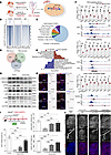 Figure 6
Hand2 coordinated TGF-β MAPK/p38 pathways control EC migration and artery formation. (A) Schematic illustration of strategies for EC collection and CUT&Tag assay. (B) Heatmap showing decreased binding of HAND2 target genes in Hand2-EKO mice. (C) Annotation of HAND2 binding peaks in endothelial cells. (D) Intersection plot showing the overlap of RNA-seq and CUT&Tag data analysis. (E) GO-KEGG analysis of gene signatures from overlapped genes. (F) qPCR analysis and genome browser tracks of CUT&Tag signals at the Tgfbr3, Fbn1, Taok3, Gab1, Rps6ka2, and Ddit3 loci. n = 3 samples per group. (G) Western blotting analysis of TGF-β target proteins, including SMAD2/3, p-SMAD2/3, ERK, p-ERK, p38, p-p38, JNK, and p-JNK, in control and Hand2 mutant hearts. n = 3 samples per group. (H) Immunostaining of TGF-β2, TAB1, TGF-BR3, and p-SAMD2/3 showing decreased TGF-β signaling in Hand2-EKO hearts. n = 3 hearts per group. (I and J) Schematic illustration of strategies for tamoxifen and TGF-β2 recombinant protein injection. Confocal images showing restored coronary artery formation by treatment with TGF-β2 recombinant protein in E15.5 Hand2-EKO hearts and quantification analysis. n = 4 hearts per group. Data represent mean ± SEM. Statistical significance was assessed using an unpaired 2-tailed Student’s t test (F) or 1-way ANOVA (I) followed by Tukey’s multiple-comparison test. *P < 0.05, **P < 0.01, ***P < 0.001. Scale bars: 100 μm (H and J).
Integration of RNA-seq and CUT&Tag datasets identified 304 directly upregulated genes and 351 directly downregulated genes in Hand2-EKO mice (Figure 6D). KEGG combined with GO term analysis revealed that gene signatures of p53, HIF-1 pathway, and glycolysis were upregulated, whereas those of MAPK and TGF/SMAD pathway were downregulated. These downregulated genes were highly relative to heart morphogenesis and mesenchymal cell differentiation and are crucial for coronary vessel development (Figure 6E). CUT&Tag and qPCR analysis confirmed these changes (Figure 6F). Next, we examined the phosphorylation level of SMAD2/3, ERK, p38, and JNK, the downstream effectors of TGF-β pathway by Western blotting analysis, and the results demonstrated decreased levels in Hand2-EKO hearts. IF staining for TGF-β pathway components (TGF-β2 and TGF-BR3, TAB1 and p-SMAD2/3) showed reduced levels in the Endo of Hand2-EKO mice (Figure 6, G and H). Notably, treatment of Hand2-EKO embryos with recombinant TGF-β2 protein could restore the defects of myocardium and coronary vessels in a dose-dependent manner (Figure 6, I and J). ECs from Hand2 mutants treated with TGF-β2 regained expression of arterial-venous and EMT/ECM-associated gene programs, concomitant with a reduction in tip cell–enriched gene expression (Supplemental Figure 7, J–O). Additionally, tip cells from the Endo could migrate into the myocardium as a result of increased Endo-MT (Supplemental Figure 7, P–Q). Since TGF-β–induced activation of p38MAP kinase is critical for EMT (38), we also found recovery of TGF-β and p38MAPK activity when suppressing the HIF pathway, indicating that HIF was also involved in TGF-β and p38MAPK-associated Endo-MT, mediated by HAND2 (Supplemental Figure 7, H and I).
We also uncovered, in the isolated Hand2-deficient endocardial ECs, substantially reduced expression levels of multiple key cardiac TFs, including Gata4, Gata5, Gata6, Nfatc1, Foxc1, and Foxc2, which were confirmed by qPCR and IF staining analysis (Supplemental Figure 8, A–C). Strikingly, our CUT&Tag analysis revealed a direct binding of HAND2 on the promoters of these genes to downregulate their transcription (Supplemental Figure 8D). Interestingly, CUT&Tag analysis also showed that HAND2 modulates HIFs and glycolytic-associated gene transcription (Supplemental Figure 8E), which is consistent with ChIP-seq data in Hand2-deficient atrioventricular canals (34) (Supplemental Figure 6B), and Hand2 deficiency resulted in upregulation of these genes (Figure 5B). To confirm the upstream feature of Hand2, we found that endothelial/endocardial Gata4 deletion exhibited similar defective coronary development to that obtained in Hand2-EKO hearts (Supplemental Figure 9, A–K).
To further investigate whether HAND2 participates in EC migration and arterial specification in vitro, we induced HAND2 expression in HUVECs (HAND2-OE) (Supplemental Figure 10, A–C). Ectopic expression of HAND2 increased cell migration and showed a high tube-forming ability (Supplemental Figure 10, D and E). Notably, HAND2-OE HUVECs showed upregulated tip cell (ESM1, ADM, ANGPT2, PDGFB, and FLT4) and arterial (CD44, CXCR4, JAG1, SOX17, EPHB2, VEGFA, NRP1, and SEMA3G) marker genes, indicating that high HAND2 expression promotes cell migration and arterial EC fate in vitro and could uncover an insight into artery formation during injury (Supplemental Figure 10F).
Collectively, these results demonstrate that HAND2 acts as an upstream regulator via transcriptional activation of signaling pathways, pinpointing the crucial role of HAND2 to establish the endocardial GRN for cardiac morphogenesis and coronary arterialization.
-
Discussion
In the developing heart, coronary arterial ECs differentiate and migrate from Endo in response to hypoxia and other factors secreted from cardiomyocytes. However, the underlying transcriptional regulatory mechanisms of Endo-derived arterialization remain to be elucidated. We found large differences in gene expression profiling between E14.5 SV-derived and Endo-derived ECs, which exhibited distinct gene signatures and functions that precisely regulate coronary angiogenesis and cardiac morphology separately.
We identified the cardiac TF HAND2 and its partner GATA4 (34, 39, 40), specifically expressed in the Endo; they are pivotal regulators of gene transcriptional regulatory network for coronary artery formation and myocardial development. We demonstrate that EC-specific deletion of Hand2 causes severe defects, including abnormal coronary artery formation and a ventricular noncompaction phenotype, leading to embryonic lethality. Transcriptomic analysis revealed that the spatiotemporal gene expression profiles were disrupted in Endo-derived and SV-derived ECs of Hand2-deleted hearts, which were also accompanied by decreases in arterial and venous gene expression. We found that Hand2 loss upregulates hypoxic and metabolic genes while downregulating TGF-β/p38MAPK–dependent Endo-MT and ECM pathways. Mechanistically, HAND2 directly binds to gene promoters to suppress the hypoxic pathway while activating TGF-β/p38MAPK–dependent Endo-MT and ECM remodeling. This dual regulation facilitates the migration of Endo-derived ECs and their differentiation into arterial cells. Consequently, Hand2 deletion disrupts this balance, upregulating metabolic genes but inhibiting essential migration signals, leading to defective coronary arteries and a noncompaction cardiomyopathy-like phenotype (Figure 7). Finally, we showed that modulating the hypoxia pathway or administering TGF-β2 can partially rescue these defects. Our study establishes endocardial Hand2 as an essential upstream regulator of coronary development and suggests its therapeutic potential for promoting neovasculogenesis.
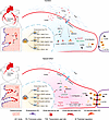 Figure 7
Proposed transcriptional regulatory mechanisms of Hand2 for coronary artery development. Endocardium is a major contributor to coronary arteries during early heart development, which is spatiotemporally regulated by multiple signaling pathways that sequentially control endocardial angiogenesis (Endo-MT) and endocardial-to-coronary vessel fate conversion. Hand2 functions as an essential upstream transcription factor that coordinates the endocardial gene regulatory network (GRN) in the developing heart. HAND2 binds to gene promoters to modulate signaling for normal cardiovascular development (top). Endocardial loss of Hand2 disrupts this complex transcriptional orchestration, impairs TGF-β–SMAD2/3 and p38 MAPK–mediated Endo-MT, upregulates hypoxia-induced glycolysis and tip cell-enriched gene expression, and eventually results in defective coronary artery formation (bottom). AO, aorta; PA, pulmonary artery; TSS, transcription start site.
The role of Hand2 in early heart development has been well-characterized in numerous studies (23). Both Hand2 systemic knockout and Hand2 cell type–specific deletion resulted in severe cardiac morphological defects, including hypoplasia of the RV, dilated aortic sac, ventricular septal defects, disorganized endocardial cushions, valvular defects, and reduced trabeculation due to abnormal cell proliferation, differentiation, and migration, indicating that HAND2 plays a vital role in regulating cardiomyocytes, cardiac neural crest cells, ECs, and epicardial precursor cells during cardiac morphogenesis (24, 25, 34, 41–47). In turn, enforced HAND2 expression in cardiomyocytes resulted in the absence of interventricular septal in the developing heart and caused cardiac hypertrophy in the adult heart (42, 48). Hand2 overexpression also enhanced cardiomyocyte production by increasing SHF-derived progenitor cell proliferation, which resulted in an enlarged heart with an increased size of outflow tract in the zebrafish model (49). Endocardial cell–specific enhancement of HAND2 within Nfatc1-Cre did not show any obvious cardiac phenotypes but improved cardiac ventricular trabeculation at early stage (24). Recently, heterozygous sequence variants in HAND2 have been reported in some patients with congenital heart defects, such as tetralogy of Fallot, patent ductus arteriosus, pulmonary atresia, pulmonary stenosis, atrial or ventricular septal defects, double outlet right ventricle, and dilated cardiomyopathy (50–53).
We observed that the expression of HAND2 in cardiac endothelium is selectively located in endocardial ECs using a HAND2-specific antibody generated recently (46), suggesting a potential role of Hand2 in the Endo in the regulation of coronary vessel formation. Previous studies have shown that valve phenotypes in Hand2-null and Hand2; Nfatc1-Cre–deleted mice were not observed in our Hand2 endocardial deletion mice, indicating that HAND2 may regulate valve formation before coronary angiogenesis initiation (24, 34). Meanwhile, apart from the well-characterized functions of HAND2 in trabeculae and valve formation, our study identified a different role of Hand2 in regulating coronary artery formation, which parallelly mediated by hypoxic/metabolic changes and TGF-β–induced EMT. This finding indicates that endocardial HAND2 timely controls key cardiac developmental events to maintain cardiac structure and function.
Previous studies have reported that Endo and coronary endothelium can compensate for each other during development. Deletion of the SV contributes to ECs by ablating Aplnr to block ELA-APJ signaling, which can increase the expansion of Endo-derived coronary endothelium to compensate for the defect in SV-derived vessel formation (54). Recently, Zhang and colleagues illustrated that elimination of endocardial cells leads to expansion of coronary vessels from the SV to compensate for the loss of coronary vessels derived from the Endo (55). Although this phenomenon of reciprocal compensation has been observed at the cellular level, the distinction between these two populations at the molecular level remains unclear. In our study, we found that Hand2 ablation downregulated the expression of numerous Endo-specific genes and TFs and upregulated the expression of coronary endothelium-enriched genes in the region of Endo, indicating that genes from these two sources inhibit each other at the molecular level to adopt cell fate commitment beyond cell heterogeneity during coronary development.
Our results also demonstrated that HAND2 acts upstream of hypoxic and glycolytic pathways and negatively regulates them. HAND2 directly binds the promoters of key regulators in these pathways to control endothelial metabolism and behavior. In Hand2-EKO mice, hypoxic pathway upregulation increased endocardial sprouting tip cells, but these cells remained confined to the Endo and failed to invade the intramyocardial wall to form arterial ECs. These might be attributable in part to Endo-MT and ECM defects caused by HAND2-mediated TGF-β and p38MAPK alteration.
In summary, our results illustrated the molecular regulatory mechanism of Endo for coronary angiogenesis and arterialization and identified an essential endocardial TF, Hand2, which controls the dynamics of endocardial ECs and coronary artery formation by coordinating the GRN, and may be a potential therapeutic target to promote neovasculogenesis in patients with myocardial infarction through reprogramming of the regulatory network for the treatment of coronary artery disease.


Copyright © 2026 American Society for Clinical Investigation
ISSN: 0021-9738 (print), 1558-8238 (online)