
Advertisement
Research ArticleNeuroscienceOtology
Open Access |  10.1172/JCI202988
Kv12.1 (Kcnh8) maintains hair cell potassium homeostasis and protects against noise-induced hearing loss in mice
Yaoqian Liu,1,2 Yanyan Jia,1,2,3,4,5 Jiaoyao Ma,1,2 Xian Gao,1,2 Yunjie Li,1,2 Yu Dong,1,2 Mingxuan Wu,1,2 Gaogan Jia,1,2 Yikang Huang,1,2 Yiyun Lou,1,2 Mengrou Xu,1,2 Tianshi Sun,6 Yuqiang Ding,1,7 Huawei Li,1,2,3,4,5,8 Mingyu Xia,1,2,3 and Wenyan Li1,2,3,4,5,8
1ENT institute and Otorhinolaryngology Department of Eye & ENT Hospital, State Key Laboratory of Medical Neurobiology and MOE Frontiers Center for Brain Science;
2Institutes of Biomedical Sciences;
3NHC Key Laboratory of Hearing Medicine; and
4The Institutes of Brain Science and the Collaborative Innovation Center for Brain Science, Fudan University, Shanghai, China.
5Shanghai Key Laboratory of Gene Editing and Cell Therapy for Rare Diseases, Shanghai, China.
6School of Life Sciences, Zhejiang Chinese Medical University, Hangzhou, China.
7Laboratory Animal Center, Fudan University, Shanghai, China.
8Shanghai Engineering Research Centre of Cochlear Implant, Shanghai, China.
Address for correspondence. Wenyan Li, Mingyu Xia, or Huawei Li, Eye & ENT Hospital of Fudan University, 83 Fenyang Road, Xuhui District, Shanghai, 200031, China. Telephone: 86.21.64377134; Email: wenyan_li@fudan.edu.cn. (WL); Xiamy17@fudan.edu.cn (MX); hwli@shmu.edu.cn (HL).
Authorship note: Y Liu and YJ contributed equally to this work.
Find articles by Liu, Y. in: PubMed | Google Scholar
1ENT institute and Otorhinolaryngology Department of Eye & ENT Hospital, State Key Laboratory of Medical Neurobiology and MOE Frontiers Center for Brain Science;
2Institutes of Biomedical Sciences;
3NHC Key Laboratory of Hearing Medicine; and
4The Institutes of Brain Science and the Collaborative Innovation Center for Brain Science, Fudan University, Shanghai, China.
5Shanghai Key Laboratory of Gene Editing and Cell Therapy for Rare Diseases, Shanghai, China.
6School of Life Sciences, Zhejiang Chinese Medical University, Hangzhou, China.
7Laboratory Animal Center, Fudan University, Shanghai, China.
8Shanghai Engineering Research Centre of Cochlear Implant, Shanghai, China.
Address for correspondence. Wenyan Li, Mingyu Xia, or Huawei Li, Eye & ENT Hospital of Fudan University, 83 Fenyang Road, Xuhui District, Shanghai, 200031, China. Telephone: 86.21.64377134; Email: wenyan_li@fudan.edu.cn. (WL); Xiamy17@fudan.edu.cn (MX); hwli@shmu.edu.cn (HL).
Authorship note: Y Liu and YJ contributed equally to this work.
Find articles by Jia, Y. in: PubMed | Google Scholar
1ENT institute and Otorhinolaryngology Department of Eye & ENT Hospital, State Key Laboratory of Medical Neurobiology and MOE Frontiers Center for Brain Science;
2Institutes of Biomedical Sciences;
3NHC Key Laboratory of Hearing Medicine; and
4The Institutes of Brain Science and the Collaborative Innovation Center for Brain Science, Fudan University, Shanghai, China.
5Shanghai Key Laboratory of Gene Editing and Cell Therapy for Rare Diseases, Shanghai, China.
6School of Life Sciences, Zhejiang Chinese Medical University, Hangzhou, China.
7Laboratory Animal Center, Fudan University, Shanghai, China.
8Shanghai Engineering Research Centre of Cochlear Implant, Shanghai, China.
Address for correspondence. Wenyan Li, Mingyu Xia, or Huawei Li, Eye & ENT Hospital of Fudan University, 83 Fenyang Road, Xuhui District, Shanghai, 200031, China. Telephone: 86.21.64377134; Email: wenyan_li@fudan.edu.cn. (WL); Xiamy17@fudan.edu.cn (MX); hwli@shmu.edu.cn (HL).
Authorship note: Y Liu and YJ contributed equally to this work.
Find articles by
Ma, J.
in:
PubMed
|
Google Scholar
|

1ENT institute and Otorhinolaryngology Department of Eye & ENT Hospital, State Key Laboratory of Medical Neurobiology and MOE Frontiers Center for Brain Science;
2Institutes of Biomedical Sciences;
3NHC Key Laboratory of Hearing Medicine; and
4The Institutes of Brain Science and the Collaborative Innovation Center for Brain Science, Fudan University, Shanghai, China.
5Shanghai Key Laboratory of Gene Editing and Cell Therapy for Rare Diseases, Shanghai, China.
6School of Life Sciences, Zhejiang Chinese Medical University, Hangzhou, China.
7Laboratory Animal Center, Fudan University, Shanghai, China.
8Shanghai Engineering Research Centre of Cochlear Implant, Shanghai, China.
Address for correspondence. Wenyan Li, Mingyu Xia, or Huawei Li, Eye & ENT Hospital of Fudan University, 83 Fenyang Road, Xuhui District, Shanghai, 200031, China. Telephone: 86.21.64377134; Email: wenyan_li@fudan.edu.cn. (WL); Xiamy17@fudan.edu.cn (MX); hwli@shmu.edu.cn (HL).
Authorship note: Y Liu and YJ contributed equally to this work.
Find articles by Gao, X. in: PubMed | Google Scholar
1ENT institute and Otorhinolaryngology Department of Eye & ENT Hospital, State Key Laboratory of Medical Neurobiology and MOE Frontiers Center for Brain Science;
2Institutes of Biomedical Sciences;
3NHC Key Laboratory of Hearing Medicine; and
4The Institutes of Brain Science and the Collaborative Innovation Center for Brain Science, Fudan University, Shanghai, China.
5Shanghai Key Laboratory of Gene Editing and Cell Therapy for Rare Diseases, Shanghai, China.
6School of Life Sciences, Zhejiang Chinese Medical University, Hangzhou, China.
7Laboratory Animal Center, Fudan University, Shanghai, China.
8Shanghai Engineering Research Centre of Cochlear Implant, Shanghai, China.
Address for correspondence. Wenyan Li, Mingyu Xia, or Huawei Li, Eye & ENT Hospital of Fudan University, 83 Fenyang Road, Xuhui District, Shanghai, 200031, China. Telephone: 86.21.64377134; Email: wenyan_li@fudan.edu.cn. (WL); Xiamy17@fudan.edu.cn (MX); hwli@shmu.edu.cn (HL).
Authorship note: Y Liu and YJ contributed equally to this work.
Find articles by Li, Y. in: PubMed | Google Scholar
1ENT institute and Otorhinolaryngology Department of Eye & ENT Hospital, State Key Laboratory of Medical Neurobiology and MOE Frontiers Center for Brain Science;
2Institutes of Biomedical Sciences;
3NHC Key Laboratory of Hearing Medicine; and
4The Institutes of Brain Science and the Collaborative Innovation Center for Brain Science, Fudan University, Shanghai, China.
5Shanghai Key Laboratory of Gene Editing and Cell Therapy for Rare Diseases, Shanghai, China.
6School of Life Sciences, Zhejiang Chinese Medical University, Hangzhou, China.
7Laboratory Animal Center, Fudan University, Shanghai, China.
8Shanghai Engineering Research Centre of Cochlear Implant, Shanghai, China.
Address for correspondence. Wenyan Li, Mingyu Xia, or Huawei Li, Eye & ENT Hospital of Fudan University, 83 Fenyang Road, Xuhui District, Shanghai, 200031, China. Telephone: 86.21.64377134; Email: wenyan_li@fudan.edu.cn. (WL); Xiamy17@fudan.edu.cn (MX); hwli@shmu.edu.cn (HL).
Authorship note: Y Liu and YJ contributed equally to this work.
Find articles by Dong, Y. in: PubMed | Google Scholar
1ENT institute and Otorhinolaryngology Department of Eye & ENT Hospital, State Key Laboratory of Medical Neurobiology and MOE Frontiers Center for Brain Science;
2Institutes of Biomedical Sciences;
3NHC Key Laboratory of Hearing Medicine; and
4The Institutes of Brain Science and the Collaborative Innovation Center for Brain Science, Fudan University, Shanghai, China.
5Shanghai Key Laboratory of Gene Editing and Cell Therapy for Rare Diseases, Shanghai, China.
6School of Life Sciences, Zhejiang Chinese Medical University, Hangzhou, China.
7Laboratory Animal Center, Fudan University, Shanghai, China.
8Shanghai Engineering Research Centre of Cochlear Implant, Shanghai, China.
Address for correspondence. Wenyan Li, Mingyu Xia, or Huawei Li, Eye & ENT Hospital of Fudan University, 83 Fenyang Road, Xuhui District, Shanghai, 200031, China. Telephone: 86.21.64377134; Email: wenyan_li@fudan.edu.cn. (WL); Xiamy17@fudan.edu.cn (MX); hwli@shmu.edu.cn (HL).
Authorship note: Y Liu and YJ contributed equally to this work.
Find articles by Wu, M. in: PubMed | Google Scholar
1ENT institute and Otorhinolaryngology Department of Eye & ENT Hospital, State Key Laboratory of Medical Neurobiology and MOE Frontiers Center for Brain Science;
2Institutes of Biomedical Sciences;
3NHC Key Laboratory of Hearing Medicine; and
4The Institutes of Brain Science and the Collaborative Innovation Center for Brain Science, Fudan University, Shanghai, China.
5Shanghai Key Laboratory of Gene Editing and Cell Therapy for Rare Diseases, Shanghai, China.
6School of Life Sciences, Zhejiang Chinese Medical University, Hangzhou, China.
7Laboratory Animal Center, Fudan University, Shanghai, China.
8Shanghai Engineering Research Centre of Cochlear Implant, Shanghai, China.
Address for correspondence. Wenyan Li, Mingyu Xia, or Huawei Li, Eye & ENT Hospital of Fudan University, 83 Fenyang Road, Xuhui District, Shanghai, 200031, China. Telephone: 86.21.64377134; Email: wenyan_li@fudan.edu.cn. (WL); Xiamy17@fudan.edu.cn (MX); hwli@shmu.edu.cn (HL).
Authorship note: Y Liu and YJ contributed equally to this work.
Find articles by Jia, G. in: PubMed | Google Scholar
1ENT institute and Otorhinolaryngology Department of Eye & ENT Hospital, State Key Laboratory of Medical Neurobiology and MOE Frontiers Center for Brain Science;
2Institutes of Biomedical Sciences;
3NHC Key Laboratory of Hearing Medicine; and
4The Institutes of Brain Science and the Collaborative Innovation Center for Brain Science, Fudan University, Shanghai, China.
5Shanghai Key Laboratory of Gene Editing and Cell Therapy for Rare Diseases, Shanghai, China.
6School of Life Sciences, Zhejiang Chinese Medical University, Hangzhou, China.
7Laboratory Animal Center, Fudan University, Shanghai, China.
8Shanghai Engineering Research Centre of Cochlear Implant, Shanghai, China.
Address for correspondence. Wenyan Li, Mingyu Xia, or Huawei Li, Eye & ENT Hospital of Fudan University, 83 Fenyang Road, Xuhui District, Shanghai, 200031, China. Telephone: 86.21.64377134; Email: wenyan_li@fudan.edu.cn. (WL); Xiamy17@fudan.edu.cn (MX); hwli@shmu.edu.cn (HL).
Authorship note: Y Liu and YJ contributed equally to this work.
Find articles by Huang, Y. in: PubMed | Google Scholar
1ENT institute and Otorhinolaryngology Department of Eye & ENT Hospital, State Key Laboratory of Medical Neurobiology and MOE Frontiers Center for Brain Science;
2Institutes of Biomedical Sciences;
3NHC Key Laboratory of Hearing Medicine; and
4The Institutes of Brain Science and the Collaborative Innovation Center for Brain Science, Fudan University, Shanghai, China.
5Shanghai Key Laboratory of Gene Editing and Cell Therapy for Rare Diseases, Shanghai, China.
6School of Life Sciences, Zhejiang Chinese Medical University, Hangzhou, China.
7Laboratory Animal Center, Fudan University, Shanghai, China.
8Shanghai Engineering Research Centre of Cochlear Implant, Shanghai, China.
Address for correspondence. Wenyan Li, Mingyu Xia, or Huawei Li, Eye & ENT Hospital of Fudan University, 83 Fenyang Road, Xuhui District, Shanghai, 200031, China. Telephone: 86.21.64377134; Email: wenyan_li@fudan.edu.cn. (WL); Xiamy17@fudan.edu.cn (MX); hwli@shmu.edu.cn (HL).
Authorship note: Y Liu and YJ contributed equally to this work.
Find articles by Lou, Y. in: PubMed | Google Scholar
1ENT institute and Otorhinolaryngology Department of Eye & ENT Hospital, State Key Laboratory of Medical Neurobiology and MOE Frontiers Center for Brain Science;
2Institutes of Biomedical Sciences;
3NHC Key Laboratory of Hearing Medicine; and
4The Institutes of Brain Science and the Collaborative Innovation Center for Brain Science, Fudan University, Shanghai, China.
5Shanghai Key Laboratory of Gene Editing and Cell Therapy for Rare Diseases, Shanghai, China.
6School of Life Sciences, Zhejiang Chinese Medical University, Hangzhou, China.
7Laboratory Animal Center, Fudan University, Shanghai, China.
8Shanghai Engineering Research Centre of Cochlear Implant, Shanghai, China.
Address for correspondence. Wenyan Li, Mingyu Xia, or Huawei Li, Eye & ENT Hospital of Fudan University, 83 Fenyang Road, Xuhui District, Shanghai, 200031, China. Telephone: 86.21.64377134; Email: wenyan_li@fudan.edu.cn. (WL); Xiamy17@fudan.edu.cn (MX); hwli@shmu.edu.cn (HL).
Authorship note: Y Liu and YJ contributed equally to this work.
Find articles by Xu, M. in: PubMed | Google Scholar
1ENT institute and Otorhinolaryngology Department of Eye & ENT Hospital, State Key Laboratory of Medical Neurobiology and MOE Frontiers Center for Brain Science;
2Institutes of Biomedical Sciences;
3NHC Key Laboratory of Hearing Medicine; and
4The Institutes of Brain Science and the Collaborative Innovation Center for Brain Science, Fudan University, Shanghai, China.
5Shanghai Key Laboratory of Gene Editing and Cell Therapy for Rare Diseases, Shanghai, China.
6School of Life Sciences, Zhejiang Chinese Medical University, Hangzhou, China.
7Laboratory Animal Center, Fudan University, Shanghai, China.
8Shanghai Engineering Research Centre of Cochlear Implant, Shanghai, China.
Address for correspondence. Wenyan Li, Mingyu Xia, or Huawei Li, Eye & ENT Hospital of Fudan University, 83 Fenyang Road, Xuhui District, Shanghai, 200031, China. Telephone: 86.21.64377134; Email: wenyan_li@fudan.edu.cn. (WL); Xiamy17@fudan.edu.cn (MX); hwli@shmu.edu.cn (HL).
Authorship note: Y Liu and YJ contributed equally to this work.
Find articles by Sun, T. in: PubMed | Google Scholar
1ENT institute and Otorhinolaryngology Department of Eye & ENT Hospital, State Key Laboratory of Medical Neurobiology and MOE Frontiers Center for Brain Science;
2Institutes of Biomedical Sciences;
3NHC Key Laboratory of Hearing Medicine; and
4The Institutes of Brain Science and the Collaborative Innovation Center for Brain Science, Fudan University, Shanghai, China.
5Shanghai Key Laboratory of Gene Editing and Cell Therapy for Rare Diseases, Shanghai, China.
6School of Life Sciences, Zhejiang Chinese Medical University, Hangzhou, China.
7Laboratory Animal Center, Fudan University, Shanghai, China.
8Shanghai Engineering Research Centre of Cochlear Implant, Shanghai, China.
Address for correspondence. Wenyan Li, Mingyu Xia, or Huawei Li, Eye & ENT Hospital of Fudan University, 83 Fenyang Road, Xuhui District, Shanghai, 200031, China. Telephone: 86.21.64377134; Email: wenyan_li@fudan.edu.cn. (WL); Xiamy17@fudan.edu.cn (MX); hwli@shmu.edu.cn (HL).
Authorship note: Y Liu and YJ contributed equally to this work.
Find articles by
Ding, Y.
in:
PubMed
|
Google Scholar
|
1ENT institute and Otorhinolaryngology Department of Eye & ENT Hospital, State Key Laboratory of Medical Neurobiology and MOE Frontiers Center for Brain Science;
2Institutes of Biomedical Sciences;
3NHC Key Laboratory of Hearing Medicine; and
4The Institutes of Brain Science and the Collaborative Innovation Center for Brain Science, Fudan University, Shanghai, China.
5Shanghai Key Laboratory of Gene Editing and Cell Therapy for Rare Diseases, Shanghai, China.
6School of Life Sciences, Zhejiang Chinese Medical University, Hangzhou, China.
7Laboratory Animal Center, Fudan University, Shanghai, China.
8Shanghai Engineering Research Centre of Cochlear Implant, Shanghai, China.
Address for correspondence. Wenyan Li, Mingyu Xia, or Huawei Li, Eye & ENT Hospital of Fudan University, 83 Fenyang Road, Xuhui District, Shanghai, 200031, China. Telephone: 86.21.64377134; Email: wenyan_li@fudan.edu.cn. (WL); Xiamy17@fudan.edu.cn (MX); hwli@shmu.edu.cn (HL).
Authorship note: Y Liu and YJ contributed equally to this work.
Find articles by Li, H. in: PubMed | Google Scholar
1ENT institute and Otorhinolaryngology Department of Eye & ENT Hospital, State Key Laboratory of Medical Neurobiology and MOE Frontiers Center for Brain Science;
2Institutes of Biomedical Sciences;
3NHC Key Laboratory of Hearing Medicine; and
4The Institutes of Brain Science and the Collaborative Innovation Center for Brain Science, Fudan University, Shanghai, China.
5Shanghai Key Laboratory of Gene Editing and Cell Therapy for Rare Diseases, Shanghai, China.
6School of Life Sciences, Zhejiang Chinese Medical University, Hangzhou, China.
7Laboratory Animal Center, Fudan University, Shanghai, China.
8Shanghai Engineering Research Centre of Cochlear Implant, Shanghai, China.
Address for correspondence. Wenyan Li, Mingyu Xia, or Huawei Li, Eye & ENT Hospital of Fudan University, 83 Fenyang Road, Xuhui District, Shanghai, 200031, China. Telephone: 86.21.64377134; Email: wenyan_li@fudan.edu.cn. (WL); Xiamy17@fudan.edu.cn (MX); hwli@shmu.edu.cn (HL).
Authorship note: Y Liu and YJ contributed equally to this work.
Find articles by Xia, M. in: PubMed | Google Scholar
1ENT institute and Otorhinolaryngology Department of Eye & ENT Hospital, State Key Laboratory of Medical Neurobiology and MOE Frontiers Center for Brain Science;
2Institutes of Biomedical Sciences;
3NHC Key Laboratory of Hearing Medicine; and
4The Institutes of Brain Science and the Collaborative Innovation Center for Brain Science, Fudan University, Shanghai, China.
5Shanghai Key Laboratory of Gene Editing and Cell Therapy for Rare Diseases, Shanghai, China.
6School of Life Sciences, Zhejiang Chinese Medical University, Hangzhou, China.
7Laboratory Animal Center, Fudan University, Shanghai, China.
8Shanghai Engineering Research Centre of Cochlear Implant, Shanghai, China.
Address for correspondence. Wenyan Li, Mingyu Xia, or Huawei Li, Eye & ENT Hospital of Fudan University, 83 Fenyang Road, Xuhui District, Shanghai, 200031, China. Telephone: 86.21.64377134; Email: wenyan_li@fudan.edu.cn. (WL); Xiamy17@fudan.edu.cn (MX); hwli@shmu.edu.cn (HL).
Authorship note: Y Liu and YJ contributed equally to this work.
Find articles by Li, W. in: PubMed | Google Scholar
Published November 16, 2026 - More info
J Clin Invest. 2026;136(22):e202988. https://doi.org/10.1172/JCI202988.
© 2026 Liu et al. This work is licensed under the Creative Commons Attribution 4.0 International License. To view a copy of this license, visit http://creativecommons.org/licenses/by/4.0/.
Received: December 2, 2025; Accepted: September 24, 2026


-
Results
The evolutionarily conserved and exclusive expression of Kcnh8 in cochlear HCs. To determine the cochlear expression pattern of KCNH family genes, we analyzed inner ear snRNA-seq data from adult mice (13). Among the 50 potassium channel genes that were differentially expressed across all cochlear cell types, Kcnh8 exhibited the highest and most specific expression in hair cells, and it was preferentially expressed in IHCs and OHCs (Figure 1, A and B). Kcnh8 encodes the Kv12.1 potassium channel, which belongs to the ELK subfamily of the KCNH channel family (Figure 1C). To comprehensively determine the spatiotemporal expression pattern of Kcnh8 in the cochlea, we performed RNAscope in situ hybridization on cochlear sections from wild-type (WT) mice at embryonic days (E) 13.5 and 18.5, as well as postnatal days (P) 1 and 14, and 1 month (M) (Figure 1, D and E). At E13.5, Kcnh8 expression was diffusely distributed throughout the entire cochlear duct (Figure 1E). From E18.5 to P14, the signals in HCs, especially in IHCs, increased in expression and gradually intensified, but signals were also detected in cells surrounding the HCs, reaching relative exclusivity by 1 M (Figure 1, E and F). Furthermore, analysis of both cochlear sections and whole-mount preparations further confirmed the abundant and specific expression of Kcnh8 in HCs, which was consistent along the base-to-apex turns of the 1 M cochlea (Figure 1D and Supplemental Figure 1, A and B; supplemental material available online with this article; https://doi.org/10.1172/JCI202988DS1). This nonspecific HC expression pattern of Kcnh8 during development was further confirmed by single-cell RNA-seq (scRNA-seq) analysis from P1–P7 mouse data (27) (Supplemental Figure 1C). Together, these data reveal a dynamic yet specific expression profile for Kcnh8, which is the predominant KCNH family member in cochlear HCs and whose localization is scattered throughout development, culminating in a stable pattern in adulthood.
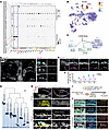 Figure 1
Expression and subcellular localization of Kcnh8 (Kv12.1) in the mouse and human cochlea. (A) Dot plot depicting the expression of all KCNH family members across distinct cell types in the adult mouse cochlea. (B) Feature plot illustrating the restricted expression of Kcnh8 within IHCs and OHCs. (C) Schematic of the KCNH channel family, highlighting the classification of Kv12.1 (encoded by Kcnh8) within the ELK subfamily. (D) Spatial localization of Kcnh8 RNA (shown in magenta) in mouse cochlea at 1 M, as detected by RNAscope. Gray dashed lines outline IHCs and OHCs. (E) Developmental time course of Kcnh8 RNA (magenta) expression in the mouse cochlea at E13.5, E18.5, P1, P14, and 1 M. Sections were costained for HC marker MYO7A (cyan) and DAPI (nuclei, gray). (F) Quantification of Kcnh8 RNA puncta per IHC (green) and OHC (magenta) from the experiments shown in E. (G) Phylogenetic analysis of Kcnh8 from representative species, based on amino acid sequence alignment. Percentage identity to the human protein is indicated. (H) Expression of Kcnh8 RNA in human fetal cochlea at PCW 11 and 17. Left panels show merged channels (Kcnh8, magenta; MYO7A, cyan; SOX2, yellow; DAPI, gray); right panels show colabeling of Kcnh8 and DAPI. Upper panels, low-magnification views with white boxes indicating regions of interest; lower panels, higher magnification views of individual channels. (I) Schematic of the HiUGE strategy for knockin of an HA-epitope tag to label the Kv12.1 (Kcnh8-encoding protein) in vitro. DIV, days in vitro; IF, immunofluorescence. (J) Representative confocal images showing HA-tagged Kv12.1 following AAV transduction. EGFP indicates transfected cells. Left panels show merged channels (HA, magenta; MYO7A, cyan; EGFP, yellow; DAPI, gray); middle panels show colabeling of HA and MYO7A; right panels show colabeling of HA and EGFP. Scale bars are provided in D, E, H, and J.
To investigate the evolutionary conservation of Kcnh8, we compared its amino acid sequence across several representative species used in auditory research. The sequences were found to be highly conserved (Figure 1G). Specifically, Macaca mulatta shared 99.19% sequence identity with humans, while mice and rats shared 91.92% and 91.73% identity with humans, respectively. Using RNAscope on cochlear sections from donated human fetuses, we detected a faint and scattered KCNH8 mRNA signal within the SOX2-positive prosensory domain at 11 postconception weeks (PCW), prior to HC formation. By 17 PCW, KCNH8 was robustly expressed in the differentiated IHCs and OHCs of the sensory domain, with additional expression in surrounding SOX2-positive supporting cells (Figure 1H). This spatiotemporal pattern in human fetuses recapitulates the expression pattern observed during HC development in mice. To determine the subcellular localization of the endogenous Kv12.1 protein, we employed an adeno-associated virus–mediated (AAV-mediated) homology-independent universal genome engineering (HiUGE) knock-in system (28). To tag the endogenous Kv12.1 protein encoded by Kcnh8 with an HA-epitope, we employed a CRISPR/Cas9-mediated knock-in strategy using a dual-orientation payload design (Supplemental Figure 1D). To achieve this, cultured cochlear explants from Cas9-expressing mice were infected with the dual-AAV system (Figure 1I). Whereas no HA signal was detected in the absence of the system, transduced explants exhibited a robust HA signal in some HCs, confirming successful insertion (Figure 1J and Supplemental Figure 1E). The HA signal was localized to both the HC plasma membrane and the cytoplasm, indicating that the Kv12.1 protein is present in these cellular compartments (Figure 1J). Collectively, our study unveils Kcnh8 as a highly specific potassium channel in cochlear hair cells and delineates its evolutionarily conserved spatiotemporal expression profile.
Kcnh8 deficiency leads to progressive hearing loss. To investigate the biological function of Kcnh8 in the auditory system, we knocked out the gene by deleting its exon 4 using CRISPR/Cas9 technology (Figure 2A). Kcnh8-deficient mice were viable with normal survival across genotypes (data not shown). We confirmed decreased Kcnh8 mRNA expression in homozygous and heterozygous knockout (KO) mice by qRT-PCR (Figure 2B). We further verified this knockout effect at the protein level, where we observed a genotype-dependent dosage effect of Kcnh8-encoded Kv12.1 expression (Figure 2C).
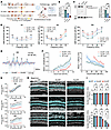 Figure 2
Progressive hearing loss in the absence of Kcnh8. (A) Strategy for generating Kcnh8-knockout mice via CRISPR/Cas9. Exon 4 of the Kcnh8 gene was deleted to generate a null allele. (B and C) Validation of Kcnh8 knockout at RNA and protein levels. (B) qRT-PCR and (C) Western blot analysis of Kcnh8 transcript and protein expression in WT, Kcnh8+/–, and Kcnh8–/– mice. GAPDH served as the internal control. (D) ABR thresholds across 4–32 kHz frequencies in WT, Kcnh8+/–, and Kcnh8–/– mice at 1, 3, and 6 M. (E) ABR waveforms evoked by a 32 kHz tone at 90 dB sound pressure level (SPL) of 3 M WT, Kcnh8+/–, and Kcnh8–/– mice. Arrowheads indicate peaks I–V. (F) Quantification of wave I peak amplitude (left) and latency (right) from the recordings in E. (G) DPOAE intensity in WT and Kcnh8–/– mice at 1, 3, and 6 M. (H) Representative confocal images of the basal cochlear turn from WT and Kcnh8–/– mice at 1, 3, and 6 M, immunolabeled for Myosin7a (MYO7A) (cyan) and DAPI (nuclei, gray). Scale bar, 50 μm. (I) Quantification of MYO7A-positive IHCs (upper panel) and OHCs (lower panel) per 100 μm cochlear length from the samples shown in H. n = 8. Statistical significance was determined using 2-way ANOVA with Tukey’s post hoc test or unpaired 2-tailed t test, as appropriate (*P < 0.05, **P < 0.01). The number (n) of mice was indicated in each group.
Next, we assessed the auditory function of Kcnh8–/– and Kcnh8+/– mice using auditory brain stem response (ABR) measurements, as well as distortion product otoacoustic emissions (DPOAE) tests. As shown in Figure 2D, hearing loss was observed in Kcnh8-deficient mice at 1 M of age, evidenced by significantly elevated ABR thresholds in response to 24 and 32 kHz tone bursts (Figure 2D). The thresholds in both Kcnh8–/– and Kcnh8+/– groups were approximately 10–20 dB higher than those in WT controls. This deficit worsened by 3 M, with Kcnh8–/– mice showing the most pronounced threshold shifts while Kcnh8+/– mice exhibited an intermediate phenotype, indicating a genotype-dependent dosage effect (Figure 2D). Although ABR thresholds in response to clicks showed no significant difference at 1 M and 3 M, a significant hearing impairment emerged in Kcnh8-deficient mice by 6 M (Supplemental Figure 2A). Across specific frequencies (4–24 kHz), ABR thresholds were further elevated in both Kcnh8–/– and Kcnh8+/– mice, with statistically significant differences observed between the Kcnh8–/– and WT groups (Figure 2D). We further analyzed the features of wave I of the ABR evoked by a 16 kHz tone at 90 dB SPL, which revealed that both Kcnh8–/– and Kcnh8+/– mice exhibited reduced amplitude and increased latency compared with controls (Figure 2, E and F). At 1 M, no DPOAE abnormality was detected in Kcnh8-deficient mice, suggesting an initial selective impairment of IHC function. However, progressively worsening DPOAE response, indicative of OHC dysfunction, emerged at 3 M and 6 M of age (Figure 2G and Supplemental Figure 2B).
Given the observed hearing deficits, we next assessed whether there were corresponding pathological changes in the cochlea of Kcnh8-deficient mice. Staining with the HC marker MYO7A revealed no significant difference in HC counts at 1 M. However, progressive OHC loss was observed in Kcnh8–/– mice over time. Severe loss was first evident in the basal turn, where only a small fraction of OHCs remained by 6 M (Figure 2, H and I). The middle turn also showed significantly greater OHC loss in Kcnh8–/– mice compared with WT at 6 M (Supplemental Figure 2, E and F). In contrast, no significant difference in HC counts was detected in the apical turn, nor were IHCs lost, in Kcnh8-deficient mice at any age (Figure 2, H and I, and Supplemental Figure 2, C–F). Collectively, these results demonstrate that Kcnh8 plays an essential role in maintaining auditory function, and its deficiency underlies a progressive hearing loss.
Kcnh8–/– mice exhibit stereocilia loss and ribbon synapse degeneration in HCs. To further characterize the ultrastructure in HCs affected by Kcnh8 deficiency, we examined HC stereocilia and synaptic morphology. Scanning electron microscopy (SEM) revealed pronounced loss of OHC stereocilia in the basal turn of Kcnh8–/– mice at 3 M and 6 M (Figure 3A). A milder stereocilia loss was also observed in the middle turn with Kcnh8 deficiency at 6 M (Supplemental Figure 3A). We next characterized the stereocilia morphology on the remaining HCs. Unlike the orderly 3 rows of stereocilia seen in WT OHCs, we observed stereocilia loss in the basal turn of WT mice at 3 and 6 months of age (Figure 3A). This loss predominantly targeted the shortest row, a phenotype that was markedly exacerbated in the basal turn OHCs of Kcnh8–/– mice at 3 M and 6 M (Figure 3, A and B). Similarly, significant stereocilia loss was observed in the middle turn of Kcnh8–/– mice at these time points. In contrast, no obvious stereocilia loss was observed in IHCs of Kcnh8–/– mice compared with controls (Figure 3, A and B, and Supplemental Figure 3, A and B). Our findings implicate the role of Kcnh8 in maintaining stereocilia integrity, as its loss causes progressive degeneration, with OHCs being particularly vulnerable.
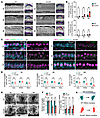 Figure 3
Stereocilia loss and ribbon synapse degeneration in Kcnh8-deficient HCs. (A) SEM images of the cochlear sensory epithelium (left) and magnified views of stereocilia bundles on IHCs and OHCs (right) from the basal turn of WT and Kcnh8–/– mice at 1, 3, and 6 M. The 3 rows of OHC stereocilia are color-coded. Red arrowheads indicate missing stereocilia. (B) Quantification of the percentage of stereocilia loss in OHCs (upper panel) and IHCs (lower panel) in WT and Kcnh8–/– mice (n = 5) from images in A. (C) CtBP2 (presynaptic ribbons, magenta), HOMER1 (postsynaptic terminals, green), MYO7A (HCs, cyan), and DAPI (nuclei, gray). Left panels show merged channels; right panels show CtBP2 and HOMER1 colabeling. IHC outlines are demarcated by gray dashed lines. (D) Quantification of the number of synaptic pairs per IHC from the apex, middle, and basal turns in WT and Kcnh8–/– mice. n = 25 IHCs per group from 5 mice. (E) Representative TEM images of IHC ribbon synapses of the basal turn from WT and Kcnh8–/– mice at 1, 3, and 6 M. (F) Quantification of the number of synaptic vesicles per ribbon (left panel) and the distance from the synaptic vesicle to ribbon (right panel) in IHCs of WT and Kcnh8–/– mice from images in E. n = 8 ribbon synapses from 5 mice for each group. (G) Schematic model summarizing the progressive structural deterioration of IHC ribbon synapses in Kcnh8–/– mice from 1 to 6 M. Statistical significance was determined using 2-way ANOVA with Tukey’s post hoc test, as appropriate (*P < 0.05, **P < 0.01). Scale bars are provided in A, C, and E.
We next assessed the IHC ribbon synapses in the cochlea by immunostaining, using CtBP2 to label the presynaptic ribbons and HOMER1 for the postsynaptic densities. Quantification of synaptic puncta revealed no significant difference in the number of synaptic pairs between Kcnh8–/– and WT mice across all cochlear turns at 1 M of age (Figure 3, C and D, and Supplemental Figure 3C). At 3 M, the number of synaptic pairs was significantly reduced in the basal turn of Kcnh8-deficient mice (Figure 3D). With advancing age, this synaptic loss progressed, and by 6 M, CtBP2/HOMER1 pairs were markedly decreased throughout the entire cochlea, from the base to the apex, compared with WT mice. We further used transmission electron microscopy (TEM) to examine the morphology of the ribbon synapses. In the basal turn of IHCs in 1 M WT mice, the synaptic vesicles (SVs) exhibited the characteristic garland-like arrangement around the synaptic ribbons (Figure 3E). Abnormal SV organization was already evident at 1 M, with SVs appearing disordered and irregularly dispersed around the ribbons in basal IHCs of Kcnh8–/– mice. We quantified the number of SVs per synaptic ribbon and the distance of SVs from the ribbons. This analysis revealed that at 3 and 6 months, Kcnh8–/– mice exhibited not only disorganized SVs but also a reduced SV count and an increased SV-to-ribbon distance compared with WT mice (Figure 3, E and F). This pathological pattern underscores the critical importance of Kcnh8 in maintaining synaptic structural stability (Figure 3G).
Kcnh8 disruption elevates early IHC activity and compromises mature synaptic function. HC activation follows a sequence of events: K+ influx depolarizes the membrane to initiate vesicle release, a process balanced by K+ efflux that repolarizes the cell to maintain resting potential (Figure 4A). Kv12.1 has been proposed to contribute to the outward K+ conductance that regulates IHC receptor potentials (26). To directly evaluate its function, we performed whole-cell patch-clamp recordings on IHCs acutely isolated from P10 mouse cochleae (Supplemental Figure 4A). Cells were held at –90 mV and depolarized in 10 mV steps to +40 mV, followed by repolarization to –110 mV (Figure 4B). The results show that Kcnh8-KO IHCs displayed markedly reduced outward K+ currents compared with WT cells across –30 to +50 mV (Figure 4, B and C), indicating that Kcnh8 deficiency substantially impairs outward K+ efflux during depolarization. Analysis of activation gating further showed a rightward shift of the conductance-voltage relationship in Kcnh8-KO IHCs (Figure 4D). These results indicate that Kcnh8 initiates outward K+ efflux at the onset of IHC depolarization and becomes progressively more prominent as depolarization proceeds. To verify the bidirectional influence of Kcnh8 in cochlear HCs, we overexpressed (OE) Kcnh8 in HCs of P3 WT mice via AAVanc80L65 delivery and assessed the outcome at P10. As expected, the Kcnh8-OE IHCs exhibited significantly enhanced outward K+ currents compared with WT controls (Figure 4, E and F). Furthermore, the activation curve displayed a pronounced leftward shift (Figure 4G), and kinetic fitting revealed a reduced Vhalf together with an increased slope factor (Figure 4H). These properties indicate that elevated Kcnh8 expression promotes earlier and steeper activation, consistent with its role as a major outward rectifier during early depolarization.
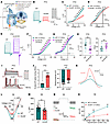 Figure 4
Kcnh8 regulates IHC excitability and synaptic transmission. (A) Model of HC auditory transduction and the K+-initiated cascade. (B) Representative whole-cell K+ currents recorded from P10 WT and Kcnh8–/– IHCs. Currents were elicited by stepping from a holding potential of –80 mV to test potentials ranging from –90 mV to +40 mV, followed by a step to –110 mV. (C) Steady-state K+ currents (IK) from P10 WT and Kcnh8–/– IHCs from 5 mice. (D) Voltage dependence of activation derived from currents in B. Solid lines indicate Boltzmann fits. (E) Representative whole-cell K+ currents from P10 WT and Kcnh8-overexpressing (Kcnh8-OE) IHCs using the same protocol as in B. (F) Steady-state K+ currents from P10 WT and Kcnh8-OE IHCs from 5 mice. (G) Activation curves and Boltzmann fits from WT and Kcnh8-OE recordings. (H) Half-activation voltage (Vhalf, left) and slope factor (right) derived from fits in G. (I) Representative current-evoked action potentials (APs) recorded from P10 WT (top) and Kcnh8–/– (bottom) IHCs. The 80 pA trace is highlighted in red. (J) Quantification of the number of evoked APs during a 250 ms current injection and resting membrane potential from P10 WT and Kcnh8–/– IHCs (n = 5 mice). (K) Representative single AP waveforms extracted from the 80 pA injection traces in I. (L) Representative I–V curves of Ca2+ currents recorded from IHCs of WT and Kcnh8–/– mice at 1 month old. Currents were evoked by a voltage ramp from –90 to +70 mV under voltage clamp and subsequently leak-subtracted. (M) Comparison of the peak Ca2+ current from 1-month-old WT and Kcnh8–/– IHCs from 8 mice. (N) A representative diagram of ICa and the corresponding changes in membrane capacitance (ΔCm) in WT and Kcnh8–/– IHCs at 1 M of stimulation of duration 500 ms. fF, femtofarads. (O) ΔCm, reflecting vesicle exocytosis, evoked by depolarizing pulses (10–500 ms) in 1 M WT and Kcnh8–/– IHCs from 8 mice. Data are reported as mean ± SEM. Statistical significance was determined by unpaired 2-tailed t test or 2-way ANOVA with Tukey’s post hoc test (*P < 0.05, **P < 0.01). The number of patched IHCs was indicated in each group.
As established in developing IHCs, outward K+ currents are the primary determinants of receptor potential amplitude and play a central role in regulating voltage-gated Ca2+ channel activation and action potential firing (29). We next recorded action potentials evoked by depolarizing current injections. Kcnh8 deficiency IHCs generated significantly more action potentials than WT cells at equivalent current steps and exhibited a more depolarized resting membrane potential (Figure 4, I and J), reflecting heightened excitability. Single-spike analysis revealed faster depolarization rates and larger spike amplitudes in Kcnh8 deficiency IHCs (Figure 4K and Supplemental Figure 4F), demonstrating that Kcnh8 normally restrains IHC firing dynamics during pre–hearing development. Elevated action potential firing is expected to enhance voltage-gated Ca2+ entry and synaptic release. Indeed, Ca2+ currents were significantly larger in Kcnh8-KO P10 IHCs than in WT controls (Supplemental Figure 4, B and C), and sustained depolarizations evoked markedly greater membrane capacitance increments, indicating increased vesicle exocytosis (Supplemental Figure 4, D and E). Together, these findings indicate that impaired outward K+ efflux at P10 causes IHC hyperexcitability, thereby increasing Ca2+ influx and promoting excessive transmitter release during the critical period of synapse maturation.
Given that prolonged or excessive glutamate release can induce excitotoxic damage to IHC ribbon synapses and downstream auditory neurons (30, 31), we asked whether the early increase in excitability and transmitter release seen at P10 in Kcnh8-KO mice results in long-lasting synaptic dysfunction. We therefore evaluated Ca2+ currents and exocytic capacitance changes in IHCs from 1-month-old mice. In contrast with P10, Kcnh8-KO adult IHCs exhibited significantly reduced Ca2+ currents and smaller ΔCm responses compared with WT cells (Figure 4, L–O). We further examined K+ currents, evoked action potentials, Ca2+ currents, and vesicle release in heterozygous Kcnh8-deficient mice and found that, compared to WT mice, the results were consistent with those observed in homozygous mice (Supplemental Figure 5, A–H). Reduced Ca2+ currents and smaller ΔCm responses were also found in heterozygous Kcnh8-deficient HCs compared with WT HCs, indicating a dose-dependent effect of Kcnh8 deficiency (Supplemental Figure 5, I–L). This functional decline suggests that early-life hyperexcitability and excessive synaptic vesicle release may impose lasting excitotoxic stress, ultimately impairing synaptic function in the mature cochlea.
Kcnh8 deficiency increases susceptibility to noise-induced HC damage. Given that hereditary deafness data do not implicate this gene and the phenotype is progressive, we sought to determine whether environmental stressors exacerbate the hearing loss caused by Kcnh8 deficiency. WT and Kcnh8–/– mice at 1 M were exposed to a moderate noise paradigm of 97 dB SPL for 2 hours to an 8–16 kHz narrow-band noise to induce threshold shifts (Figure 5A). ABR testing performed 1 day postexposure revealed a marked elevation in thresholds across 8–32 kHz in both genotypes compared with preexposure baselines, confirming successful induction of temporary threshold shift (Figure 5B). Notably, at 16 and 24 kHz, the threshold shifts in Kcnh8–/– mice were significantly greater than those in WT littermates, by 14.37 ± 4.76 dB and 24.5 ± 4.04 dB SPL, respectively. As recovery progressed, a significant intergroup difference emerged at 32 kHz by day 3, with Kcnh8–/– mice exhibiting thresholds 13.12 ± 3.65 dB SPL higher than WT controls. These differences between genotypes persisted from 16 to 32 kHz through day 14 (Figure 5B). Although WT mice showed substantial recovery across the 8–32 kHz range over time, the Kcnh8–/– mice exhibited pronouncedly poorer recovery. Together, these results indicate that Kcnh8 deficiency increases susceptibility to noise-induced hearing loss and impairs functional restoration after acoustic trauma.
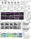 Figure 5
Kcnh8 deficiency exacerbates the susceptibility to noise exposure. (A) Schematic of the noise exposure experimental workflow. (B) ABR threshold in WT and Kcnh8–/– mice before and after noise exposure. Dashed lines indicate prenoise thresholds for each genotype. (C) Representative confocal images of IHC ribbon synapses from the middle turns of WT and Kcnh8–/– mice before and after noise exposure. Tissues were immunolabeled for CtBP2 (magenta), HOMER1 (green), and DAPI (nuclei, gray). (D) Quantification of the number of synaptic pairs per IHC from images in C. 50 IHCs from 8 mice per group. (E) Representative TEM images of IHC ribbon synapses from the middle turns of WT and Kcnh8–/– mice before and after noise exposure. White arrows indicate free vesicles at postnoise day 14. (F) Quantification of the number of synaptic vesicles tethered to each ribbon (upper panels) and the number of free vesicles in the surrounding 1 μm2 area of ribbons (lower panel) from the TEM images in E. 25 ribbons from 7 mice per group. (G) Representative fluorescence and pseudocolor images showing intracellular K+ concentration in IHCs of the middle turns of WT and Kcnh8–/– mice, measured using the EPG-2AM K+ indicator. IHCs are outlined by red dashed lines. (H) Quantification of the mean fluorescence intensity of EPG-2AM in IHCs from WT and Kcnh8–/– mice (n = 5) before and after noise exposure from images in G. Data are presented as means ± SEMs. Statistical significance was determined using 2-way ANOVA with Tukey’s post hoc test or unpaired 2-tailed t test, as appropriate (*P < 0.05, **P < 0.01). Scale bars are provided in C, E, and G.
We next examined the morphological differences in synaptic structure after noise exposure. On postnoise day 1, the number of synaptic pairs in the middle turn of IHCs was significantly reduced in both groups compared with their preexposure baselines (Figure 5, C and D). Furthermore, throughout the 14-day recovery period, Kcnh8–/– mice consistently exhibited fewer CtBP2/HOMER1-labeled synaptic pairs in IHCs compared with WT mice at all time points checked, despite a gradual recovery trend in both genotypes. Ultrastructural analysis by TEM further identified exacerbated ribbon synapse degeneration in noise-exposed Kcnh8–/– mice (Figure 5E). Specifically, from day 1 to day 14 postexposure, the synapses showed improperly aligned vesicles and a lower vesicle count around ribbons compared with WT (Figure 5, E and F). Corroborating this observation, further analysis of the vesicle reserve showed a significant decrease in the number of free vesicles in Kcnh8–/– mice relative to WT controls (Figure 5F).
To determine if the noise vulnerability of Kcnh8–/– HCs results from defective K+ efflux, we loaded dissected cochlear tissues with the K+ indicator EPG-2AM. On day 1 postexposure, both genotypes exhibited marked K+ accumulation in IHCs, with a nonsignificant increasing trend in Kcnh8–/– versus WT (Figure 5, G and H). By day 14, however, WT IHCs had recovered to baseline K+ levels, while Kcnh8–/– IHCs maintained a significantly higher K+ load. This persistent dysregulation demonstrates that Kcnh8 deficiency causes hearing damage by disrupting hair cell K+ homeostasis. Compared with WT mice, Kcnh8–/– mice had higher K+ concentration on day 7 and 14 after noise (Figure 5H). These results indicate that the auditory deficits in Kcnh8–/– mice are due to a failure to restore K+ homeostasis in HCs.
AAV-mediated delivery of Kcnh8 protects against noise-induced HC injury and preserves hearing function in WT mice. Having established that Kv12.1 is essential for K+ efflux in hair cells, we next explored its therapeutic potential by targeting this channel to treat noise-induced hearing loss (Figure 6A). Given the delayed onset of AAV-mediated transgene expression in vivo, we verified the presence of exogenous Kcnh8-encoding protein in HCs to define the appropriate time point for subsequent noise exposure. Detection of the FLAG fusion tag indicated that robust exogenous protein expression appeared by day 7 postinjection and remained stable up to day 7 in Kcnh8 delivery WT mice (Figure 6B). Furthermore, FLAG signal was detected in HCs across the apex, middle, and base cochlear turns on day 7 postinjection (Figure 6C). The absence of fluorescence in contralateral ears, together with no hearing loss on day 7 postinjection, confirms the targeted expression and safety of the AAV delivery procedure (Supplemental Figure 6, A and B). We confirmed successful Kcnh8 overexpression in HCs by RT-PCR, Western blot, and RNAscope, which all showed elevated Kcnh8 mRNA and protein levels in the delivery ears relative to the contralateral ears (Supplemental Figure 6, C–F). Moreover, under noise-free conditions, we compared hearing before and 7 days after Kcnh8 delivery in 1-month-old mice and observed no difference (Supplemental Figure 6, G and H). We then subjected the WT mice to a 2-hour exposure of 8–16 kHz narrow-band noise at 97 dB SPL on day 6 after AAV injection, using the noninjected contralateral ears as internal controls. ABR assessment revealed that AAV-mediated Kcnh8 delivery alleviated noise-induced hearing loss (Figure 6D). Notably, on postnoise day 1, the treated ears showed significant threshold improvements of 36 ± 2.6 dB (16 kHz), 26.5 ± 5.05 dB (24 kHz), and 16.5 ± 4.52 dB (32 kHz) over the contralateral controls. This protective effect persisted throughout the 14-day recovery period, as treated ears maintained lower thresholds by 16.11 ± 5.58 dB (16 kHz), 17.78 ± 5.71 dB (24 kHz), and 22.22 ± 6.78 dB (32 kHz) on day 14. These results demonstrate that exogenous Kcnh8 gene delivery effectively mitigates noise-induced hearing loss in mice.
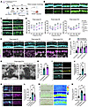 Figure 6
AAV-mediated Kcnh8 overexpression protects against noise-induced damage in WT mice. (A) Schematic diagram of the experimental timeline for noise exposure and AAV delivery. (B) Time course of AAV-Kcnh8 transduction efficiency in the middle turn of the cochlea, assessed by FLAG (Kcnh8-encoding protein, green) and MYO7A (cyan) colabeling. (C) Representative confocal images showing successful transduction of AAV-Kcnh8 (FLAG, green) in HCs throughout the apex, middle, and base turns of the cochlea. (D) ABR threshold from AAV-Kcnh8–treated ears and contralateral ears after noise exposure days 1, 5, and 14 (n = 13). Dashed lines indicate prenoise thresholds for treated and contralateral ears. (E) Representative confocal images of IHC ribbon synapses in the middle turns of treated and contralateral ears at postnoise days 1, 5, and 14. Tissues were costained with MYO7A (cyan), CtBP2 (presynaptic ribbons, magenta), HOMER1 (green), and DAPI (gray). (F) Quantification of the number of synaptic pairs per IHC from the images in E. 50 IHCs from 5 mice per group. (G) Representative TEM images of IHC ribbon synapses in the middle turns of treated and contralateral ears at postnoise day 14. (H) Quantification of the number of synaptic vesicles per ribbon from the TEM images in G. 28 ribbons from 6 mice per group. (I–L) Representative confocal images of MitoSOX (yellow) and DCFH-DA (cyan) in IHCs of the middle turns of treated, contralateral, and prenoise ears at postnoise day 7. Fluorescence intensities of MitoSOX and DCFH-DA are shown in J and L. (M) Representative fluorescence images of intracellular K+ concentration, measured using the EPG-2AM indicator, in IHCs of the middle turns of treated, contralateral, and control ears at postnoise day 7. (N) Fluorescence intensity of EPG-2AM from the images in M. 28 IHCs from 6 mice per group in I, L, and N. Data are presented as means ± SEMs. Statistical significance was determined using 2-way ANOVA with Tukey’s post hoc test or unpaired 2-tailed t test (**P < 0.01).
We further evaluated the therapeutic efficacy on HC morphology. Throughout the postnoise recovery period, no significant difference was observed in the number of MYO7A+ HCs between groups (Figure 6E). In contrast, analysis of synaptic pairs revealed a marked increase in CtBP2/HOMER1-positive puncta in AAV-treated ears compared with contralateral controls (Figure 6, E and F). Consistent with this synaptic preservation, TEM analysis demonstrated substantial ultrastructural improvement in treated HCs, characterized by more regularly organized vesicles and more abundant vesicles anchored at the synaptic ribbons (Figure 6, G and H). On day 7 after noise exposure, cochlear HCs were stained with MitoSOX and DCFH-DA to assess mitochondrial superoxide and intracellular ROS levels, respectively (Figure 6, I–L). The results showed that AAV-Kcnh8 treatment significantly alleviated noise-induced HC damage. This protective effect corresponded with a marked reduction in noise-induced intracellular K+ accumulation (Figure 6, M and N). Collectively, these findings demonstrate that targeting Kcnh8 effectively prevents noise-induced HC and synaptic degeneration, thereby preserving auditory function.
-
Discussion
In this study, we demonstrate that Kcnh8, which encodes the voltage-gated potassium channel Kv12.1, is specifically expressed in cochlear HCs and is essential for maintaining intracellular potassium homeostasis (Figure 7). Notably, the in vivo function of Kcnh8 had not previously been investigated in any mammalian system to our knowledge. Our work addresses this fundamental gap by establishing that Kcnh8 deficiency leads to progressive hearing loss in mice, thereby revealing Kcnh8’s critical role in the auditory system. Mechanistically, we show that loss of Kcnh8 impairs potassium efflux, resulting in HC hyperexcitability and ultimately leading to synaptic vesicle dysfunction. Furthermore, we found that targeted delivery of a Kcnh8-based construct ameliorates noise-induced HC damage and hearing loss by mitigating intracellular potassium accumulation. Collectively, our findings not only fill a key knowledge gap regarding the KCNH family in auditory research but also identify Kcnh8 as a potential therapeutic target for the treatment of noise-induced hearing loss.
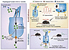 Figure 7
Role of the Kv12.1 (Kcnh8) K+ efflux channel in hearing and therapy for noise-induced loss. Under physiological conditions, Kcnh8-encoded Kv12.1 channel provides a crucial K+ efflux pathway that stabilizes the membrane potential of HCs during sound stimulation, ensuring precise synaptic transmission. Kcnh8 deficiency disrupts K+ homeostasis, causing IHC hyperexcitability, excitotoxic synaptic damage, and progressive hearing loss — a pathology exacerbated by noise exposure. Conversely, in noise-exposed WT mice, AAV-mediated Kcnh8 delivery into the cochlea promotes K+ efflux, prevents ribbon synapse degeneration, and accelerates hearing recovery, highlighting its therapeutic potential.
Notably, Kcnh8 is the sole KCNH family gene we found to be specifically and highly expressed in both adult mouse IHCs and OHCs. Its high conservation and expression in human fetal HCs provide the necessary foundation for its function. This unique expression profile thus positions Kcnh8 as a prime functional candidate representing the entire KCNH family in auditory processing. Prior evidence, though limited, has implicated Kcnh8 in the excitability of thalamic neurons (32). A key in vitro study demonstrated that Kcnh8 regulates the daytime/nighttime rhythms in repetitive firing rates of suprachiasmatic nucleus neurons (16). Despite its known roles, no study has directly linked Kcnh8 to an organism-level phenotype through in vivo evidence. Our research fills this void by employing genetic intervention to definitively establish its requirement in maintaining auditory function.
We have identified Kcnh8-encoded Kv12.1 as a mediator of outward K+ currents in cochlear HCs, thereby exerting a functionally opposing effect to the excitatory K+ influx mediated by the nonselective mechanotransduction channel (33, 34). HCs act as the units for mechanoelectrical transduction; consequently, their excitability is closely linked to the function of K+ channels (35, 36). Previous studies have highlighted the crucial role of several outward K+ channels, with the most prominent examples being Kv7.4 (encoded by Kcnq4) (35, 37) and BK channels (38, 39). In terms of expression pattern, Kcnq4 is specific to OHCs (37), while the BK channels are predominantly expressed in IHCs (40, 41). In contrast, Kcnh8 is abundantly expressed in both IHCs and OHCs, distinguishing it from both. The distinct expression patterns and functions of these 3 types of outward K+ channels underlie their differential contributions to auditory function. By serving as a major determinant of the OHC resting membrane potential, the voltage-gated Kv7.4 channel mediates a noninactivating, slow, linear K+ current that is critical for K+ efflux (37, 42). Mutations in KCNQ4 impair this efflux, resulting in chronic K+ overload, which in turn causes the severe, early-onset hereditary deafness observed in patients (19, 37). The loss of Kcnh8 results in a progressive hearing impairment in mice, with a phenotypic similarity to that caused by BK channel dysfunction (43). The rapid repolarization mediated by BK channels, enabled by their fast activation kinetics and large conductance, is essential for tracking high-frequency stimuli (44, 45). Kv12.1 in IHCs operates on a different timescale. It mediates a component of the slowly activating IK, s with intermediate kinetics, which contributes substantially to the outward conductance (26). Rather than driving the most rapid repolarization phases, this activity helps control the membrane potential during sound stimulation, thereby regulating IHC excitability. We found that Kcnh8 deletion causes abnormal evoked action potentials and markedly increased IHC excitability at an early stage. Thus, we identify Kcnh8 as a richly expressed K+ channel in both IHCs and OHCs, establishing its function as a key regulator of HC excitability.
We reveal that Kcnh8 deficiency causes HC hyperexcitability at P10 because of impaired K+ efflux, paralleling the pathogenic excitability caused by other KCNH family members, such as KCNH2 variants in long-QT syndrome 2 and epilepsy (46). By 1 M of age, this auditory function declines, suggesting that sustained hyperexcitability ultimately leads to functional loss, much like the hyperexcitability in Kcnh7-deficient Purkinje cells that causes behavioral deficits (10). The progressive delayed hearing loss by Kcnh8 deficiency with advancing age might be partly attributed to a chronic intracellular K+ overload in the HCs, which may trigger a series of stress-induced injury responses. On the other hand, increased excitability upon acoustic stimulation led to excessive glutamate release, which may be responsible for part of the synaptic damage and hearing loss outcome. Similarly, neuronal hyperexcitability is crucially linked to neurodegeneration and synaptic abnormalities (6, 7), and excessive glutamate release is implicated in the pathogenesis of synapse damage and neurological disorders (8). Our findings suggest that impairment of the Kv12.1 channel causes progressive auditory decline. However, the mechanism by which intracellular K+ overload and glutamate excitotoxicity lead to hair cell degeneration requires further investigation.
The role of Kcnh8 in maintaining K+ homeostasis in HCs provides a solid rationale and identifies it as a promising therapeutic target for treating hearing loss caused by environmental insults. Recent studies have shown that BK channel deficiency or pharmacological blockade ameliorates noise-induced HC degeneration (47, 48). Other reports conflictingly suggest that the lack of BK channels could instead increase noise vulnerability (24, 49). Our results provide compelling evidence that AAV-mediated delivery of Kcnh8 mitigates noise-induced IHC damage by facilitating potassium efflux. In future studies, we will employ mouse models of noise-induced injury to both IHCs and OHCs at different intensities to evaluate Kcnh8 effects on OHCs and its overall protective role. Notably, this gene delivery strategy proved effective in mice without employing a specific channel agonist, as none is currently available. This limitation stems from the unresolved protein structure of Kv12.1. Like other KCNH channels, Kv12.1 features intracellular N- and C-terminal regions containing a cyclic nucleotide-binding homology domain (50). These domains, serving as potential hubs for posttranslational modifications and molecular interactions, provide key structural insights. This knowledge thereby guides our future research aimed at solving the Kv12.1 structure, with the ultimate goal of developing specific small-molecule agonists.
In summary, we have identified Kv12.1 as a K+ channel highly enriched in auditory HCs and vital for auditory function. We further show that its role in K+ homeostasis is so critical that its restoration via gene delivery can alleviate noise-induced hearing damage. This work, therefore, progresses from the discovery of a fundamental auditory mechanism to the proposal of a viable therapeutic strategy, highlighting Kcnh8 as a highly promising target in the fight against sensorineural hearing loss.


Copyright © 2026 American Society for Clinical Investigation
ISSN: 0021-9738 (print), 1558-8238 (online)