
Advertisement
Research ArticleCell biologyReproductive biology
Open Access |  10.1172/JCI201124
Single-cell analysis of fetal testis reveals dysfunction of human Leydig cells in Klinefelter syndrome
Tong Yan,1 Guancheng Chen,1 Jie Zhang,2 Wenjing Jia,3 Nan Lu,4 Shuping Jin,5 Haotian Zhang,1 Yichen Zhao,1 Lu Jiang,5 Jing Wu,6 Qing Liu,6 Chenghao Situ,1 Hui Zhu,1 Yan Li,7 Quan Wang,3 Xiaoyu Yang,4 Chao Qin,8 Xiaofeng Song,3 Qing Cheng,5 and Xuejiang Guo1
1Department of Histology and Embryology, State Key Laboratory of Reproductive Medicine and Offspring Health, Nanjing Medical University, Nanjing, Jiangsu, China.
2Department of Gynecology, The First Affiliated Hospital of Nanjing Medical University, Nanjing, Jiangsu, China.
3Department of Biomedical Engineering, Nanjing University of Aeronautics and Astronautics, Nanjing, Jiangsu, China.
4State Key Laboratory of Reproductive Medicine and Offspring Health, Clinical Center of Reproductive Medicine, First Affiliated Hospital, Nanjing Medical University, Nanjing, Jiangsu, China.
5Department of Obstetrics and Gynecology, Women’s Hospital of Nanjing Medical University, Nanjing Women and Children’s Healthcare Hospital, Nanjing, Jiangsu, China.
6School of Biomedical Engineering and Informatics and
7Department of Clinical Laboratory, Sir Run Run Hospital, Nanjing Medical University, Nanjing, Jiangsu, China.
8State Key Laboratory of Reproductive Medicine and Offspring Health, Department of Urology, The First Affiliated Hospital of Nanjing Medical University, Nanjing, Jiangsu, China.
Address correspondence to: Xuejiang Guo, State Key Laboratory of Reproductive Medicine and Offspring Health, Nanjing Medical University, 101 Longmian Avenue, Nanjing 211166, China. Phone: 86.25.86869383; Email: guo_xuejiang@njmu.edu.cn. Or to: Qing Cheng, Department of Obstetrics and Gynecology,Women’s Hospital of Nanjing Medical University, 123 Tianfei Lane, Mochou Road, Nanjing 210004, China. Phone: 86.25.52226846; Email: chengq@njmu.edu.cn. Or to: Xiaofeng Song, Department of Biomedical Engineering, Nanjing University of Aeronautics and Astronautics, No. 28 Jiangjun Avenue, Moling Subdistrict, Nanjing 211100, China. Phone: 86-25-84891938; Email: xfsong@nuaa.edu.cn. Or to: Chao Qin, Department of Urology, The First Affiliated Hospital of Nanjing Medical University, No. 300 Guangzhou Road, Nanjing 211000, China. Phone: 86.25.83714511; Email: qinchao@njmu.edu.cn.
Authorship note: TY, GC, JZ, WJ, and NL contributed equally to this work.
Find articles by Yan, T. in: PubMed | Google Scholar
1Department of Histology and Embryology, State Key Laboratory of Reproductive Medicine and Offspring Health, Nanjing Medical University, Nanjing, Jiangsu, China.
2Department of Gynecology, The First Affiliated Hospital of Nanjing Medical University, Nanjing, Jiangsu, China.
3Department of Biomedical Engineering, Nanjing University of Aeronautics and Astronautics, Nanjing, Jiangsu, China.
4State Key Laboratory of Reproductive Medicine and Offspring Health, Clinical Center of Reproductive Medicine, First Affiliated Hospital, Nanjing Medical University, Nanjing, Jiangsu, China.
5Department of Obstetrics and Gynecology, Women’s Hospital of Nanjing Medical University, Nanjing Women and Children’s Healthcare Hospital, Nanjing, Jiangsu, China.
6School of Biomedical Engineering and Informatics and
7Department of Clinical Laboratory, Sir Run Run Hospital, Nanjing Medical University, Nanjing, Jiangsu, China.
8State Key Laboratory of Reproductive Medicine and Offspring Health, Department of Urology, The First Affiliated Hospital of Nanjing Medical University, Nanjing, Jiangsu, China.
Address correspondence to: Xuejiang Guo, State Key Laboratory of Reproductive Medicine and Offspring Health, Nanjing Medical University, 101 Longmian Avenue, Nanjing 211166, China. Phone: 86.25.86869383; Email: guo_xuejiang@njmu.edu.cn. Or to: Qing Cheng, Department of Obstetrics and Gynecology,Women’s Hospital of Nanjing Medical University, 123 Tianfei Lane, Mochou Road, Nanjing 210004, China. Phone: 86.25.52226846; Email: chengq@njmu.edu.cn. Or to: Xiaofeng Song, Department of Biomedical Engineering, Nanjing University of Aeronautics and Astronautics, No. 28 Jiangjun Avenue, Moling Subdistrict, Nanjing 211100, China. Phone: 86-25-84891938; Email: xfsong@nuaa.edu.cn. Or to: Chao Qin, Department of Urology, The First Affiliated Hospital of Nanjing Medical University, No. 300 Guangzhou Road, Nanjing 211000, China. Phone: 86.25.83714511; Email: qinchao@njmu.edu.cn.
Authorship note: TY, GC, JZ, WJ, and NL contributed equally to this work.
Find articles by Chen, G. in: PubMed | Google Scholar
1Department of Histology and Embryology, State Key Laboratory of Reproductive Medicine and Offspring Health, Nanjing Medical University, Nanjing, Jiangsu, China.
2Department of Gynecology, The First Affiliated Hospital of Nanjing Medical University, Nanjing, Jiangsu, China.
3Department of Biomedical Engineering, Nanjing University of Aeronautics and Astronautics, Nanjing, Jiangsu, China.
4State Key Laboratory of Reproductive Medicine and Offspring Health, Clinical Center of Reproductive Medicine, First Affiliated Hospital, Nanjing Medical University, Nanjing, Jiangsu, China.
5Department of Obstetrics and Gynecology, Women’s Hospital of Nanjing Medical University, Nanjing Women and Children’s Healthcare Hospital, Nanjing, Jiangsu, China.
6School of Biomedical Engineering and Informatics and
7Department of Clinical Laboratory, Sir Run Run Hospital, Nanjing Medical University, Nanjing, Jiangsu, China.
8State Key Laboratory of Reproductive Medicine and Offspring Health, Department of Urology, The First Affiliated Hospital of Nanjing Medical University, Nanjing, Jiangsu, China.
Address correspondence to: Xuejiang Guo, State Key Laboratory of Reproductive Medicine and Offspring Health, Nanjing Medical University, 101 Longmian Avenue, Nanjing 211166, China. Phone: 86.25.86869383; Email: guo_xuejiang@njmu.edu.cn. Or to: Qing Cheng, Department of Obstetrics and Gynecology,Women’s Hospital of Nanjing Medical University, 123 Tianfei Lane, Mochou Road, Nanjing 210004, China. Phone: 86.25.52226846; Email: chengq@njmu.edu.cn. Or to: Xiaofeng Song, Department of Biomedical Engineering, Nanjing University of Aeronautics and Astronautics, No. 28 Jiangjun Avenue, Moling Subdistrict, Nanjing 211100, China. Phone: 86-25-84891938; Email: xfsong@nuaa.edu.cn. Or to: Chao Qin, Department of Urology, The First Affiliated Hospital of Nanjing Medical University, No. 300 Guangzhou Road, Nanjing 211000, China. Phone: 86.25.83714511; Email: qinchao@njmu.edu.cn.
Authorship note: TY, GC, JZ, WJ, and NL contributed equally to this work.
Find articles by Zhang, J. in: PubMed | Google Scholar
1Department of Histology and Embryology, State Key Laboratory of Reproductive Medicine and Offspring Health, Nanjing Medical University, Nanjing, Jiangsu, China.
2Department of Gynecology, The First Affiliated Hospital of Nanjing Medical University, Nanjing, Jiangsu, China.
3Department of Biomedical Engineering, Nanjing University of Aeronautics and Astronautics, Nanjing, Jiangsu, China.
4State Key Laboratory of Reproductive Medicine and Offspring Health, Clinical Center of Reproductive Medicine, First Affiliated Hospital, Nanjing Medical University, Nanjing, Jiangsu, China.
5Department of Obstetrics and Gynecology, Women’s Hospital of Nanjing Medical University, Nanjing Women and Children’s Healthcare Hospital, Nanjing, Jiangsu, China.
6School of Biomedical Engineering and Informatics and
7Department of Clinical Laboratory, Sir Run Run Hospital, Nanjing Medical University, Nanjing, Jiangsu, China.
8State Key Laboratory of Reproductive Medicine and Offspring Health, Department of Urology, The First Affiliated Hospital of Nanjing Medical University, Nanjing, Jiangsu, China.
Address correspondence to: Xuejiang Guo, State Key Laboratory of Reproductive Medicine and Offspring Health, Nanjing Medical University, 101 Longmian Avenue, Nanjing 211166, China. Phone: 86.25.86869383; Email: guo_xuejiang@njmu.edu.cn. Or to: Qing Cheng, Department of Obstetrics and Gynecology,Women’s Hospital of Nanjing Medical University, 123 Tianfei Lane, Mochou Road, Nanjing 210004, China. Phone: 86.25.52226846; Email: chengq@njmu.edu.cn. Or to: Xiaofeng Song, Department of Biomedical Engineering, Nanjing University of Aeronautics and Astronautics, No. 28 Jiangjun Avenue, Moling Subdistrict, Nanjing 211100, China. Phone: 86-25-84891938; Email: xfsong@nuaa.edu.cn. Or to: Chao Qin, Department of Urology, The First Affiliated Hospital of Nanjing Medical University, No. 300 Guangzhou Road, Nanjing 211000, China. Phone: 86.25.83714511; Email: qinchao@njmu.edu.cn.
Authorship note: TY, GC, JZ, WJ, and NL contributed equally to this work.
Find articles by Jia, W. in: PubMed | Google Scholar
1Department of Histology and Embryology, State Key Laboratory of Reproductive Medicine and Offspring Health, Nanjing Medical University, Nanjing, Jiangsu, China.
2Department of Gynecology, The First Affiliated Hospital of Nanjing Medical University, Nanjing, Jiangsu, China.
3Department of Biomedical Engineering, Nanjing University of Aeronautics and Astronautics, Nanjing, Jiangsu, China.
4State Key Laboratory of Reproductive Medicine and Offspring Health, Clinical Center of Reproductive Medicine, First Affiliated Hospital, Nanjing Medical University, Nanjing, Jiangsu, China.
5Department of Obstetrics and Gynecology, Women’s Hospital of Nanjing Medical University, Nanjing Women and Children’s Healthcare Hospital, Nanjing, Jiangsu, China.
6School of Biomedical Engineering and Informatics and
7Department of Clinical Laboratory, Sir Run Run Hospital, Nanjing Medical University, Nanjing, Jiangsu, China.
8State Key Laboratory of Reproductive Medicine and Offspring Health, Department of Urology, The First Affiliated Hospital of Nanjing Medical University, Nanjing, Jiangsu, China.
Address correspondence to: Xuejiang Guo, State Key Laboratory of Reproductive Medicine and Offspring Health, Nanjing Medical University, 101 Longmian Avenue, Nanjing 211166, China. Phone: 86.25.86869383; Email: guo_xuejiang@njmu.edu.cn. Or to: Qing Cheng, Department of Obstetrics and Gynecology,Women’s Hospital of Nanjing Medical University, 123 Tianfei Lane, Mochou Road, Nanjing 210004, China. Phone: 86.25.52226846; Email: chengq@njmu.edu.cn. Or to: Xiaofeng Song, Department of Biomedical Engineering, Nanjing University of Aeronautics and Astronautics, No. 28 Jiangjun Avenue, Moling Subdistrict, Nanjing 211100, China. Phone: 86-25-84891938; Email: xfsong@nuaa.edu.cn. Or to: Chao Qin, Department of Urology, The First Affiliated Hospital of Nanjing Medical University, No. 300 Guangzhou Road, Nanjing 211000, China. Phone: 86.25.83714511; Email: qinchao@njmu.edu.cn.
Authorship note: TY, GC, JZ, WJ, and NL contributed equally to this work.
Find articles by Lu, N. in: PubMed | Google Scholar
1Department of Histology and Embryology, State Key Laboratory of Reproductive Medicine and Offspring Health, Nanjing Medical University, Nanjing, Jiangsu, China.
2Department of Gynecology, The First Affiliated Hospital of Nanjing Medical University, Nanjing, Jiangsu, China.
3Department of Biomedical Engineering, Nanjing University of Aeronautics and Astronautics, Nanjing, Jiangsu, China.
4State Key Laboratory of Reproductive Medicine and Offspring Health, Clinical Center of Reproductive Medicine, First Affiliated Hospital, Nanjing Medical University, Nanjing, Jiangsu, China.
5Department of Obstetrics and Gynecology, Women’s Hospital of Nanjing Medical University, Nanjing Women and Children’s Healthcare Hospital, Nanjing, Jiangsu, China.
6School of Biomedical Engineering and Informatics and
7Department of Clinical Laboratory, Sir Run Run Hospital, Nanjing Medical University, Nanjing, Jiangsu, China.
8State Key Laboratory of Reproductive Medicine and Offspring Health, Department of Urology, The First Affiliated Hospital of Nanjing Medical University, Nanjing, Jiangsu, China.
Address correspondence to: Xuejiang Guo, State Key Laboratory of Reproductive Medicine and Offspring Health, Nanjing Medical University, 101 Longmian Avenue, Nanjing 211166, China. Phone: 86.25.86869383; Email: guo_xuejiang@njmu.edu.cn. Or to: Qing Cheng, Department of Obstetrics and Gynecology,Women’s Hospital of Nanjing Medical University, 123 Tianfei Lane, Mochou Road, Nanjing 210004, China. Phone: 86.25.52226846; Email: chengq@njmu.edu.cn. Or to: Xiaofeng Song, Department of Biomedical Engineering, Nanjing University of Aeronautics and Astronautics, No. 28 Jiangjun Avenue, Moling Subdistrict, Nanjing 211100, China. Phone: 86-25-84891938; Email: xfsong@nuaa.edu.cn. Or to: Chao Qin, Department of Urology, The First Affiliated Hospital of Nanjing Medical University, No. 300 Guangzhou Road, Nanjing 211000, China. Phone: 86.25.83714511; Email: qinchao@njmu.edu.cn.
Authorship note: TY, GC, JZ, WJ, and NL contributed equally to this work.
Find articles by Jin, S. in: PubMed | Google Scholar
1Department of Histology and Embryology, State Key Laboratory of Reproductive Medicine and Offspring Health, Nanjing Medical University, Nanjing, Jiangsu, China.
2Department of Gynecology, The First Affiliated Hospital of Nanjing Medical University, Nanjing, Jiangsu, China.
3Department of Biomedical Engineering, Nanjing University of Aeronautics and Astronautics, Nanjing, Jiangsu, China.
4State Key Laboratory of Reproductive Medicine and Offspring Health, Clinical Center of Reproductive Medicine, First Affiliated Hospital, Nanjing Medical University, Nanjing, Jiangsu, China.
5Department of Obstetrics and Gynecology, Women’s Hospital of Nanjing Medical University, Nanjing Women and Children’s Healthcare Hospital, Nanjing, Jiangsu, China.
6School of Biomedical Engineering and Informatics and
7Department of Clinical Laboratory, Sir Run Run Hospital, Nanjing Medical University, Nanjing, Jiangsu, China.
8State Key Laboratory of Reproductive Medicine and Offspring Health, Department of Urology, The First Affiliated Hospital of Nanjing Medical University, Nanjing, Jiangsu, China.
Address correspondence to: Xuejiang Guo, State Key Laboratory of Reproductive Medicine and Offspring Health, Nanjing Medical University, 101 Longmian Avenue, Nanjing 211166, China. Phone: 86.25.86869383; Email: guo_xuejiang@njmu.edu.cn. Or to: Qing Cheng, Department of Obstetrics and Gynecology,Women’s Hospital of Nanjing Medical University, 123 Tianfei Lane, Mochou Road, Nanjing 210004, China. Phone: 86.25.52226846; Email: chengq@njmu.edu.cn. Or to: Xiaofeng Song, Department of Biomedical Engineering, Nanjing University of Aeronautics and Astronautics, No. 28 Jiangjun Avenue, Moling Subdistrict, Nanjing 211100, China. Phone: 86-25-84891938; Email: xfsong@nuaa.edu.cn. Or to: Chao Qin, Department of Urology, The First Affiliated Hospital of Nanjing Medical University, No. 300 Guangzhou Road, Nanjing 211000, China. Phone: 86.25.83714511; Email: qinchao@njmu.edu.cn.
Authorship note: TY, GC, JZ, WJ, and NL contributed equally to this work.
Find articles by Zhang, H. in: PubMed | Google Scholar
1Department of Histology and Embryology, State Key Laboratory of Reproductive Medicine and Offspring Health, Nanjing Medical University, Nanjing, Jiangsu, China.
2Department of Gynecology, The First Affiliated Hospital of Nanjing Medical University, Nanjing, Jiangsu, China.
3Department of Biomedical Engineering, Nanjing University of Aeronautics and Astronautics, Nanjing, Jiangsu, China.
4State Key Laboratory of Reproductive Medicine and Offspring Health, Clinical Center of Reproductive Medicine, First Affiliated Hospital, Nanjing Medical University, Nanjing, Jiangsu, China.
5Department of Obstetrics and Gynecology, Women’s Hospital of Nanjing Medical University, Nanjing Women and Children’s Healthcare Hospital, Nanjing, Jiangsu, China.
6School of Biomedical Engineering and Informatics and
7Department of Clinical Laboratory, Sir Run Run Hospital, Nanjing Medical University, Nanjing, Jiangsu, China.
8State Key Laboratory of Reproductive Medicine and Offspring Health, Department of Urology, The First Affiliated Hospital of Nanjing Medical University, Nanjing, Jiangsu, China.
Address correspondence to: Xuejiang Guo, State Key Laboratory of Reproductive Medicine and Offspring Health, Nanjing Medical University, 101 Longmian Avenue, Nanjing 211166, China. Phone: 86.25.86869383; Email: guo_xuejiang@njmu.edu.cn. Or to: Qing Cheng, Department of Obstetrics and Gynecology,Women’s Hospital of Nanjing Medical University, 123 Tianfei Lane, Mochou Road, Nanjing 210004, China. Phone: 86.25.52226846; Email: chengq@njmu.edu.cn. Or to: Xiaofeng Song, Department of Biomedical Engineering, Nanjing University of Aeronautics and Astronautics, No. 28 Jiangjun Avenue, Moling Subdistrict, Nanjing 211100, China. Phone: 86-25-84891938; Email: xfsong@nuaa.edu.cn. Or to: Chao Qin, Department of Urology, The First Affiliated Hospital of Nanjing Medical University, No. 300 Guangzhou Road, Nanjing 211000, China. Phone: 86.25.83714511; Email: qinchao@njmu.edu.cn.
Authorship note: TY, GC, JZ, WJ, and NL contributed equally to this work.
Find articles by Zhao, Y. in: PubMed | Google Scholar
1Department of Histology and Embryology, State Key Laboratory of Reproductive Medicine and Offspring Health, Nanjing Medical University, Nanjing, Jiangsu, China.
2Department of Gynecology, The First Affiliated Hospital of Nanjing Medical University, Nanjing, Jiangsu, China.
3Department of Biomedical Engineering, Nanjing University of Aeronautics and Astronautics, Nanjing, Jiangsu, China.
4State Key Laboratory of Reproductive Medicine and Offspring Health, Clinical Center of Reproductive Medicine, First Affiliated Hospital, Nanjing Medical University, Nanjing, Jiangsu, China.
5Department of Obstetrics and Gynecology, Women’s Hospital of Nanjing Medical University, Nanjing Women and Children’s Healthcare Hospital, Nanjing, Jiangsu, China.
6School of Biomedical Engineering and Informatics and
7Department of Clinical Laboratory, Sir Run Run Hospital, Nanjing Medical University, Nanjing, Jiangsu, China.
8State Key Laboratory of Reproductive Medicine and Offspring Health, Department of Urology, The First Affiliated Hospital of Nanjing Medical University, Nanjing, Jiangsu, China.
Address correspondence to: Xuejiang Guo, State Key Laboratory of Reproductive Medicine and Offspring Health, Nanjing Medical University, 101 Longmian Avenue, Nanjing 211166, China. Phone: 86.25.86869383; Email: guo_xuejiang@njmu.edu.cn. Or to: Qing Cheng, Department of Obstetrics and Gynecology,Women’s Hospital of Nanjing Medical University, 123 Tianfei Lane, Mochou Road, Nanjing 210004, China. Phone: 86.25.52226846; Email: chengq@njmu.edu.cn. Or to: Xiaofeng Song, Department of Biomedical Engineering, Nanjing University of Aeronautics and Astronautics, No. 28 Jiangjun Avenue, Moling Subdistrict, Nanjing 211100, China. Phone: 86-25-84891938; Email: xfsong@nuaa.edu.cn. Or to: Chao Qin, Department of Urology, The First Affiliated Hospital of Nanjing Medical University, No. 300 Guangzhou Road, Nanjing 211000, China. Phone: 86.25.83714511; Email: qinchao@njmu.edu.cn.
Authorship note: TY, GC, JZ, WJ, and NL contributed equally to this work.
Find articles by Jiang, L. in: PubMed | Google Scholar
1Department of Histology and Embryology, State Key Laboratory of Reproductive Medicine and Offspring Health, Nanjing Medical University, Nanjing, Jiangsu, China.
2Department of Gynecology, The First Affiliated Hospital of Nanjing Medical University, Nanjing, Jiangsu, China.
3Department of Biomedical Engineering, Nanjing University of Aeronautics and Astronautics, Nanjing, Jiangsu, China.
4State Key Laboratory of Reproductive Medicine and Offspring Health, Clinical Center of Reproductive Medicine, First Affiliated Hospital, Nanjing Medical University, Nanjing, Jiangsu, China.
5Department of Obstetrics and Gynecology, Women’s Hospital of Nanjing Medical University, Nanjing Women and Children’s Healthcare Hospital, Nanjing, Jiangsu, China.
6School of Biomedical Engineering and Informatics and
7Department of Clinical Laboratory, Sir Run Run Hospital, Nanjing Medical University, Nanjing, Jiangsu, China.
8State Key Laboratory of Reproductive Medicine and Offspring Health, Department of Urology, The First Affiliated Hospital of Nanjing Medical University, Nanjing, Jiangsu, China.
Address correspondence to: Xuejiang Guo, State Key Laboratory of Reproductive Medicine and Offspring Health, Nanjing Medical University, 101 Longmian Avenue, Nanjing 211166, China. Phone: 86.25.86869383; Email: guo_xuejiang@njmu.edu.cn. Or to: Qing Cheng, Department of Obstetrics and Gynecology,Women’s Hospital of Nanjing Medical University, 123 Tianfei Lane, Mochou Road, Nanjing 210004, China. Phone: 86.25.52226846; Email: chengq@njmu.edu.cn. Or to: Xiaofeng Song, Department of Biomedical Engineering, Nanjing University of Aeronautics and Astronautics, No. 28 Jiangjun Avenue, Moling Subdistrict, Nanjing 211100, China. Phone: 86-25-84891938; Email: xfsong@nuaa.edu.cn. Or to: Chao Qin, Department of Urology, The First Affiliated Hospital of Nanjing Medical University, No. 300 Guangzhou Road, Nanjing 211000, China. Phone: 86.25.83714511; Email: qinchao@njmu.edu.cn.
Authorship note: TY, GC, JZ, WJ, and NL contributed equally to this work.
Find articles by Wu, J. in: PubMed | Google Scholar
1Department of Histology and Embryology, State Key Laboratory of Reproductive Medicine and Offspring Health, Nanjing Medical University, Nanjing, Jiangsu, China.
2Department of Gynecology, The First Affiliated Hospital of Nanjing Medical University, Nanjing, Jiangsu, China.
3Department of Biomedical Engineering, Nanjing University of Aeronautics and Astronautics, Nanjing, Jiangsu, China.
4State Key Laboratory of Reproductive Medicine and Offspring Health, Clinical Center of Reproductive Medicine, First Affiliated Hospital, Nanjing Medical University, Nanjing, Jiangsu, China.
5Department of Obstetrics and Gynecology, Women’s Hospital of Nanjing Medical University, Nanjing Women and Children’s Healthcare Hospital, Nanjing, Jiangsu, China.
6School of Biomedical Engineering and Informatics and
7Department of Clinical Laboratory, Sir Run Run Hospital, Nanjing Medical University, Nanjing, Jiangsu, China.
8State Key Laboratory of Reproductive Medicine and Offspring Health, Department of Urology, The First Affiliated Hospital of Nanjing Medical University, Nanjing, Jiangsu, China.
Address correspondence to: Xuejiang Guo, State Key Laboratory of Reproductive Medicine and Offspring Health, Nanjing Medical University, 101 Longmian Avenue, Nanjing 211166, China. Phone: 86.25.86869383; Email: guo_xuejiang@njmu.edu.cn. Or to: Qing Cheng, Department of Obstetrics and Gynecology,Women’s Hospital of Nanjing Medical University, 123 Tianfei Lane, Mochou Road, Nanjing 210004, China. Phone: 86.25.52226846; Email: chengq@njmu.edu.cn. Or to: Xiaofeng Song, Department of Biomedical Engineering, Nanjing University of Aeronautics and Astronautics, No. 28 Jiangjun Avenue, Moling Subdistrict, Nanjing 211100, China. Phone: 86-25-84891938; Email: xfsong@nuaa.edu.cn. Or to: Chao Qin, Department of Urology, The First Affiliated Hospital of Nanjing Medical University, No. 300 Guangzhou Road, Nanjing 211000, China. Phone: 86.25.83714511; Email: qinchao@njmu.edu.cn.
Authorship note: TY, GC, JZ, WJ, and NL contributed equally to this work.
Find articles by Liu, Q. in: PubMed | Google Scholar
1Department of Histology and Embryology, State Key Laboratory of Reproductive Medicine and Offspring Health, Nanjing Medical University, Nanjing, Jiangsu, China.
2Department of Gynecology, The First Affiliated Hospital of Nanjing Medical University, Nanjing, Jiangsu, China.
3Department of Biomedical Engineering, Nanjing University of Aeronautics and Astronautics, Nanjing, Jiangsu, China.
4State Key Laboratory of Reproductive Medicine and Offspring Health, Clinical Center of Reproductive Medicine, First Affiliated Hospital, Nanjing Medical University, Nanjing, Jiangsu, China.
5Department of Obstetrics and Gynecology, Women’s Hospital of Nanjing Medical University, Nanjing Women and Children’s Healthcare Hospital, Nanjing, Jiangsu, China.
6School of Biomedical Engineering and Informatics and
7Department of Clinical Laboratory, Sir Run Run Hospital, Nanjing Medical University, Nanjing, Jiangsu, China.
8State Key Laboratory of Reproductive Medicine and Offspring Health, Department of Urology, The First Affiliated Hospital of Nanjing Medical University, Nanjing, Jiangsu, China.
Address correspondence to: Xuejiang Guo, State Key Laboratory of Reproductive Medicine and Offspring Health, Nanjing Medical University, 101 Longmian Avenue, Nanjing 211166, China. Phone: 86.25.86869383; Email: guo_xuejiang@njmu.edu.cn. Or to: Qing Cheng, Department of Obstetrics and Gynecology,Women’s Hospital of Nanjing Medical University, 123 Tianfei Lane, Mochou Road, Nanjing 210004, China. Phone: 86.25.52226846; Email: chengq@njmu.edu.cn. Or to: Xiaofeng Song, Department of Biomedical Engineering, Nanjing University of Aeronautics and Astronautics, No. 28 Jiangjun Avenue, Moling Subdistrict, Nanjing 211100, China. Phone: 86-25-84891938; Email: xfsong@nuaa.edu.cn. Or to: Chao Qin, Department of Urology, The First Affiliated Hospital of Nanjing Medical University, No. 300 Guangzhou Road, Nanjing 211000, China. Phone: 86.25.83714511; Email: qinchao@njmu.edu.cn.
Authorship note: TY, GC, JZ, WJ, and NL contributed equally to this work.
Find articles by Situ, C. in: PubMed | Google Scholar
1Department of Histology and Embryology, State Key Laboratory of Reproductive Medicine and Offspring Health, Nanjing Medical University, Nanjing, Jiangsu, China.
2Department of Gynecology, The First Affiliated Hospital of Nanjing Medical University, Nanjing, Jiangsu, China.
3Department of Biomedical Engineering, Nanjing University of Aeronautics and Astronautics, Nanjing, Jiangsu, China.
4State Key Laboratory of Reproductive Medicine and Offspring Health, Clinical Center of Reproductive Medicine, First Affiliated Hospital, Nanjing Medical University, Nanjing, Jiangsu, China.
5Department of Obstetrics and Gynecology, Women’s Hospital of Nanjing Medical University, Nanjing Women and Children’s Healthcare Hospital, Nanjing, Jiangsu, China.
6School of Biomedical Engineering and Informatics and
7Department of Clinical Laboratory, Sir Run Run Hospital, Nanjing Medical University, Nanjing, Jiangsu, China.
8State Key Laboratory of Reproductive Medicine and Offspring Health, Department of Urology, The First Affiliated Hospital of Nanjing Medical University, Nanjing, Jiangsu, China.
Address correspondence to: Xuejiang Guo, State Key Laboratory of Reproductive Medicine and Offspring Health, Nanjing Medical University, 101 Longmian Avenue, Nanjing 211166, China. Phone: 86.25.86869383; Email: guo_xuejiang@njmu.edu.cn. Or to: Qing Cheng, Department of Obstetrics and Gynecology,Women’s Hospital of Nanjing Medical University, 123 Tianfei Lane, Mochou Road, Nanjing 210004, China. Phone: 86.25.52226846; Email: chengq@njmu.edu.cn. Or to: Xiaofeng Song, Department of Biomedical Engineering, Nanjing University of Aeronautics and Astronautics, No. 28 Jiangjun Avenue, Moling Subdistrict, Nanjing 211100, China. Phone: 86-25-84891938; Email: xfsong@nuaa.edu.cn. Or to: Chao Qin, Department of Urology, The First Affiliated Hospital of Nanjing Medical University, No. 300 Guangzhou Road, Nanjing 211000, China. Phone: 86.25.83714511; Email: qinchao@njmu.edu.cn.
Authorship note: TY, GC, JZ, WJ, and NL contributed equally to this work.
Find articles by Zhu, H. in: PubMed | Google Scholar
1Department of Histology and Embryology, State Key Laboratory of Reproductive Medicine and Offspring Health, Nanjing Medical University, Nanjing, Jiangsu, China.
2Department of Gynecology, The First Affiliated Hospital of Nanjing Medical University, Nanjing, Jiangsu, China.
3Department of Biomedical Engineering, Nanjing University of Aeronautics and Astronautics, Nanjing, Jiangsu, China.
4State Key Laboratory of Reproductive Medicine and Offspring Health, Clinical Center of Reproductive Medicine, First Affiliated Hospital, Nanjing Medical University, Nanjing, Jiangsu, China.
5Department of Obstetrics and Gynecology, Women’s Hospital of Nanjing Medical University, Nanjing Women and Children’s Healthcare Hospital, Nanjing, Jiangsu, China.
6School of Biomedical Engineering and Informatics and
7Department of Clinical Laboratory, Sir Run Run Hospital, Nanjing Medical University, Nanjing, Jiangsu, China.
8State Key Laboratory of Reproductive Medicine and Offspring Health, Department of Urology, The First Affiliated Hospital of Nanjing Medical University, Nanjing, Jiangsu, China.
Address correspondence to: Xuejiang Guo, State Key Laboratory of Reproductive Medicine and Offspring Health, Nanjing Medical University, 101 Longmian Avenue, Nanjing 211166, China. Phone: 86.25.86869383; Email: guo_xuejiang@njmu.edu.cn. Or to: Qing Cheng, Department of Obstetrics and Gynecology,Women’s Hospital of Nanjing Medical University, 123 Tianfei Lane, Mochou Road, Nanjing 210004, China. Phone: 86.25.52226846; Email: chengq@njmu.edu.cn. Or to: Xiaofeng Song, Department of Biomedical Engineering, Nanjing University of Aeronautics and Astronautics, No. 28 Jiangjun Avenue, Moling Subdistrict, Nanjing 211100, China. Phone: 86-25-84891938; Email: xfsong@nuaa.edu.cn. Or to: Chao Qin, Department of Urology, The First Affiliated Hospital of Nanjing Medical University, No. 300 Guangzhou Road, Nanjing 211000, China. Phone: 86.25.83714511; Email: qinchao@njmu.edu.cn.
Authorship note: TY, GC, JZ, WJ, and NL contributed equally to this work.
Find articles by Li, Y. in: PubMed | Google Scholar
1Department of Histology and Embryology, State Key Laboratory of Reproductive Medicine and Offspring Health, Nanjing Medical University, Nanjing, Jiangsu, China.
2Department of Gynecology, The First Affiliated Hospital of Nanjing Medical University, Nanjing, Jiangsu, China.
3Department of Biomedical Engineering, Nanjing University of Aeronautics and Astronautics, Nanjing, Jiangsu, China.
4State Key Laboratory of Reproductive Medicine and Offspring Health, Clinical Center of Reproductive Medicine, First Affiliated Hospital, Nanjing Medical University, Nanjing, Jiangsu, China.
5Department of Obstetrics and Gynecology, Women’s Hospital of Nanjing Medical University, Nanjing Women and Children’s Healthcare Hospital, Nanjing, Jiangsu, China.
6School of Biomedical Engineering and Informatics and
7Department of Clinical Laboratory, Sir Run Run Hospital, Nanjing Medical University, Nanjing, Jiangsu, China.
8State Key Laboratory of Reproductive Medicine and Offspring Health, Department of Urology, The First Affiliated Hospital of Nanjing Medical University, Nanjing, Jiangsu, China.
Address correspondence to: Xuejiang Guo, State Key Laboratory of Reproductive Medicine and Offspring Health, Nanjing Medical University, 101 Longmian Avenue, Nanjing 211166, China. Phone: 86.25.86869383; Email: guo_xuejiang@njmu.edu.cn. Or to: Qing Cheng, Department of Obstetrics and Gynecology,Women’s Hospital of Nanjing Medical University, 123 Tianfei Lane, Mochou Road, Nanjing 210004, China. Phone: 86.25.52226846; Email: chengq@njmu.edu.cn. Or to: Xiaofeng Song, Department of Biomedical Engineering, Nanjing University of Aeronautics and Astronautics, No. 28 Jiangjun Avenue, Moling Subdistrict, Nanjing 211100, China. Phone: 86-25-84891938; Email: xfsong@nuaa.edu.cn. Or to: Chao Qin, Department of Urology, The First Affiliated Hospital of Nanjing Medical University, No. 300 Guangzhou Road, Nanjing 211000, China. Phone: 86.25.83714511; Email: qinchao@njmu.edu.cn.
Authorship note: TY, GC, JZ, WJ, and NL contributed equally to this work.
Find articles by Wang, Q. in: PubMed | Google Scholar
1Department of Histology and Embryology, State Key Laboratory of Reproductive Medicine and Offspring Health, Nanjing Medical University, Nanjing, Jiangsu, China.
2Department of Gynecology, The First Affiliated Hospital of Nanjing Medical University, Nanjing, Jiangsu, China.
3Department of Biomedical Engineering, Nanjing University of Aeronautics and Astronautics, Nanjing, Jiangsu, China.
4State Key Laboratory of Reproductive Medicine and Offspring Health, Clinical Center of Reproductive Medicine, First Affiliated Hospital, Nanjing Medical University, Nanjing, Jiangsu, China.
5Department of Obstetrics and Gynecology, Women’s Hospital of Nanjing Medical University, Nanjing Women and Children’s Healthcare Hospital, Nanjing, Jiangsu, China.
6School of Biomedical Engineering and Informatics and
7Department of Clinical Laboratory, Sir Run Run Hospital, Nanjing Medical University, Nanjing, Jiangsu, China.
8State Key Laboratory of Reproductive Medicine and Offspring Health, Department of Urology, The First Affiliated Hospital of Nanjing Medical University, Nanjing, Jiangsu, China.
Address correspondence to: Xuejiang Guo, State Key Laboratory of Reproductive Medicine and Offspring Health, Nanjing Medical University, 101 Longmian Avenue, Nanjing 211166, China. Phone: 86.25.86869383; Email: guo_xuejiang@njmu.edu.cn. Or to: Qing Cheng, Department of Obstetrics and Gynecology,Women’s Hospital of Nanjing Medical University, 123 Tianfei Lane, Mochou Road, Nanjing 210004, China. Phone: 86.25.52226846; Email: chengq@njmu.edu.cn. Or to: Xiaofeng Song, Department of Biomedical Engineering, Nanjing University of Aeronautics and Astronautics, No. 28 Jiangjun Avenue, Moling Subdistrict, Nanjing 211100, China. Phone: 86-25-84891938; Email: xfsong@nuaa.edu.cn. Or to: Chao Qin, Department of Urology, The First Affiliated Hospital of Nanjing Medical University, No. 300 Guangzhou Road, Nanjing 211000, China. Phone: 86.25.83714511; Email: qinchao@njmu.edu.cn.
Authorship note: TY, GC, JZ, WJ, and NL contributed equally to this work.
Find articles by Yang, X. in: PubMed | Google Scholar
1Department of Histology and Embryology, State Key Laboratory of Reproductive Medicine and Offspring Health, Nanjing Medical University, Nanjing, Jiangsu, China.
2Department of Gynecology, The First Affiliated Hospital of Nanjing Medical University, Nanjing, Jiangsu, China.
3Department of Biomedical Engineering, Nanjing University of Aeronautics and Astronautics, Nanjing, Jiangsu, China.
4State Key Laboratory of Reproductive Medicine and Offspring Health, Clinical Center of Reproductive Medicine, First Affiliated Hospital, Nanjing Medical University, Nanjing, Jiangsu, China.
5Department of Obstetrics and Gynecology, Women’s Hospital of Nanjing Medical University, Nanjing Women and Children’s Healthcare Hospital, Nanjing, Jiangsu, China.
6School of Biomedical Engineering and Informatics and
7Department of Clinical Laboratory, Sir Run Run Hospital, Nanjing Medical University, Nanjing, Jiangsu, China.
8State Key Laboratory of Reproductive Medicine and Offspring Health, Department of Urology, The First Affiliated Hospital of Nanjing Medical University, Nanjing, Jiangsu, China.
Address correspondence to: Xuejiang Guo, State Key Laboratory of Reproductive Medicine and Offspring Health, Nanjing Medical University, 101 Longmian Avenue, Nanjing 211166, China. Phone: 86.25.86869383; Email: guo_xuejiang@njmu.edu.cn. Or to: Qing Cheng, Department of Obstetrics and Gynecology,Women’s Hospital of Nanjing Medical University, 123 Tianfei Lane, Mochou Road, Nanjing 210004, China. Phone: 86.25.52226846; Email: chengq@njmu.edu.cn. Or to: Xiaofeng Song, Department of Biomedical Engineering, Nanjing University of Aeronautics and Astronautics, No. 28 Jiangjun Avenue, Moling Subdistrict, Nanjing 211100, China. Phone: 86-25-84891938; Email: xfsong@nuaa.edu.cn. Or to: Chao Qin, Department of Urology, The First Affiliated Hospital of Nanjing Medical University, No. 300 Guangzhou Road, Nanjing 211000, China. Phone: 86.25.83714511; Email: qinchao@njmu.edu.cn.
Authorship note: TY, GC, JZ, WJ, and NL contributed equally to this work.
Find articles by Qin, C. in: PubMed | Google Scholar
1Department of Histology and Embryology, State Key Laboratory of Reproductive Medicine and Offspring Health, Nanjing Medical University, Nanjing, Jiangsu, China.
2Department of Gynecology, The First Affiliated Hospital of Nanjing Medical University, Nanjing, Jiangsu, China.
3Department of Biomedical Engineering, Nanjing University of Aeronautics and Astronautics, Nanjing, Jiangsu, China.
4State Key Laboratory of Reproductive Medicine and Offspring Health, Clinical Center of Reproductive Medicine, First Affiliated Hospital, Nanjing Medical University, Nanjing, Jiangsu, China.
5Department of Obstetrics and Gynecology, Women’s Hospital of Nanjing Medical University, Nanjing Women and Children’s Healthcare Hospital, Nanjing, Jiangsu, China.
6School of Biomedical Engineering and Informatics and
7Department of Clinical Laboratory, Sir Run Run Hospital, Nanjing Medical University, Nanjing, Jiangsu, China.
8State Key Laboratory of Reproductive Medicine and Offspring Health, Department of Urology, The First Affiliated Hospital of Nanjing Medical University, Nanjing, Jiangsu, China.
Address correspondence to: Xuejiang Guo, State Key Laboratory of Reproductive Medicine and Offspring Health, Nanjing Medical University, 101 Longmian Avenue, Nanjing 211166, China. Phone: 86.25.86869383; Email: guo_xuejiang@njmu.edu.cn. Or to: Qing Cheng, Department of Obstetrics and Gynecology,Women’s Hospital of Nanjing Medical University, 123 Tianfei Lane, Mochou Road, Nanjing 210004, China. Phone: 86.25.52226846; Email: chengq@njmu.edu.cn. Or to: Xiaofeng Song, Department of Biomedical Engineering, Nanjing University of Aeronautics and Astronautics, No. 28 Jiangjun Avenue, Moling Subdistrict, Nanjing 211100, China. Phone: 86-25-84891938; Email: xfsong@nuaa.edu.cn. Or to: Chao Qin, Department of Urology, The First Affiliated Hospital of Nanjing Medical University, No. 300 Guangzhou Road, Nanjing 211000, China. Phone: 86.25.83714511; Email: qinchao@njmu.edu.cn.
Authorship note: TY, GC, JZ, WJ, and NL contributed equally to this work.
Find articles by Song, X. in: PubMed | Google Scholar
1Department of Histology and Embryology, State Key Laboratory of Reproductive Medicine and Offspring Health, Nanjing Medical University, Nanjing, Jiangsu, China.
2Department of Gynecology, The First Affiliated Hospital of Nanjing Medical University, Nanjing, Jiangsu, China.
3Department of Biomedical Engineering, Nanjing University of Aeronautics and Astronautics, Nanjing, Jiangsu, China.
4State Key Laboratory of Reproductive Medicine and Offspring Health, Clinical Center of Reproductive Medicine, First Affiliated Hospital, Nanjing Medical University, Nanjing, Jiangsu, China.
5Department of Obstetrics and Gynecology, Women’s Hospital of Nanjing Medical University, Nanjing Women and Children’s Healthcare Hospital, Nanjing, Jiangsu, China.
6School of Biomedical Engineering and Informatics and
7Department of Clinical Laboratory, Sir Run Run Hospital, Nanjing Medical University, Nanjing, Jiangsu, China.
8State Key Laboratory of Reproductive Medicine and Offspring Health, Department of Urology, The First Affiliated Hospital of Nanjing Medical University, Nanjing, Jiangsu, China.
Address correspondence to: Xuejiang Guo, State Key Laboratory of Reproductive Medicine and Offspring Health, Nanjing Medical University, 101 Longmian Avenue, Nanjing 211166, China. Phone: 86.25.86869383; Email: guo_xuejiang@njmu.edu.cn. Or to: Qing Cheng, Department of Obstetrics and Gynecology,Women’s Hospital of Nanjing Medical University, 123 Tianfei Lane, Mochou Road, Nanjing 210004, China. Phone: 86.25.52226846; Email: chengq@njmu.edu.cn. Or to: Xiaofeng Song, Department of Biomedical Engineering, Nanjing University of Aeronautics and Astronautics, No. 28 Jiangjun Avenue, Moling Subdistrict, Nanjing 211100, China. Phone: 86-25-84891938; Email: xfsong@nuaa.edu.cn. Or to: Chao Qin, Department of Urology, The First Affiliated Hospital of Nanjing Medical University, No. 300 Guangzhou Road, Nanjing 211000, China. Phone: 86.25.83714511; Email: qinchao@njmu.edu.cn.
Authorship note: TY, GC, JZ, WJ, and NL contributed equally to this work.
Find articles by Cheng, Q. in: PubMed | Google Scholar
1Department of Histology and Embryology, State Key Laboratory of Reproductive Medicine and Offspring Health, Nanjing Medical University, Nanjing, Jiangsu, China.
2Department of Gynecology, The First Affiliated Hospital of Nanjing Medical University, Nanjing, Jiangsu, China.
3Department of Biomedical Engineering, Nanjing University of Aeronautics and Astronautics, Nanjing, Jiangsu, China.
4State Key Laboratory of Reproductive Medicine and Offspring Health, Clinical Center of Reproductive Medicine, First Affiliated Hospital, Nanjing Medical University, Nanjing, Jiangsu, China.
5Department of Obstetrics and Gynecology, Women’s Hospital of Nanjing Medical University, Nanjing Women and Children’s Healthcare Hospital, Nanjing, Jiangsu, China.
6School of Biomedical Engineering and Informatics and
7Department of Clinical Laboratory, Sir Run Run Hospital, Nanjing Medical University, Nanjing, Jiangsu, China.
8State Key Laboratory of Reproductive Medicine and Offspring Health, Department of Urology, The First Affiliated Hospital of Nanjing Medical University, Nanjing, Jiangsu, China.
Address correspondence to: Xuejiang Guo, State Key Laboratory of Reproductive Medicine and Offspring Health, Nanjing Medical University, 101 Longmian Avenue, Nanjing 211166, China. Phone: 86.25.86869383; Email: guo_xuejiang@njmu.edu.cn. Or to: Qing Cheng, Department of Obstetrics and Gynecology,Women’s Hospital of Nanjing Medical University, 123 Tianfei Lane, Mochou Road, Nanjing 210004, China. Phone: 86.25.52226846; Email: chengq@njmu.edu.cn. Or to: Xiaofeng Song, Department of Biomedical Engineering, Nanjing University of Aeronautics and Astronautics, No. 28 Jiangjun Avenue, Moling Subdistrict, Nanjing 211100, China. Phone: 86-25-84891938; Email: xfsong@nuaa.edu.cn. Or to: Chao Qin, Department of Urology, The First Affiliated Hospital of Nanjing Medical University, No. 300 Guangzhou Road, Nanjing 211000, China. Phone: 86.25.83714511; Email: qinchao@njmu.edu.cn.
Authorship note: TY, GC, JZ, WJ, and NL contributed equally to this work.
Find articles by
Guo, X.
in:
PubMed
|
Google Scholar
|

Published June 9, 2026 - More info
J Clin Invest. 2026;136(14):e201124. https://doi.org/10.1172/JCI201124.
© 2026 Yan et al. This work is licensed under the Creative Commons Attribution 4.0 International License. To view a copy of this license, visit http://creativecommons.org/licenses/by/4.0/.
Received: October 20, 2025; Accepted: June 2, 2026

-
Results
Single-cell transcriptome analysis of fetal testis with KS. We isolated single cells from 4 fetal testis samples at 20–21 weeks postfertilization (Supplemental Table 1; supplemental material available online with this article; https://doi.org/10.1172/JCI201124DS1) and performed scRNA-seq using microfluidic chip–based microwell technology (19) (Figure 1A and Supplemental Figure 1A). After standard quality control, we obtained a total of 38,952 high-quality cells retained for downstream analysis, with 1,501–32,391 reads/cell and 670–5,988 genes/cell (Supplemental Figure 1, B and D). When all scRNA-seq cells were projected into low-dimensional subspaces, we found 20 clusters and annotated them into 6 cell types (Figure 1B). Based on marker genes, 6 major cell types were designated as Leydig cells (DLK1), Sertoli cells (SOX9), germ cells (POU5F1), endothelial cells (VWF), immune cells (TYROBP), and peritubular myoid cells (RGS5) (Figure 1C and Supplemental Figure 1F). To validate the accuracy of our cell type annotation, we performed the enrichment analysis of Gene Ontology (GO) terms on these clusters (Figure 1D and Supplemental Table 2). These enrichment results were consistent with the cell types annotated in marker genes.
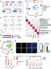 Figure 1
Single-cell transcriptome profiling and analysis of KS and loss of prespermatogonia in KS testis. (A) Schematic diagram of experimental workflow. Fetal samples (KS n = 2, control n = 2) and adult samples (KS n = 2, control n = 1). (B) Uniform manifold approximation and projection (UMAP) plot illustrating the major cell types (n = 38,952 cells) from KS samples (n = 2) and samples from aborted male fetuses (n = 2). Each dot corresponds to an individual cell, and the colors denote different cell types. (C) Expression patterns of marker genes displayed on the UMAP plot. (D) Heatmap of marker genes in each cluster and GO term enrichment. (E) Fraction of each cell types isolated from KS and normal samples. (F) UMAP plot of all germ cells from the embryonic sample (n = 1,866 cells, 4 samples). Each dot corresponds to an individual cell, and the colors denote different cell types. (G) Immunofluorescence costaining of DDX4 (green) and MAGEA4 (red). Scale bars, 50 μm. (H) The number of DDX4+MAGEA4– cells and MAGEA4+ cells in the fetal gonads of male control fetuses and KS fetuses. Data are mean ± SEM. Statistical analysis was performed using unpaired 2-sided t tests. n = 15 regions from 3 independent samples or 20 regions from 3 independent samples in each group. (I) Dot plot showing the expression of selected gene markers for different stages of germ cells. (J) The expression level of 4 selected genes located on the X chromosome with significant difference between control and KS samples. Statistical analysis was performed using 2-sided Wilcoxon’s rank-sum test; ****P < 0.0001.
To characterize major cell type differences between fetal KS and normal samples, we further analyzed their cell type proportions. Notably, the ratio of primordial germ cells was reduced in KS fetal testis, while Leydig cells exhibited a higher proportion than normal (Figure 1E and Supplemental Figure 1G). To reinforce the robustness of our findings, we performed H&E and immunofluorescence (IF) staining of fetal KS and control testes and observed a consistently significant decrease in the number of DDX4-positive cells (indicative of germ cells) in the KS group (Supplemental Figure 1E). Primordial germ cells differentiate into gonocytes in the first trimester of pregnancy and further differentiate into MAGEA4-positive prespermatogonia in the second and third trimesters of pregnancy (20, 21). According to cell markers, we observed 2 types of germ cells, gonocytes (POU5F1) and prespermatogonia (MAGEA4), in the control and KS testis (Figure 1, F and I, and Supplemental Figure 2, A and B). To confirm the development change of KS germ cells, we performed IF analysis, and we observed a significantly decreased number of MAGEA4-positive prespermatogonia and increased number of MAGEA4-negative germ cells in the KS testis (Figure 1, G and H), consistent with Lu et al.’s findings of differentiation abnormalities of fetal germ cells (17, 18). These results suggest that the differentiation potential of germ cells in KS fetal testis is severely impaired.
We assessed the gene expression changes between KS and control samples by analyzing differentially expressed genes (DEGs). We found 1,125 DEGs in germ cells (Supplemental Figure 2C). GO enrichment analysis showed enrichment of gonadal development and endoplasmic reticulum processes in upregulated genes, and translation-related and apoptosis-related processes in downregulated genes, suggesting a general dysfunction of the fetal germ cells in KS (Supplemental Figure 2, D and E). One-eighth (78/609) of the upregulated genes in the KS germ cells, including ATRX, DMD, EIF1AX, and HUWE1, was located on the X chromosome. This finding suggests that the extra X chromosome leads to upregulation of X-linked genes, which may be associated with germ cell development abnormality (Figure 1J).
Sertoli cell heterogeneity and defects in fetal KS testis. It was reported that inactivation of the extra X chromosome is necessary for Sertoli cell maturation and focal spermatogenesis in adult testis (10), but the effect of the extra X chromosome on KS fetal Sertoli cells is still not well known. To evaluate the effects of the extra X chromosome on Sertoli cells at the fetal stage, we characterized the heterogeneity of the Sertoli cell subset by clustering, and we observed 2 clusters of Sertoli cells (Figure 2A). Specifically, cluster 1 contained both KS and normal Sertoli cells. Normal Sertoli cells with XY chromosomes were negative for XIST, while KS Sertoli cells with an extra X chromosome were positive for XIST, indicating important roles of suppression of the extra X chromosome by XIST. Cluster 2 was composed of only KS Sertoli cells negative for XIST. It seems that XIST activation is important for the normal development of KS Sertoli cells. In addition, we found that the expression of AP-1 complex members, such as FOSB, increased in XIST-negative cells (Figure 2B). We performed IF and observed both FOSB-positive and FOSB-negative Sertoli cells (Figure 2C), consistent with the 2 clusters of KS Sertoli cells identified by UMAP analysis. XIST-negative KS Sertoli cells were different from the normal Sertoli cells and showed induced expression of FOSB. FOSB stimulates S phase entry and induces cyclin D1 expression (22). We observed an abnormal increase in the proportion of G2/M-phase cells in XIST-negative KS Sertoli cells by cell cycle analysis, indicating abnormal cell proliferation (Figure 2D).
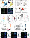 Figure 2
Defect and heterogeneity of fetal Sertoli cells in KS. (A) UMAP plot of all Sertoli cells (4 samples). Each dot corresponds to an individual cell, and colors represent different clusters and samples. (B) XIST and FOSB expression patterns drawn in UMAP. (C) Immunofluorescence costaining of SOX9 (green) and FOSB (red). Scale bars, 50 μm. White arrow indicates FOSB– Sertoli cell, and red arrow indicates FOSB+ Sertoli cell. (D) Ratios of cells in G0, S, and G2/M phases. (E) The expression levels of SOX9 and AMH were significantly different among control, XIST-positive, and XIST-negative KS samples. Statistical analysis was performed using 2-sided Wilcoxon’s rank-sum test; ****P < 0.0001. (F) GO terms enriched in fNorm Sertoli cells. The x axis is –log10(P value), and the size of the dots represents count. (G) GO terms enriched in fKS XIST+ Sertoli cells. The x axis is –log10(P value), and the size of the dots represents count. (H) GO terms enriched in fKS XIST– Sertoli cells. The x axis is –log10(P value), and the size of the dots represents count. (I) Immunofluorescence staining of ZO-1, β-catenin, and occludin. Scale bars, 50 μm. Insets original magnification, ×600. Fluorescence signal intensities of 3 proteins are shown as mean ± SEM. Statistical analysis was performed using unpaired 2-sided t tests (n = 4).
To elucidate the regulatory role of XIST expression on KS Sertoli cell function, we analyzed scRNA-seq data using 3 types of Sertoli cells. The expression of SOX9 and AMH was significantly reduced in XIST-negative KS Sertoli cells (Figure 2E). GO enrichment analysis revealed that both control and XIST-positive KS Sertoli cells were enriched in pathways related to cytoskeletal and cell adhesion, suggesting that they shared similar molecular characteristics and retained key functions of normal Sertoli cells (Figure 2, F and G). In contrast, XIST-negative KS Sertoli cells were enriched in unfolded protein reactions, misfolded protein reactions, and apoptosis, indicating that these cells were more severely damaged (Figure 2H and Supplemental Table 3). β-Catenin is a marker for cell adherens junctions, while occludin and ZO-1 are markers for tight junctions (23–26). During the fetal period, the expression of certain junctional proteins can already be detected in the testis, and focal tight junctions begin to form between Sertoli cells (27, 28). To evaluate the function of KS Sertoli cells, we examined the expression of these 3 proteins by IF. All 3 proteins showed reduced expression in KS testis (Figure 2I). Together, our study reveals that XIST expression plays an important role in the KS Sertoli cell development defect.
Abnormal intercellular communication network in fetal KS testis. To evaluate which cells might affect the Sertoli cells, we analyzed the interactions among different cell populations using CellChat (29). The results showed that both the number and strength of interactions among testicular cell populations were generally reduced in KS testis (Figure 3A). The cell-cell communication patterns identified Leydig cells as the primary senders both in KS and in control samples (Figure 3B). Given the close anatomical proximity between Leydig cells and Sertoli cells, and that androgens secreted by Leydig cells regulate Sertoli cell functions via their androgen receptors to support spermatogenesis, we further analyzed the intercellular communication between these 2 cell types and found that the midkine (MK) pathway exhibited the most marked alteration in KS testis (Figure 3C and Supplemental Figure 2, F and G). Notably, our cell-cell communication analysis identified the MK and PTN signaling pathways as the most differentially activated pathways in KS, which strongly replicates the prior findings by Mahyari et al. (30), where MK and PTN signaling were also ranked as the top upregulated pathways in adult KS testis. Although the intensity of the major sender of MK pathway signals did not differ between control and KS Leydig cells, the intensity of signals received by Sertoli cells as receivers changed (Figure 3D). Detailed ligand-receptor pair analysis of the MK pathway showed attenuated MDK-NCL, MDK-LRP, and MDK-(ITGA6+ITGB1) (Figure 3E). Among these interactions, MDK-NCL and MDK-LRP pairs were mainly restricted to the Leydig, Sertoli, and peritubular myoid cells. MDK, the ligand of the MK signaling pathway, was predominantly expressed in control Leydig cells (Figure 3F) but was significantly reduced in KS Leydig cells (Figure 3G). We performed qRT-PCR quantification and observed consistent lower expression in KS Leydig cells (Figure 3H). Studies have shown that MDK could activate prosurvival signaling through LRP receptor internalization and nucleolin-dependent translocation to the nucleus (31, 32). The MK signaling pathway has been also observed in species such as sheep, buffalo, and mice (33, 34). As for receptors of the MK signaling pathway, LRP1 was reduced in KS Sertoli cells, but NCL remained unchanged (Figure 3I). The downregulation of LRP1 was verified by qRT-PCR analysis of control and KS Sertoli cells (Figure 3J). The reduced MK pathway signaling from Leydig cells to Sertoli cells indicates possible important roles of the Leydig cell defects in KS testis.
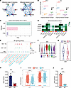 Figure 3
Intercellular interactions between Sertoli and other cell types in KS. (A) The circle diagrams depicting the relative number (left) or strength (right) of interactions in the cell-cell communication network between KS and control group. Red or blue edges represent an increase or decrease trend in signaling in KS group compared with control group, respectively. (B) Dot plot showing the relative strength of signaling pathways sent or received by each cell type (Endothelial, Sertoli, Peritubular myoid, Immune, Germ, Leydig). (C) Signaling pathways with the greatest variation between Sertoli cells and Leydig cells. PTN, pleiotrophin. (D) Heatmap shows the roles of different cell types in the MK signaling pathway. (E) Dot plot of ligand-receptor interactions in the MK pathway from different cell types to Sertoli in KS and control fetal testes. Commun. Prob., communication probability. (F) Expression of ligand MDK in testicular cell populations. (G) The expression of MDK in Leydig cells of KS and control. Statistical analysis was performed using 2-sided Wilcoxon’s rank-sum test; ****P < 0.0001. (H) The expression of MDK in control and KS primary Leydig cells using qRT-PCR. Data are presented as the mean ± SEM from 3 independent experiments. **P < 0.01. GAPDH was used as a loading control (n = 3). (I) The expression of LRP1 and NCL in Sertoli cells of KS and control. Statistical analysis was performed using 2-sided Wilcoxon’s rank-sum test; ****P < 0.0001. The triangle (▲) represents the arithmetic mean in each group. (J) The expression of LRP1 in control and KS primary Sertoli cells using qRT-PCR. Data are presented as the mean ± SEM from 3 independent experiments. ****P < 0.0001. GAPDH was used as a loading control (n = 3).
Delayed development of Leydig cells in KS fetal and adult testis. Previous studies have reported that adult KS testis exhibits a reduction or even absence of germ cells, along with hyperplasia of Leydig cells and impaired androgen secretory function (35). To further explore these findings, we performed scRNA-seq analysis of adult KS testes, and we annotated 6 major cell types, including Leydig cells (DLK1), Sertoli cells (SOX9), germ cells (DAZL), endothelial cells (VWF), immune cells (TYROBP), and peritubular myoid cells (RGS5) (Figure 4A and Supplemental Figure 3, A and B). The proportions of cell types showed reduction in germ cells and increase in Leydig cells in adult KS testis (Figure 4B). H&E staining revealed a substantial loss of seminiferous tubules in adult KS testis, with germ cell depletion and extensive interstitial hyperplasia (Figure 4C).
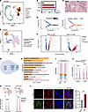 Figure 4
Delayed development of fetal Leydig cells in patients with KS. (A) UMAP plot of all cells in adults (3 samples). Dots represent individual cells, and colors represent different cell populations. (B) Cell ratio of cell types from A. (C) H&E staining of testicular biopsy samples from adult control (left) and KS (right) patients. (D) Pseudotime analysis of Leydig cells in adults. (E) UMAP plot of all Leydig cells in fetal stages (4 samples). Dots represent individual cells, and colors represent different cell populations. (F) Volcano plots compare control and KS Leydig cells for DEGs in fetuses (left) and adults (right). Genes with log2 (fold-change) above 0.25 or below –0.25 with P value lower than 0.05 were considered as having significantly differential expression. (G) Venn diagram of the 472 shared DEGs identified in both adult and fetal stages. (H) Bar plot showing GO terms enriched in shared DEGs. (I) Cell ratio of cells in the G1, S, and G2/M phases. (J) The expression level of MYC and BCL2 with significant difference between control and KS samples. Statistical analysis was performed using 2-sided Wilcoxon’s rank-sum test; ****P < 0.0001. (K) The expression level of CYP17A1 and CYP11A1 with significant difference between control and KS samples. Statistical analysis was performed using 2-sided Wilcoxon’s rank-sum test; ****P < 0.0001. (L) Left: Immunofluorescence costaining of NR2F2 (green) and CYP11A1 (red). Scale bars, 50 μm. Right: Data are presented as the mean ± SEM from 3 independent experiments. ***P < 0.001 (n = 3).
Pseudotime analysis of scRNA-seq of adult KS testis demonstrated that KS Leydig cells appeared at the beginning of the developmental trajectory, whereas control Leydig cells were located mainly at the end (Figure 4D). Meanwhile, compared with the control group, adult KS testis contained 4 additional clusters of Leydig cells, which coincidentally also existed at the fetal stage (Supplemental Figure 3C). All of the above suggest that Leydig cells in adult patients with KS exhibit immature developmental state. We further analyzed the molecular markers of Leydig cells during the fetal period and identified 4 subpopulations, including embryonic Leydig progenitor cells (MKI67), fetal Leydig progenitor cells (DLK1), fetal Leydig cells (CYP17A1), and KS Leydig cells (Figure 4E and Supplemental Figure 3, D and E). We examined 472 shared DEGs in both adult and fetal stages (Figure 4, F and G, and Supplemental Tables 4 and 5), which were enriched in biological processes of cell cycle regulation, apoptosis, and response to steroid hormone (Figure 4H and Supplemental Table 6). This finding suggests that the impairment of Leydig cells in KS may originate from fetal development, with abnormal gene expression patterns established early and potentially exacerbated in adulthood.
One of the major manifestations of KS is the massive proliferation of Leydig cells and even Leydig cell tumors in some patients in adulthood (36). We performed cell cycle analysis, and we found that KS Leydig cells exhibited cell cycle changes, with an increase in G2/M-phase cells (Figure 4I). This indicated that the cells rapidly enter the division phase. We also observed abnormally elevated expression of MYC and BCL2 genes in KS Leydig cells (Figure 4J and Supplemental Figure 3F). Previous studies showed that MYC and BCL2 were abnormally expressed and led to abnormal cell proliferation in many cancers, suggesting that these 2 genes may play an important role in the phenomenon of carcinogenesis of Leydig cells observed in patients with KS (37, 38).
Of particular note, another major phenotype of KS testis is impaired testosterone synthesis. CYP17A1 and CYP11A1 are key enzymes in the steroid hormone synthesis pathway (39), and they regulate the production of testosterone. The expression of CYP11A1 was significantly reduced in KS Leydig cells (P < 0.05), but CYP17A1 showed no difference (P > 0.05) (Figure 4K). We further analyzed fetal Leydig progenitor cells and fetal Leydig cells in KS testes by IF staining, using NR2F2 to mark progenitors and CYP11A1 to mark fetal Leydig cells. The results showed increased percentage of fetal Leydig progenitor cells and decreased percentage of fetal Leydig cells in KS testis (Figure 4L). The gene set enrichment analysis (GSEA) further indicated enrichment of adipocytokine signaling pathways and steroid hormone synthesis pathways in KS Leydig cells (Figure 5A and Supplemental Table 7). This finding suggests disturbances of fat metabolism and hormone regulation in Leydig cells. Taken together, these results support abnormal cell proliferation and differentiation in KS Leydig cells, characterized by impaired maturation and hormone synthesis pathway defects.
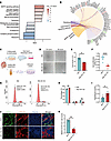 Figure 5
Inhibition of the MAPK pathway prevents overproliferation of KS Leydig cells and promotes testosterone production. (A) Bar plot showing the upregulated and downregulated Kyoto Encyclopedia of Genes and Genomes (KEGG) terms enriched in Leydig cells in the KS group compared with control. The length of the bar represents normalized enrichment score (NES). The color represents P value. (B) Chord diagram displaying the MAPK pathway and its associated genes. (C) Schematic diagram of fetal testis as well as primary Leydig cell culture. (D) Light micrographs of KS primary Leydig cells cultured with BSJ-04-122 or DMSO for 24 and 48 hours. Scale bar: 50 μm. (E) Cell count of D. Data are expressed as the mean ± SEM from triplicate experiments. *P < 0.05 (n = 3). (F) Cell proliferation analysis of KS primary Leydig cells cultured with BSJ-04-122 or DMSO for 48 hours. Data are expressed as the mean ± SEM from 5 experiments. ****P < 0.0001. (G) Cell cycle analysis of KS primary Leydig cells cultured with BSJ-04-122 or DMSO for 48 hours by flow cytometry (n = 6). (H) Statistical data on the cell cycle of 2 groups. Data are presented as the mean ± SEM from 3 independent experiments. **P < 0.01 (n = 3). (I) Testosterone content in the supernatant of KS primary Leydig cells cultured with BSJ-04-122 or DMSO for 48 hours. Data are expressed as the mean ± SEM from 5 experiments. *P < 0.05 (n = 5). (J) Immunofluorescence costaining of NR2F2 (green) and CYP11A1 (red). Scale bars, 50 μm. (K) Statistical data of J. Data are presented as the mean ± SEM from 3 independent experiments. **P < 0.01.
MAPK inhibition promotes the differentiation of KS Leydig cells. To further investigate the proliferation and differentiation capacity of KS Leydig cells, we performed GSEA and found that the MAPK pathway was significantly upregulated in the KS group, including genes such as MKK4, JUN, and FOS (Figure 5, A and B, and Supplemental Figure 4, A and B).
KS Leydig cells exhibited defects in proliferation and testosterone secretion. To explore the roles of the MAPK pathway on the proliferation and differentiation of KS Leydig cells, we first isolated Leydig cells from KS fetal testis (Figure 5C). Morphological analysis and NR2F2 staining showed the purity of the isolated cells exceeded 86% (Supplemental Figure 4C). With KS Leydig cells, we used the inhibitor BSJ-04-122 to inhibit the MAPK pathway, and we evaluated cell number changes after 24 and 48 hours (Figure 5, D and E). The results showed that compared with the control group, the BSJ-04-122–treated group exhibited a significant reduction in cell numbers, indicating important roles of MAPK pathway in Leydig cell proliferation. The cell counting kit-8 (CCK-8) assay was also used to assess the proliferative activity, and the results demonstrated consistently reduced proliferative activity in the BSJ-04-122–treated group (Figure 5F). Additionally, we analyzed the cell cycle distribution of KS Leydig cells in the BSJ-04-122–treated and control groups using flow cytometry. The results revealed a significant decrease in the proportion of G2/M cells in the BSJ-04-122–treated group compared with the control group (Figure 5, G and H). The G2/M stage involves completion of DNA replication and preparation for mitosis. The reduction in G2/M cells suggests that the MAPK pathway inhibition interfered with the cell cycle progression, thereby inhibiting Leydig cell proliferation.
Fetal Leydig cells are major hormone-synthesizing cells in the fetal testis and play a key role in male sex development and reproductive system formation. We collected the culture medium of primary KS Leydig cells with and without BSJ-04-122 treatment, and we measured the levels of testosterone. We found that the concentration of medium testosterone increased significantly in the BSJ-04-122 treatment group, and inhibition of the upregulated MAPK pathway rescued the testosterone secretion disorder in KS Leydig cells (Figure 5I). To assess the testosterone synthesis capacity of KS Leydig cells, we performed qRT-PCR for CYP11A1, CYP17A1, STAR, and 3B-HSD. Results revealed a significant upregulation in the BSJ-04-122–treated group for all targets except CYP17A1 (Supplemental Figure 4D). The upregulated MAPK pathway impaired proliferation and differentiation of KS Leydig cells. Meanwhile, we examined the effects of MAPK inhibitors in normal Leydig cells and reached the same conclusion: adding inhibitors reduced cell proliferation and increased testosterone secretion. This indicates that Leydig cell progenitor fate is balanced between proliferation and differentiation (Supplemental Figure 4, E–G).
To study ex vivo functions of the MAPK pathway, we utilized a gonad tissue culture system (Figure 5C) and treated KS fetal testis tissue with the inhibitor BSJ-04-122. After 7 days of culture, the ratio of NR2F2-positive fetal Leydig progenitor cells to CYP11A1-positive fetal Leydig cells in KS fetal testis significantly decreased (Figure 5, J and K). These results collectively support that upregulation of the MAPK pathway is an important cause of differentiation impairment of KS Leydig cells.
X-linked EIF1AX promotes proliferation of Leydig cell line. Among 472 DEGs between KS and control Leydig cells in both adult and fetal stages (Figure 4G), we found that EIF1AX, a gene located on the X chromosome, was abnormally elevated in KS Leydig cells (Figure 6A). To evaluate the expression changes of EIF1AX at the protein level, we performed Western blot analysis. The results showed that EIF1AX was higher in fetal KS Leydig cells than in normal Leydig cells at protein levels (Figure 6B). Previous studies have reported upregulation of EIF1AX in KS blood samples (14). Furthermore, EIF1AX overexpression was reported to cause cell cycle dysregulation and increase cell proliferation in breast cancer (40). To further explore the function of EIF1AX in Leydig cells, we measured proliferation and hormone synthesis capacity in Eif1ax-overexpressing TM3 cells (Figure 6C and Supplemental Figure 4H). We found that EIF1AX overexpression upregulated the cell proliferation based on the CCK-8 assay (Figure 6F). Flow cytometry analysis showed that Eif1ax overexpression increased the transition from G0/G1 to S+G2/M phases, especially G2/M phase (Figure 6, D and E). To test the testosterone synthesis ability of TM3 cells, we performed qRT-PCR for Cyp11a1, StAR, and 3b-Hsd, and we found a general decrease in gene expression (Figure 6G). Subsequently, we performed Western blot assay for CYP11A1 and found that Eif1ax overexpression resulted in a decrease in its protein expression in TM3 cells (Figure 6H). With an extra X chromosome in KS Leydig cells, upregulation of X-linked EIF1AX may be responsible for the aberrant proliferation of Leydig cells by facilitating the transition from G0/G1 to S+G2/M phases, which affects their hormone synthesis capacity.
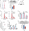 Figure 6
EIF1AX overexpression promotes TM3 cell proliferation and suppresses testosterone synthesis capacity. (A) The expression level of EIF1AX with significant difference between control and KS samples. Statistical analysis was performed using 2-sided Wilcoxon’s rank-sum test; ****P < 0.0001. (B) Western blot analysis of EIF1AX protein in KS fetal testis and control. Data are presented as the mean ± SEM from 3 independent experiments. *P < 0.05 (n = 3). (C) Western blot analysis of HA protein in control and EIF1AX-overexpressing TM3 cells. (D) Cell cycle analysis of control and EIF1AX-overexpressing TM3 cells by flow cytometry. (E) Statistical data on the cell cycle of 2 groups. Data are presented as the mean ± SEM from 3 independent experiments. *P < 0.05 (n = 3). (F) Cell proliferation analysis of control and EIF1AX-overexpressing TM3 cells. Data are expressed as the mean ± SEM from 10 experiments. ****P < 0.0001. (G) The expression of Cyp11a1, StAR, and 3b-Hsd in control and EIF1AX-overexpressing TM3 cells using qRT-PCR. Data are presented as the mean ± SEM from 3 independent experiments. *P < 0.05. **P < 0.01. GAPDH was used as a loading control (n = 3). (H) Western blot analysis of CYP11A1 protein in control and EIF1AX-overexpressing TM3 cells. Data are presented as the mean ± SEM from 3 independent experiments. *P < 0.05 (n = 3).


Copyright © 2026 American Society for Clinical Investigation
ISSN: 0021-9738 (print), 1558-8238 (online)