
Advertisement
Research ArticleGeneticsNephrology
Open Access |  10.1172/JCI196814
Cilia–to–basement membrane signaling is a biomechanical driver in models of autosomal dominant polycystic kidney disease
Manal Mazloum,1,2 Brice Lapin,3 Rushdi Alghamdi,1,4 Jessica Vandensteen,1,3 Martine Burtin,1 Pascal Houillier,5,6 Lydie Cheval,5 Gilles Crambert,5 Vicky Scata,1 Camille Cohen,7,8 Christoph Schell,9 Michael Rehman,10 Amandine Aka,1 Karim Ourahmoun,11 Rui Benedito,12 E. Wolfgang Kuehn,13 Stéphanie Descroix,3 Tilman Busch,13,14 Michael Köttgen,13,14 Serge Garbay,1 Marie-Christine Verpont,15 Ellie Tang,15 Brigitte Lelongt,15 Nicolas Cagnard,16 Stefan Somlo,10,17 Sylvie Coscoy,3 Fabiola Terzi,1 Amandine Viau,18 and Frank Bienaimé1,2
1Université Paris Cité, INSERM U1151, CNRS UMR 8235, Institut Necker Enfants Malades, Département Croissance et Signalisation, Paris, France.
2Assistance Publique-Hôpitaux de Paris, Hôpital Necker Enfants Malades, Service de Physiologie, Paris, France.
3Institut Curie, Université PSL, Sorbonne Université, CNRS UMR 168, Physics of Cells and Cancer, Paris, France.
4Department of Clinical Physiology, Faculty of Medicine, King Abdulaziz University, Jeddah, Saudi Arabia.
5Sorbonne Université et Université Paris Cité, Centre de Recherche des Cordeliers, INSERM, CNRS, Paris, France.
6Assistance Publique-Hôpitaux de Paris, Hôpital Européen Georges Pompidou, Service de Physiologie, Paris, France.
7Université Paris Cité, Centre de Recherche sur l’Inflammation, INSERM U1149, EMR 8252, Paris, France.
8Service de Néphrologie, Hôpital Bichat, Assistance Publique Hôpitaux de Paris, Paris, France.
9Institute of Surgical Pathology, Faculty of Medicine, Medical Center, University of Freiburg, Freiburg, Germany.
10Department of Internal Medicine, Yale School of Medicine, New Haven, Connecticut, USA.
11Institut du Cerveau et de la Moelle Épinière, INSERM U975, CNRS UMR 7225, Hôpital Pitié-Salpêtrière, Sorbonne Université, Paris, France.
12Molecular Genetics of Angiogenesis Group, Centro Nacional de Investigaciones Cardiovasculares, Madrid, Spain.
13Renal Division, Faculty of Medicine, Department of Medicine, Medical Center, University of Freiburg, Freiburg, Germany.
14Centre for Integrative Biological Signaling Studies, Freiburg, Germany.
15Sorbonne Université, CoRaKiD, INSERM UMRS 1155, Hôpital Tenon, Paris, France.
16Bioinformatic Platform, INSERM UMR 1163, Imagine Institute, Université Paris Cité, Paris, France.
17Department of Genetics, Yale School of Medicine, New Haven, Connecticut, USA.
18Université Paris Cité, Imagine Institute, Laboratory of Hereditary Kidney Diseases, INSERM UMR 1163, Paris, France.
Address correspondence to: Frank Bienaimé, INSERM U1151, Team: Mechanisms and Therapeutic Strategies of Chronic Kidney Disease, Institut Necker Enfants Malades, Bureau 606B, 160 Rue de Vaugirard, 75015 Paris, France. Phone: 33.1.40615366; Email: frank.bienaime@inserm.fr.
Authorship note: AV and FB contributed equally to this work.
Find articles by Mazloum, M. in: PubMed | Google Scholar
1Université Paris Cité, INSERM U1151, CNRS UMR 8235, Institut Necker Enfants Malades, Département Croissance et Signalisation, Paris, France.
2Assistance Publique-Hôpitaux de Paris, Hôpital Necker Enfants Malades, Service de Physiologie, Paris, France.
3Institut Curie, Université PSL, Sorbonne Université, CNRS UMR 168, Physics of Cells and Cancer, Paris, France.
4Department of Clinical Physiology, Faculty of Medicine, King Abdulaziz University, Jeddah, Saudi Arabia.
5Sorbonne Université et Université Paris Cité, Centre de Recherche des Cordeliers, INSERM, CNRS, Paris, France.
6Assistance Publique-Hôpitaux de Paris, Hôpital Européen Georges Pompidou, Service de Physiologie, Paris, France.
7Université Paris Cité, Centre de Recherche sur l’Inflammation, INSERM U1149, EMR 8252, Paris, France.
8Service de Néphrologie, Hôpital Bichat, Assistance Publique Hôpitaux de Paris, Paris, France.
9Institute of Surgical Pathology, Faculty of Medicine, Medical Center, University of Freiburg, Freiburg, Germany.
10Department of Internal Medicine, Yale School of Medicine, New Haven, Connecticut, USA.
11Institut du Cerveau et de la Moelle Épinière, INSERM U975, CNRS UMR 7225, Hôpital Pitié-Salpêtrière, Sorbonne Université, Paris, France.
12Molecular Genetics of Angiogenesis Group, Centro Nacional de Investigaciones Cardiovasculares, Madrid, Spain.
13Renal Division, Faculty of Medicine, Department of Medicine, Medical Center, University of Freiburg, Freiburg, Germany.
14Centre for Integrative Biological Signaling Studies, Freiburg, Germany.
15Sorbonne Université, CoRaKiD, INSERM UMRS 1155, Hôpital Tenon, Paris, France.
16Bioinformatic Platform, INSERM UMR 1163, Imagine Institute, Université Paris Cité, Paris, France.
17Department of Genetics, Yale School of Medicine, New Haven, Connecticut, USA.
18Université Paris Cité, Imagine Institute, Laboratory of Hereditary Kidney Diseases, INSERM UMR 1163, Paris, France.
Address correspondence to: Frank Bienaimé, INSERM U1151, Team: Mechanisms and Therapeutic Strategies of Chronic Kidney Disease, Institut Necker Enfants Malades, Bureau 606B, 160 Rue de Vaugirard, 75015 Paris, France. Phone: 33.1.40615366; Email: frank.bienaime@inserm.fr.
Authorship note: AV and FB contributed equally to this work.
Find articles by Lapin, B. in: PubMed | Google Scholar
1Université Paris Cité, INSERM U1151, CNRS UMR 8235, Institut Necker Enfants Malades, Département Croissance et Signalisation, Paris, France.
2Assistance Publique-Hôpitaux de Paris, Hôpital Necker Enfants Malades, Service de Physiologie, Paris, France.
3Institut Curie, Université PSL, Sorbonne Université, CNRS UMR 168, Physics of Cells and Cancer, Paris, France.
4Department of Clinical Physiology, Faculty of Medicine, King Abdulaziz University, Jeddah, Saudi Arabia.
5Sorbonne Université et Université Paris Cité, Centre de Recherche des Cordeliers, INSERM, CNRS, Paris, France.
6Assistance Publique-Hôpitaux de Paris, Hôpital Européen Georges Pompidou, Service de Physiologie, Paris, France.
7Université Paris Cité, Centre de Recherche sur l’Inflammation, INSERM U1149, EMR 8252, Paris, France.
8Service de Néphrologie, Hôpital Bichat, Assistance Publique Hôpitaux de Paris, Paris, France.
9Institute of Surgical Pathology, Faculty of Medicine, Medical Center, University of Freiburg, Freiburg, Germany.
10Department of Internal Medicine, Yale School of Medicine, New Haven, Connecticut, USA.
11Institut du Cerveau et de la Moelle Épinière, INSERM U975, CNRS UMR 7225, Hôpital Pitié-Salpêtrière, Sorbonne Université, Paris, France.
12Molecular Genetics of Angiogenesis Group, Centro Nacional de Investigaciones Cardiovasculares, Madrid, Spain.
13Renal Division, Faculty of Medicine, Department of Medicine, Medical Center, University of Freiburg, Freiburg, Germany.
14Centre for Integrative Biological Signaling Studies, Freiburg, Germany.
15Sorbonne Université, CoRaKiD, INSERM UMRS 1155, Hôpital Tenon, Paris, France.
16Bioinformatic Platform, INSERM UMR 1163, Imagine Institute, Université Paris Cité, Paris, France.
17Department of Genetics, Yale School of Medicine, New Haven, Connecticut, USA.
18Université Paris Cité, Imagine Institute, Laboratory of Hereditary Kidney Diseases, INSERM UMR 1163, Paris, France.
Address correspondence to: Frank Bienaimé, INSERM U1151, Team: Mechanisms and Therapeutic Strategies of Chronic Kidney Disease, Institut Necker Enfants Malades, Bureau 606B, 160 Rue de Vaugirard, 75015 Paris, France. Phone: 33.1.40615366; Email: frank.bienaime@inserm.fr.
Authorship note: AV and FB contributed equally to this work.
Find articles by Alghamdi, R. in: PubMed | Google Scholar
1Université Paris Cité, INSERM U1151, CNRS UMR 8235, Institut Necker Enfants Malades, Département Croissance et Signalisation, Paris, France.
2Assistance Publique-Hôpitaux de Paris, Hôpital Necker Enfants Malades, Service de Physiologie, Paris, France.
3Institut Curie, Université PSL, Sorbonne Université, CNRS UMR 168, Physics of Cells and Cancer, Paris, France.
4Department of Clinical Physiology, Faculty of Medicine, King Abdulaziz University, Jeddah, Saudi Arabia.
5Sorbonne Université et Université Paris Cité, Centre de Recherche des Cordeliers, INSERM, CNRS, Paris, France.
6Assistance Publique-Hôpitaux de Paris, Hôpital Européen Georges Pompidou, Service de Physiologie, Paris, France.
7Université Paris Cité, Centre de Recherche sur l’Inflammation, INSERM U1149, EMR 8252, Paris, France.
8Service de Néphrologie, Hôpital Bichat, Assistance Publique Hôpitaux de Paris, Paris, France.
9Institute of Surgical Pathology, Faculty of Medicine, Medical Center, University of Freiburg, Freiburg, Germany.
10Department of Internal Medicine, Yale School of Medicine, New Haven, Connecticut, USA.
11Institut du Cerveau et de la Moelle Épinière, INSERM U975, CNRS UMR 7225, Hôpital Pitié-Salpêtrière, Sorbonne Université, Paris, France.
12Molecular Genetics of Angiogenesis Group, Centro Nacional de Investigaciones Cardiovasculares, Madrid, Spain.
13Renal Division, Faculty of Medicine, Department of Medicine, Medical Center, University of Freiburg, Freiburg, Germany.
14Centre for Integrative Biological Signaling Studies, Freiburg, Germany.
15Sorbonne Université, CoRaKiD, INSERM UMRS 1155, Hôpital Tenon, Paris, France.
16Bioinformatic Platform, INSERM UMR 1163, Imagine Institute, Université Paris Cité, Paris, France.
17Department of Genetics, Yale School of Medicine, New Haven, Connecticut, USA.
18Université Paris Cité, Imagine Institute, Laboratory of Hereditary Kidney Diseases, INSERM UMR 1163, Paris, France.
Address correspondence to: Frank Bienaimé, INSERM U1151, Team: Mechanisms and Therapeutic Strategies of Chronic Kidney Disease, Institut Necker Enfants Malades, Bureau 606B, 160 Rue de Vaugirard, 75015 Paris, France. Phone: 33.1.40615366; Email: frank.bienaime@inserm.fr.
Authorship note: AV and FB contributed equally to this work.
Find articles by Vandensteen, J. in: PubMed | Google Scholar
1Université Paris Cité, INSERM U1151, CNRS UMR 8235, Institut Necker Enfants Malades, Département Croissance et Signalisation, Paris, France.
2Assistance Publique-Hôpitaux de Paris, Hôpital Necker Enfants Malades, Service de Physiologie, Paris, France.
3Institut Curie, Université PSL, Sorbonne Université, CNRS UMR 168, Physics of Cells and Cancer, Paris, France.
4Department of Clinical Physiology, Faculty of Medicine, King Abdulaziz University, Jeddah, Saudi Arabia.
5Sorbonne Université et Université Paris Cité, Centre de Recherche des Cordeliers, INSERM, CNRS, Paris, France.
6Assistance Publique-Hôpitaux de Paris, Hôpital Européen Georges Pompidou, Service de Physiologie, Paris, France.
7Université Paris Cité, Centre de Recherche sur l’Inflammation, INSERM U1149, EMR 8252, Paris, France.
8Service de Néphrologie, Hôpital Bichat, Assistance Publique Hôpitaux de Paris, Paris, France.
9Institute of Surgical Pathology, Faculty of Medicine, Medical Center, University of Freiburg, Freiburg, Germany.
10Department of Internal Medicine, Yale School of Medicine, New Haven, Connecticut, USA.
11Institut du Cerveau et de la Moelle Épinière, INSERM U975, CNRS UMR 7225, Hôpital Pitié-Salpêtrière, Sorbonne Université, Paris, France.
12Molecular Genetics of Angiogenesis Group, Centro Nacional de Investigaciones Cardiovasculares, Madrid, Spain.
13Renal Division, Faculty of Medicine, Department of Medicine, Medical Center, University of Freiburg, Freiburg, Germany.
14Centre for Integrative Biological Signaling Studies, Freiburg, Germany.
15Sorbonne Université, CoRaKiD, INSERM UMRS 1155, Hôpital Tenon, Paris, France.
16Bioinformatic Platform, INSERM UMR 1163, Imagine Institute, Université Paris Cité, Paris, France.
17Department of Genetics, Yale School of Medicine, New Haven, Connecticut, USA.
18Université Paris Cité, Imagine Institute, Laboratory of Hereditary Kidney Diseases, INSERM UMR 1163, Paris, France.
Address correspondence to: Frank Bienaimé, INSERM U1151, Team: Mechanisms and Therapeutic Strategies of Chronic Kidney Disease, Institut Necker Enfants Malades, Bureau 606B, 160 Rue de Vaugirard, 75015 Paris, France. Phone: 33.1.40615366; Email: frank.bienaime@inserm.fr.
Authorship note: AV and FB contributed equally to this work.
Find articles by Burtin, M. in: PubMed | Google Scholar
1Université Paris Cité, INSERM U1151, CNRS UMR 8235, Institut Necker Enfants Malades, Département Croissance et Signalisation, Paris, France.
2Assistance Publique-Hôpitaux de Paris, Hôpital Necker Enfants Malades, Service de Physiologie, Paris, France.
3Institut Curie, Université PSL, Sorbonne Université, CNRS UMR 168, Physics of Cells and Cancer, Paris, France.
4Department of Clinical Physiology, Faculty of Medicine, King Abdulaziz University, Jeddah, Saudi Arabia.
5Sorbonne Université et Université Paris Cité, Centre de Recherche des Cordeliers, INSERM, CNRS, Paris, France.
6Assistance Publique-Hôpitaux de Paris, Hôpital Européen Georges Pompidou, Service de Physiologie, Paris, France.
7Université Paris Cité, Centre de Recherche sur l’Inflammation, INSERM U1149, EMR 8252, Paris, France.
8Service de Néphrologie, Hôpital Bichat, Assistance Publique Hôpitaux de Paris, Paris, France.
9Institute of Surgical Pathology, Faculty of Medicine, Medical Center, University of Freiburg, Freiburg, Germany.
10Department of Internal Medicine, Yale School of Medicine, New Haven, Connecticut, USA.
11Institut du Cerveau et de la Moelle Épinière, INSERM U975, CNRS UMR 7225, Hôpital Pitié-Salpêtrière, Sorbonne Université, Paris, France.
12Molecular Genetics of Angiogenesis Group, Centro Nacional de Investigaciones Cardiovasculares, Madrid, Spain.
13Renal Division, Faculty of Medicine, Department of Medicine, Medical Center, University of Freiburg, Freiburg, Germany.
14Centre for Integrative Biological Signaling Studies, Freiburg, Germany.
15Sorbonne Université, CoRaKiD, INSERM UMRS 1155, Hôpital Tenon, Paris, France.
16Bioinformatic Platform, INSERM UMR 1163, Imagine Institute, Université Paris Cité, Paris, France.
17Department of Genetics, Yale School of Medicine, New Haven, Connecticut, USA.
18Université Paris Cité, Imagine Institute, Laboratory of Hereditary Kidney Diseases, INSERM UMR 1163, Paris, France.
Address correspondence to: Frank Bienaimé, INSERM U1151, Team: Mechanisms and Therapeutic Strategies of Chronic Kidney Disease, Institut Necker Enfants Malades, Bureau 606B, 160 Rue de Vaugirard, 75015 Paris, France. Phone: 33.1.40615366; Email: frank.bienaime@inserm.fr.
Authorship note: AV and FB contributed equally to this work.
Find articles by
Houillier, P.
in:
PubMed
|
Google Scholar
|

1Université Paris Cité, INSERM U1151, CNRS UMR 8235, Institut Necker Enfants Malades, Département Croissance et Signalisation, Paris, France.
2Assistance Publique-Hôpitaux de Paris, Hôpital Necker Enfants Malades, Service de Physiologie, Paris, France.
3Institut Curie, Université PSL, Sorbonne Université, CNRS UMR 168, Physics of Cells and Cancer, Paris, France.
4Department of Clinical Physiology, Faculty of Medicine, King Abdulaziz University, Jeddah, Saudi Arabia.
5Sorbonne Université et Université Paris Cité, Centre de Recherche des Cordeliers, INSERM, CNRS, Paris, France.
6Assistance Publique-Hôpitaux de Paris, Hôpital Européen Georges Pompidou, Service de Physiologie, Paris, France.
7Université Paris Cité, Centre de Recherche sur l’Inflammation, INSERM U1149, EMR 8252, Paris, France.
8Service de Néphrologie, Hôpital Bichat, Assistance Publique Hôpitaux de Paris, Paris, France.
9Institute of Surgical Pathology, Faculty of Medicine, Medical Center, University of Freiburg, Freiburg, Germany.
10Department of Internal Medicine, Yale School of Medicine, New Haven, Connecticut, USA.
11Institut du Cerveau et de la Moelle Épinière, INSERM U975, CNRS UMR 7225, Hôpital Pitié-Salpêtrière, Sorbonne Université, Paris, France.
12Molecular Genetics of Angiogenesis Group, Centro Nacional de Investigaciones Cardiovasculares, Madrid, Spain.
13Renal Division, Faculty of Medicine, Department of Medicine, Medical Center, University of Freiburg, Freiburg, Germany.
14Centre for Integrative Biological Signaling Studies, Freiburg, Germany.
15Sorbonne Université, CoRaKiD, INSERM UMRS 1155, Hôpital Tenon, Paris, France.
16Bioinformatic Platform, INSERM UMR 1163, Imagine Institute, Université Paris Cité, Paris, France.
17Department of Genetics, Yale School of Medicine, New Haven, Connecticut, USA.
18Université Paris Cité, Imagine Institute, Laboratory of Hereditary Kidney Diseases, INSERM UMR 1163, Paris, France.
Address correspondence to: Frank Bienaimé, INSERM U1151, Team: Mechanisms and Therapeutic Strategies of Chronic Kidney Disease, Institut Necker Enfants Malades, Bureau 606B, 160 Rue de Vaugirard, 75015 Paris, France. Phone: 33.1.40615366; Email: frank.bienaime@inserm.fr.
Authorship note: AV and FB contributed equally to this work.
Find articles by
Cheval, L.
in:
PubMed
|
Google Scholar
|
1Université Paris Cité, INSERM U1151, CNRS UMR 8235, Institut Necker Enfants Malades, Département Croissance et Signalisation, Paris, France.
2Assistance Publique-Hôpitaux de Paris, Hôpital Necker Enfants Malades, Service de Physiologie, Paris, France.
3Institut Curie, Université PSL, Sorbonne Université, CNRS UMR 168, Physics of Cells and Cancer, Paris, France.
4Department of Clinical Physiology, Faculty of Medicine, King Abdulaziz University, Jeddah, Saudi Arabia.
5Sorbonne Université et Université Paris Cité, Centre de Recherche des Cordeliers, INSERM, CNRS, Paris, France.
6Assistance Publique-Hôpitaux de Paris, Hôpital Européen Georges Pompidou, Service de Physiologie, Paris, France.
7Université Paris Cité, Centre de Recherche sur l’Inflammation, INSERM U1149, EMR 8252, Paris, France.
8Service de Néphrologie, Hôpital Bichat, Assistance Publique Hôpitaux de Paris, Paris, France.
9Institute of Surgical Pathology, Faculty of Medicine, Medical Center, University of Freiburg, Freiburg, Germany.
10Department of Internal Medicine, Yale School of Medicine, New Haven, Connecticut, USA.
11Institut du Cerveau et de la Moelle Épinière, INSERM U975, CNRS UMR 7225, Hôpital Pitié-Salpêtrière, Sorbonne Université, Paris, France.
12Molecular Genetics of Angiogenesis Group, Centro Nacional de Investigaciones Cardiovasculares, Madrid, Spain.
13Renal Division, Faculty of Medicine, Department of Medicine, Medical Center, University of Freiburg, Freiburg, Germany.
14Centre for Integrative Biological Signaling Studies, Freiburg, Germany.
15Sorbonne Université, CoRaKiD, INSERM UMRS 1155, Hôpital Tenon, Paris, France.
16Bioinformatic Platform, INSERM UMR 1163, Imagine Institute, Université Paris Cité, Paris, France.
17Department of Genetics, Yale School of Medicine, New Haven, Connecticut, USA.
18Université Paris Cité, Imagine Institute, Laboratory of Hereditary Kidney Diseases, INSERM UMR 1163, Paris, France.
Address correspondence to: Frank Bienaimé, INSERM U1151, Team: Mechanisms and Therapeutic Strategies of Chronic Kidney Disease, Institut Necker Enfants Malades, Bureau 606B, 160 Rue de Vaugirard, 75015 Paris, France. Phone: 33.1.40615366; Email: frank.bienaime@inserm.fr.
Authorship note: AV and FB contributed equally to this work.
Find articles by
Crambert, G.
in:
PubMed
|
Google Scholar
|
1Université Paris Cité, INSERM U1151, CNRS UMR 8235, Institut Necker Enfants Malades, Département Croissance et Signalisation, Paris, France.
2Assistance Publique-Hôpitaux de Paris, Hôpital Necker Enfants Malades, Service de Physiologie, Paris, France.
3Institut Curie, Université PSL, Sorbonne Université, CNRS UMR 168, Physics of Cells and Cancer, Paris, France.
4Department of Clinical Physiology, Faculty of Medicine, King Abdulaziz University, Jeddah, Saudi Arabia.
5Sorbonne Université et Université Paris Cité, Centre de Recherche des Cordeliers, INSERM, CNRS, Paris, France.
6Assistance Publique-Hôpitaux de Paris, Hôpital Européen Georges Pompidou, Service de Physiologie, Paris, France.
7Université Paris Cité, Centre de Recherche sur l’Inflammation, INSERM U1149, EMR 8252, Paris, France.
8Service de Néphrologie, Hôpital Bichat, Assistance Publique Hôpitaux de Paris, Paris, France.
9Institute of Surgical Pathology, Faculty of Medicine, Medical Center, University of Freiburg, Freiburg, Germany.
10Department of Internal Medicine, Yale School of Medicine, New Haven, Connecticut, USA.
11Institut du Cerveau et de la Moelle Épinière, INSERM U975, CNRS UMR 7225, Hôpital Pitié-Salpêtrière, Sorbonne Université, Paris, France.
12Molecular Genetics of Angiogenesis Group, Centro Nacional de Investigaciones Cardiovasculares, Madrid, Spain.
13Renal Division, Faculty of Medicine, Department of Medicine, Medical Center, University of Freiburg, Freiburg, Germany.
14Centre for Integrative Biological Signaling Studies, Freiburg, Germany.
15Sorbonne Université, CoRaKiD, INSERM UMRS 1155, Hôpital Tenon, Paris, France.
16Bioinformatic Platform, INSERM UMR 1163, Imagine Institute, Université Paris Cité, Paris, France.
17Department of Genetics, Yale School of Medicine, New Haven, Connecticut, USA.
18Université Paris Cité, Imagine Institute, Laboratory of Hereditary Kidney Diseases, INSERM UMR 1163, Paris, France.
Address correspondence to: Frank Bienaimé, INSERM U1151, Team: Mechanisms and Therapeutic Strategies of Chronic Kidney Disease, Institut Necker Enfants Malades, Bureau 606B, 160 Rue de Vaugirard, 75015 Paris, France. Phone: 33.1.40615366; Email: frank.bienaime@inserm.fr.
Authorship note: AV and FB contributed equally to this work.
Find articles by Scata, V. in: PubMed | Google Scholar
1Université Paris Cité, INSERM U1151, CNRS UMR 8235, Institut Necker Enfants Malades, Département Croissance et Signalisation, Paris, France.
2Assistance Publique-Hôpitaux de Paris, Hôpital Necker Enfants Malades, Service de Physiologie, Paris, France.
3Institut Curie, Université PSL, Sorbonne Université, CNRS UMR 168, Physics of Cells and Cancer, Paris, France.
4Department of Clinical Physiology, Faculty of Medicine, King Abdulaziz University, Jeddah, Saudi Arabia.
5Sorbonne Université et Université Paris Cité, Centre de Recherche des Cordeliers, INSERM, CNRS, Paris, France.
6Assistance Publique-Hôpitaux de Paris, Hôpital Européen Georges Pompidou, Service de Physiologie, Paris, France.
7Université Paris Cité, Centre de Recherche sur l’Inflammation, INSERM U1149, EMR 8252, Paris, France.
8Service de Néphrologie, Hôpital Bichat, Assistance Publique Hôpitaux de Paris, Paris, France.
9Institute of Surgical Pathology, Faculty of Medicine, Medical Center, University of Freiburg, Freiburg, Germany.
10Department of Internal Medicine, Yale School of Medicine, New Haven, Connecticut, USA.
11Institut du Cerveau et de la Moelle Épinière, INSERM U975, CNRS UMR 7225, Hôpital Pitié-Salpêtrière, Sorbonne Université, Paris, France.
12Molecular Genetics of Angiogenesis Group, Centro Nacional de Investigaciones Cardiovasculares, Madrid, Spain.
13Renal Division, Faculty of Medicine, Department of Medicine, Medical Center, University of Freiburg, Freiburg, Germany.
14Centre for Integrative Biological Signaling Studies, Freiburg, Germany.
15Sorbonne Université, CoRaKiD, INSERM UMRS 1155, Hôpital Tenon, Paris, France.
16Bioinformatic Platform, INSERM UMR 1163, Imagine Institute, Université Paris Cité, Paris, France.
17Department of Genetics, Yale School of Medicine, New Haven, Connecticut, USA.
18Université Paris Cité, Imagine Institute, Laboratory of Hereditary Kidney Diseases, INSERM UMR 1163, Paris, France.
Address correspondence to: Frank Bienaimé, INSERM U1151, Team: Mechanisms and Therapeutic Strategies of Chronic Kidney Disease, Institut Necker Enfants Malades, Bureau 606B, 160 Rue de Vaugirard, 75015 Paris, France. Phone: 33.1.40615366; Email: frank.bienaime@inserm.fr.
Authorship note: AV and FB contributed equally to this work.
Find articles by Cohen, C. in: PubMed | Google Scholar
1Université Paris Cité, INSERM U1151, CNRS UMR 8235, Institut Necker Enfants Malades, Département Croissance et Signalisation, Paris, France.
2Assistance Publique-Hôpitaux de Paris, Hôpital Necker Enfants Malades, Service de Physiologie, Paris, France.
3Institut Curie, Université PSL, Sorbonne Université, CNRS UMR 168, Physics of Cells and Cancer, Paris, France.
4Department of Clinical Physiology, Faculty of Medicine, King Abdulaziz University, Jeddah, Saudi Arabia.
5Sorbonne Université et Université Paris Cité, Centre de Recherche des Cordeliers, INSERM, CNRS, Paris, France.
6Assistance Publique-Hôpitaux de Paris, Hôpital Européen Georges Pompidou, Service de Physiologie, Paris, France.
7Université Paris Cité, Centre de Recherche sur l’Inflammation, INSERM U1149, EMR 8252, Paris, France.
8Service de Néphrologie, Hôpital Bichat, Assistance Publique Hôpitaux de Paris, Paris, France.
9Institute of Surgical Pathology, Faculty of Medicine, Medical Center, University of Freiburg, Freiburg, Germany.
10Department of Internal Medicine, Yale School of Medicine, New Haven, Connecticut, USA.
11Institut du Cerveau et de la Moelle Épinière, INSERM U975, CNRS UMR 7225, Hôpital Pitié-Salpêtrière, Sorbonne Université, Paris, France.
12Molecular Genetics of Angiogenesis Group, Centro Nacional de Investigaciones Cardiovasculares, Madrid, Spain.
13Renal Division, Faculty of Medicine, Department of Medicine, Medical Center, University of Freiburg, Freiburg, Germany.
14Centre for Integrative Biological Signaling Studies, Freiburg, Germany.
15Sorbonne Université, CoRaKiD, INSERM UMRS 1155, Hôpital Tenon, Paris, France.
16Bioinformatic Platform, INSERM UMR 1163, Imagine Institute, Université Paris Cité, Paris, France.
17Department of Genetics, Yale School of Medicine, New Haven, Connecticut, USA.
18Université Paris Cité, Imagine Institute, Laboratory of Hereditary Kidney Diseases, INSERM UMR 1163, Paris, France.
Address correspondence to: Frank Bienaimé, INSERM U1151, Team: Mechanisms and Therapeutic Strategies of Chronic Kidney Disease, Institut Necker Enfants Malades, Bureau 606B, 160 Rue de Vaugirard, 75015 Paris, France. Phone: 33.1.40615366; Email: frank.bienaime@inserm.fr.
Authorship note: AV and FB contributed equally to this work.
Find articles by Schell, C. in: PubMed | Google Scholar
1Université Paris Cité, INSERM U1151, CNRS UMR 8235, Institut Necker Enfants Malades, Département Croissance et Signalisation, Paris, France.
2Assistance Publique-Hôpitaux de Paris, Hôpital Necker Enfants Malades, Service de Physiologie, Paris, France.
3Institut Curie, Université PSL, Sorbonne Université, CNRS UMR 168, Physics of Cells and Cancer, Paris, France.
4Department of Clinical Physiology, Faculty of Medicine, King Abdulaziz University, Jeddah, Saudi Arabia.
5Sorbonne Université et Université Paris Cité, Centre de Recherche des Cordeliers, INSERM, CNRS, Paris, France.
6Assistance Publique-Hôpitaux de Paris, Hôpital Européen Georges Pompidou, Service de Physiologie, Paris, France.
7Université Paris Cité, Centre de Recherche sur l’Inflammation, INSERM U1149, EMR 8252, Paris, France.
8Service de Néphrologie, Hôpital Bichat, Assistance Publique Hôpitaux de Paris, Paris, France.
9Institute of Surgical Pathology, Faculty of Medicine, Medical Center, University of Freiburg, Freiburg, Germany.
10Department of Internal Medicine, Yale School of Medicine, New Haven, Connecticut, USA.
11Institut du Cerveau et de la Moelle Épinière, INSERM U975, CNRS UMR 7225, Hôpital Pitié-Salpêtrière, Sorbonne Université, Paris, France.
12Molecular Genetics of Angiogenesis Group, Centro Nacional de Investigaciones Cardiovasculares, Madrid, Spain.
13Renal Division, Faculty of Medicine, Department of Medicine, Medical Center, University of Freiburg, Freiburg, Germany.
14Centre for Integrative Biological Signaling Studies, Freiburg, Germany.
15Sorbonne Université, CoRaKiD, INSERM UMRS 1155, Hôpital Tenon, Paris, France.
16Bioinformatic Platform, INSERM UMR 1163, Imagine Institute, Université Paris Cité, Paris, France.
17Department of Genetics, Yale School of Medicine, New Haven, Connecticut, USA.
18Université Paris Cité, Imagine Institute, Laboratory of Hereditary Kidney Diseases, INSERM UMR 1163, Paris, France.
Address correspondence to: Frank Bienaimé, INSERM U1151, Team: Mechanisms and Therapeutic Strategies of Chronic Kidney Disease, Institut Necker Enfants Malades, Bureau 606B, 160 Rue de Vaugirard, 75015 Paris, France. Phone: 33.1.40615366; Email: frank.bienaime@inserm.fr.
Authorship note: AV and FB contributed equally to this work.
Find articles by Rehman, M. in: PubMed | Google Scholar
1Université Paris Cité, INSERM U1151, CNRS UMR 8235, Institut Necker Enfants Malades, Département Croissance et Signalisation, Paris, France.
2Assistance Publique-Hôpitaux de Paris, Hôpital Necker Enfants Malades, Service de Physiologie, Paris, France.
3Institut Curie, Université PSL, Sorbonne Université, CNRS UMR 168, Physics of Cells and Cancer, Paris, France.
4Department of Clinical Physiology, Faculty of Medicine, King Abdulaziz University, Jeddah, Saudi Arabia.
5Sorbonne Université et Université Paris Cité, Centre de Recherche des Cordeliers, INSERM, CNRS, Paris, France.
6Assistance Publique-Hôpitaux de Paris, Hôpital Européen Georges Pompidou, Service de Physiologie, Paris, France.
7Université Paris Cité, Centre de Recherche sur l’Inflammation, INSERM U1149, EMR 8252, Paris, France.
8Service de Néphrologie, Hôpital Bichat, Assistance Publique Hôpitaux de Paris, Paris, France.
9Institute of Surgical Pathology, Faculty of Medicine, Medical Center, University of Freiburg, Freiburg, Germany.
10Department of Internal Medicine, Yale School of Medicine, New Haven, Connecticut, USA.
11Institut du Cerveau et de la Moelle Épinière, INSERM U975, CNRS UMR 7225, Hôpital Pitié-Salpêtrière, Sorbonne Université, Paris, France.
12Molecular Genetics of Angiogenesis Group, Centro Nacional de Investigaciones Cardiovasculares, Madrid, Spain.
13Renal Division, Faculty of Medicine, Department of Medicine, Medical Center, University of Freiburg, Freiburg, Germany.
14Centre for Integrative Biological Signaling Studies, Freiburg, Germany.
15Sorbonne Université, CoRaKiD, INSERM UMRS 1155, Hôpital Tenon, Paris, France.
16Bioinformatic Platform, INSERM UMR 1163, Imagine Institute, Université Paris Cité, Paris, France.
17Department of Genetics, Yale School of Medicine, New Haven, Connecticut, USA.
18Université Paris Cité, Imagine Institute, Laboratory of Hereditary Kidney Diseases, INSERM UMR 1163, Paris, France.
Address correspondence to: Frank Bienaimé, INSERM U1151, Team: Mechanisms and Therapeutic Strategies of Chronic Kidney Disease, Institut Necker Enfants Malades, Bureau 606B, 160 Rue de Vaugirard, 75015 Paris, France. Phone: 33.1.40615366; Email: frank.bienaime@inserm.fr.
Authorship note: AV and FB contributed equally to this work.
Find articles by Aka, A. in: PubMed | Google Scholar
1Université Paris Cité, INSERM U1151, CNRS UMR 8235, Institut Necker Enfants Malades, Département Croissance et Signalisation, Paris, France.
2Assistance Publique-Hôpitaux de Paris, Hôpital Necker Enfants Malades, Service de Physiologie, Paris, France.
3Institut Curie, Université PSL, Sorbonne Université, CNRS UMR 168, Physics of Cells and Cancer, Paris, France.
4Department of Clinical Physiology, Faculty of Medicine, King Abdulaziz University, Jeddah, Saudi Arabia.
5Sorbonne Université et Université Paris Cité, Centre de Recherche des Cordeliers, INSERM, CNRS, Paris, France.
6Assistance Publique-Hôpitaux de Paris, Hôpital Européen Georges Pompidou, Service de Physiologie, Paris, France.
7Université Paris Cité, Centre de Recherche sur l’Inflammation, INSERM U1149, EMR 8252, Paris, France.
8Service de Néphrologie, Hôpital Bichat, Assistance Publique Hôpitaux de Paris, Paris, France.
9Institute of Surgical Pathology, Faculty of Medicine, Medical Center, University of Freiburg, Freiburg, Germany.
10Department of Internal Medicine, Yale School of Medicine, New Haven, Connecticut, USA.
11Institut du Cerveau et de la Moelle Épinière, INSERM U975, CNRS UMR 7225, Hôpital Pitié-Salpêtrière, Sorbonne Université, Paris, France.
12Molecular Genetics of Angiogenesis Group, Centro Nacional de Investigaciones Cardiovasculares, Madrid, Spain.
13Renal Division, Faculty of Medicine, Department of Medicine, Medical Center, University of Freiburg, Freiburg, Germany.
14Centre for Integrative Biological Signaling Studies, Freiburg, Germany.
15Sorbonne Université, CoRaKiD, INSERM UMRS 1155, Hôpital Tenon, Paris, France.
16Bioinformatic Platform, INSERM UMR 1163, Imagine Institute, Université Paris Cité, Paris, France.
17Department of Genetics, Yale School of Medicine, New Haven, Connecticut, USA.
18Université Paris Cité, Imagine Institute, Laboratory of Hereditary Kidney Diseases, INSERM UMR 1163, Paris, France.
Address correspondence to: Frank Bienaimé, INSERM U1151, Team: Mechanisms and Therapeutic Strategies of Chronic Kidney Disease, Institut Necker Enfants Malades, Bureau 606B, 160 Rue de Vaugirard, 75015 Paris, France. Phone: 33.1.40615366; Email: frank.bienaime@inserm.fr.
Authorship note: AV and FB contributed equally to this work.
Find articles by Ourahmoun, K. in: PubMed | Google Scholar
1Université Paris Cité, INSERM U1151, CNRS UMR 8235, Institut Necker Enfants Malades, Département Croissance et Signalisation, Paris, France.
2Assistance Publique-Hôpitaux de Paris, Hôpital Necker Enfants Malades, Service de Physiologie, Paris, France.
3Institut Curie, Université PSL, Sorbonne Université, CNRS UMR 168, Physics of Cells and Cancer, Paris, France.
4Department of Clinical Physiology, Faculty of Medicine, King Abdulaziz University, Jeddah, Saudi Arabia.
5Sorbonne Université et Université Paris Cité, Centre de Recherche des Cordeliers, INSERM, CNRS, Paris, France.
6Assistance Publique-Hôpitaux de Paris, Hôpital Européen Georges Pompidou, Service de Physiologie, Paris, France.
7Université Paris Cité, Centre de Recherche sur l’Inflammation, INSERM U1149, EMR 8252, Paris, France.
8Service de Néphrologie, Hôpital Bichat, Assistance Publique Hôpitaux de Paris, Paris, France.
9Institute of Surgical Pathology, Faculty of Medicine, Medical Center, University of Freiburg, Freiburg, Germany.
10Department of Internal Medicine, Yale School of Medicine, New Haven, Connecticut, USA.
11Institut du Cerveau et de la Moelle Épinière, INSERM U975, CNRS UMR 7225, Hôpital Pitié-Salpêtrière, Sorbonne Université, Paris, France.
12Molecular Genetics of Angiogenesis Group, Centro Nacional de Investigaciones Cardiovasculares, Madrid, Spain.
13Renal Division, Faculty of Medicine, Department of Medicine, Medical Center, University of Freiburg, Freiburg, Germany.
14Centre for Integrative Biological Signaling Studies, Freiburg, Germany.
15Sorbonne Université, CoRaKiD, INSERM UMRS 1155, Hôpital Tenon, Paris, France.
16Bioinformatic Platform, INSERM UMR 1163, Imagine Institute, Université Paris Cité, Paris, France.
17Department of Genetics, Yale School of Medicine, New Haven, Connecticut, USA.
18Université Paris Cité, Imagine Institute, Laboratory of Hereditary Kidney Diseases, INSERM UMR 1163, Paris, France.
Address correspondence to: Frank Bienaimé, INSERM U1151, Team: Mechanisms and Therapeutic Strategies of Chronic Kidney Disease, Institut Necker Enfants Malades, Bureau 606B, 160 Rue de Vaugirard, 75015 Paris, France. Phone: 33.1.40615366; Email: frank.bienaime@inserm.fr.
Authorship note: AV and FB contributed equally to this work.
Find articles by Benedito, R. in: PubMed | Google Scholar
1Université Paris Cité, INSERM U1151, CNRS UMR 8235, Institut Necker Enfants Malades, Département Croissance et Signalisation, Paris, France.
2Assistance Publique-Hôpitaux de Paris, Hôpital Necker Enfants Malades, Service de Physiologie, Paris, France.
3Institut Curie, Université PSL, Sorbonne Université, CNRS UMR 168, Physics of Cells and Cancer, Paris, France.
4Department of Clinical Physiology, Faculty of Medicine, King Abdulaziz University, Jeddah, Saudi Arabia.
5Sorbonne Université et Université Paris Cité, Centre de Recherche des Cordeliers, INSERM, CNRS, Paris, France.
6Assistance Publique-Hôpitaux de Paris, Hôpital Européen Georges Pompidou, Service de Physiologie, Paris, France.
7Université Paris Cité, Centre de Recherche sur l’Inflammation, INSERM U1149, EMR 8252, Paris, France.
8Service de Néphrologie, Hôpital Bichat, Assistance Publique Hôpitaux de Paris, Paris, France.
9Institute of Surgical Pathology, Faculty of Medicine, Medical Center, University of Freiburg, Freiburg, Germany.
10Department of Internal Medicine, Yale School of Medicine, New Haven, Connecticut, USA.
11Institut du Cerveau et de la Moelle Épinière, INSERM U975, CNRS UMR 7225, Hôpital Pitié-Salpêtrière, Sorbonne Université, Paris, France.
12Molecular Genetics of Angiogenesis Group, Centro Nacional de Investigaciones Cardiovasculares, Madrid, Spain.
13Renal Division, Faculty of Medicine, Department of Medicine, Medical Center, University of Freiburg, Freiburg, Germany.
14Centre for Integrative Biological Signaling Studies, Freiburg, Germany.
15Sorbonne Université, CoRaKiD, INSERM UMRS 1155, Hôpital Tenon, Paris, France.
16Bioinformatic Platform, INSERM UMR 1163, Imagine Institute, Université Paris Cité, Paris, France.
17Department of Genetics, Yale School of Medicine, New Haven, Connecticut, USA.
18Université Paris Cité, Imagine Institute, Laboratory of Hereditary Kidney Diseases, INSERM UMR 1163, Paris, France.
Address correspondence to: Frank Bienaimé, INSERM U1151, Team: Mechanisms and Therapeutic Strategies of Chronic Kidney Disease, Institut Necker Enfants Malades, Bureau 606B, 160 Rue de Vaugirard, 75015 Paris, France. Phone: 33.1.40615366; Email: frank.bienaime@inserm.fr.
Authorship note: AV and FB contributed equally to this work.
Find articles by Kuehn, E. in: PubMed | Google Scholar
1Université Paris Cité, INSERM U1151, CNRS UMR 8235, Institut Necker Enfants Malades, Département Croissance et Signalisation, Paris, France.
2Assistance Publique-Hôpitaux de Paris, Hôpital Necker Enfants Malades, Service de Physiologie, Paris, France.
3Institut Curie, Université PSL, Sorbonne Université, CNRS UMR 168, Physics of Cells and Cancer, Paris, France.
4Department of Clinical Physiology, Faculty of Medicine, King Abdulaziz University, Jeddah, Saudi Arabia.
5Sorbonne Université et Université Paris Cité, Centre de Recherche des Cordeliers, INSERM, CNRS, Paris, France.
6Assistance Publique-Hôpitaux de Paris, Hôpital Européen Georges Pompidou, Service de Physiologie, Paris, France.
7Université Paris Cité, Centre de Recherche sur l’Inflammation, INSERM U1149, EMR 8252, Paris, France.
8Service de Néphrologie, Hôpital Bichat, Assistance Publique Hôpitaux de Paris, Paris, France.
9Institute of Surgical Pathology, Faculty of Medicine, Medical Center, University of Freiburg, Freiburg, Germany.
10Department of Internal Medicine, Yale School of Medicine, New Haven, Connecticut, USA.
11Institut du Cerveau et de la Moelle Épinière, INSERM U975, CNRS UMR 7225, Hôpital Pitié-Salpêtrière, Sorbonne Université, Paris, France.
12Molecular Genetics of Angiogenesis Group, Centro Nacional de Investigaciones Cardiovasculares, Madrid, Spain.
13Renal Division, Faculty of Medicine, Department of Medicine, Medical Center, University of Freiburg, Freiburg, Germany.
14Centre for Integrative Biological Signaling Studies, Freiburg, Germany.
15Sorbonne Université, CoRaKiD, INSERM UMRS 1155, Hôpital Tenon, Paris, France.
16Bioinformatic Platform, INSERM UMR 1163, Imagine Institute, Université Paris Cité, Paris, France.
17Department of Genetics, Yale School of Medicine, New Haven, Connecticut, USA.
18Université Paris Cité, Imagine Institute, Laboratory of Hereditary Kidney Diseases, INSERM UMR 1163, Paris, France.
Address correspondence to: Frank Bienaimé, INSERM U1151, Team: Mechanisms and Therapeutic Strategies of Chronic Kidney Disease, Institut Necker Enfants Malades, Bureau 606B, 160 Rue de Vaugirard, 75015 Paris, France. Phone: 33.1.40615366; Email: frank.bienaime@inserm.fr.
Authorship note: AV and FB contributed equally to this work.
Find articles by Descroix, S. in: PubMed | Google Scholar
1Université Paris Cité, INSERM U1151, CNRS UMR 8235, Institut Necker Enfants Malades, Département Croissance et Signalisation, Paris, France.
2Assistance Publique-Hôpitaux de Paris, Hôpital Necker Enfants Malades, Service de Physiologie, Paris, France.
3Institut Curie, Université PSL, Sorbonne Université, CNRS UMR 168, Physics of Cells and Cancer, Paris, France.
4Department of Clinical Physiology, Faculty of Medicine, King Abdulaziz University, Jeddah, Saudi Arabia.
5Sorbonne Université et Université Paris Cité, Centre de Recherche des Cordeliers, INSERM, CNRS, Paris, France.
6Assistance Publique-Hôpitaux de Paris, Hôpital Européen Georges Pompidou, Service de Physiologie, Paris, France.
7Université Paris Cité, Centre de Recherche sur l’Inflammation, INSERM U1149, EMR 8252, Paris, France.
8Service de Néphrologie, Hôpital Bichat, Assistance Publique Hôpitaux de Paris, Paris, France.
9Institute of Surgical Pathology, Faculty of Medicine, Medical Center, University of Freiburg, Freiburg, Germany.
10Department of Internal Medicine, Yale School of Medicine, New Haven, Connecticut, USA.
11Institut du Cerveau et de la Moelle Épinière, INSERM U975, CNRS UMR 7225, Hôpital Pitié-Salpêtrière, Sorbonne Université, Paris, France.
12Molecular Genetics of Angiogenesis Group, Centro Nacional de Investigaciones Cardiovasculares, Madrid, Spain.
13Renal Division, Faculty of Medicine, Department of Medicine, Medical Center, University of Freiburg, Freiburg, Germany.
14Centre for Integrative Biological Signaling Studies, Freiburg, Germany.
15Sorbonne Université, CoRaKiD, INSERM UMRS 1155, Hôpital Tenon, Paris, France.
16Bioinformatic Platform, INSERM UMR 1163, Imagine Institute, Université Paris Cité, Paris, France.
17Department of Genetics, Yale School of Medicine, New Haven, Connecticut, USA.
18Université Paris Cité, Imagine Institute, Laboratory of Hereditary Kidney Diseases, INSERM UMR 1163, Paris, France.
Address correspondence to: Frank Bienaimé, INSERM U1151, Team: Mechanisms and Therapeutic Strategies of Chronic Kidney Disease, Institut Necker Enfants Malades, Bureau 606B, 160 Rue de Vaugirard, 75015 Paris, France. Phone: 33.1.40615366; Email: frank.bienaime@inserm.fr.
Authorship note: AV and FB contributed equally to this work.
Find articles by Busch, T. in: PubMed | Google Scholar
1Université Paris Cité, INSERM U1151, CNRS UMR 8235, Institut Necker Enfants Malades, Département Croissance et Signalisation, Paris, France.
2Assistance Publique-Hôpitaux de Paris, Hôpital Necker Enfants Malades, Service de Physiologie, Paris, France.
3Institut Curie, Université PSL, Sorbonne Université, CNRS UMR 168, Physics of Cells and Cancer, Paris, France.
4Department of Clinical Physiology, Faculty of Medicine, King Abdulaziz University, Jeddah, Saudi Arabia.
5Sorbonne Université et Université Paris Cité, Centre de Recherche des Cordeliers, INSERM, CNRS, Paris, France.
6Assistance Publique-Hôpitaux de Paris, Hôpital Européen Georges Pompidou, Service de Physiologie, Paris, France.
7Université Paris Cité, Centre de Recherche sur l’Inflammation, INSERM U1149, EMR 8252, Paris, France.
8Service de Néphrologie, Hôpital Bichat, Assistance Publique Hôpitaux de Paris, Paris, France.
9Institute of Surgical Pathology, Faculty of Medicine, Medical Center, University of Freiburg, Freiburg, Germany.
10Department of Internal Medicine, Yale School of Medicine, New Haven, Connecticut, USA.
11Institut du Cerveau et de la Moelle Épinière, INSERM U975, CNRS UMR 7225, Hôpital Pitié-Salpêtrière, Sorbonne Université, Paris, France.
12Molecular Genetics of Angiogenesis Group, Centro Nacional de Investigaciones Cardiovasculares, Madrid, Spain.
13Renal Division, Faculty of Medicine, Department of Medicine, Medical Center, University of Freiburg, Freiburg, Germany.
14Centre for Integrative Biological Signaling Studies, Freiburg, Germany.
15Sorbonne Université, CoRaKiD, INSERM UMRS 1155, Hôpital Tenon, Paris, France.
16Bioinformatic Platform, INSERM UMR 1163, Imagine Institute, Université Paris Cité, Paris, France.
17Department of Genetics, Yale School of Medicine, New Haven, Connecticut, USA.
18Université Paris Cité, Imagine Institute, Laboratory of Hereditary Kidney Diseases, INSERM UMR 1163, Paris, France.
Address correspondence to: Frank Bienaimé, INSERM U1151, Team: Mechanisms and Therapeutic Strategies of Chronic Kidney Disease, Institut Necker Enfants Malades, Bureau 606B, 160 Rue de Vaugirard, 75015 Paris, France. Phone: 33.1.40615366; Email: frank.bienaime@inserm.fr.
Authorship note: AV and FB contributed equally to this work.
Find articles by Köttgen, M. in: PubMed | Google Scholar
1Université Paris Cité, INSERM U1151, CNRS UMR 8235, Institut Necker Enfants Malades, Département Croissance et Signalisation, Paris, France.
2Assistance Publique-Hôpitaux de Paris, Hôpital Necker Enfants Malades, Service de Physiologie, Paris, France.
3Institut Curie, Université PSL, Sorbonne Université, CNRS UMR 168, Physics of Cells and Cancer, Paris, France.
4Department of Clinical Physiology, Faculty of Medicine, King Abdulaziz University, Jeddah, Saudi Arabia.
5Sorbonne Université et Université Paris Cité, Centre de Recherche des Cordeliers, INSERM, CNRS, Paris, France.
6Assistance Publique-Hôpitaux de Paris, Hôpital Européen Georges Pompidou, Service de Physiologie, Paris, France.
7Université Paris Cité, Centre de Recherche sur l’Inflammation, INSERM U1149, EMR 8252, Paris, France.
8Service de Néphrologie, Hôpital Bichat, Assistance Publique Hôpitaux de Paris, Paris, France.
9Institute of Surgical Pathology, Faculty of Medicine, Medical Center, University of Freiburg, Freiburg, Germany.
10Department of Internal Medicine, Yale School of Medicine, New Haven, Connecticut, USA.
11Institut du Cerveau et de la Moelle Épinière, INSERM U975, CNRS UMR 7225, Hôpital Pitié-Salpêtrière, Sorbonne Université, Paris, France.
12Molecular Genetics of Angiogenesis Group, Centro Nacional de Investigaciones Cardiovasculares, Madrid, Spain.
13Renal Division, Faculty of Medicine, Department of Medicine, Medical Center, University of Freiburg, Freiburg, Germany.
14Centre for Integrative Biological Signaling Studies, Freiburg, Germany.
15Sorbonne Université, CoRaKiD, INSERM UMRS 1155, Hôpital Tenon, Paris, France.
16Bioinformatic Platform, INSERM UMR 1163, Imagine Institute, Université Paris Cité, Paris, France.
17Department of Genetics, Yale School of Medicine, New Haven, Connecticut, USA.
18Université Paris Cité, Imagine Institute, Laboratory of Hereditary Kidney Diseases, INSERM UMR 1163, Paris, France.
Address correspondence to: Frank Bienaimé, INSERM U1151, Team: Mechanisms and Therapeutic Strategies of Chronic Kidney Disease, Institut Necker Enfants Malades, Bureau 606B, 160 Rue de Vaugirard, 75015 Paris, France. Phone: 33.1.40615366; Email: frank.bienaime@inserm.fr.
Authorship note: AV and FB contributed equally to this work.
Find articles by Garbay, S. in: PubMed | Google Scholar
1Université Paris Cité, INSERM U1151, CNRS UMR 8235, Institut Necker Enfants Malades, Département Croissance et Signalisation, Paris, France.
2Assistance Publique-Hôpitaux de Paris, Hôpital Necker Enfants Malades, Service de Physiologie, Paris, France.
3Institut Curie, Université PSL, Sorbonne Université, CNRS UMR 168, Physics of Cells and Cancer, Paris, France.
4Department of Clinical Physiology, Faculty of Medicine, King Abdulaziz University, Jeddah, Saudi Arabia.
5Sorbonne Université et Université Paris Cité, Centre de Recherche des Cordeliers, INSERM, CNRS, Paris, France.
6Assistance Publique-Hôpitaux de Paris, Hôpital Européen Georges Pompidou, Service de Physiologie, Paris, France.
7Université Paris Cité, Centre de Recherche sur l’Inflammation, INSERM U1149, EMR 8252, Paris, France.
8Service de Néphrologie, Hôpital Bichat, Assistance Publique Hôpitaux de Paris, Paris, France.
9Institute of Surgical Pathology, Faculty of Medicine, Medical Center, University of Freiburg, Freiburg, Germany.
10Department of Internal Medicine, Yale School of Medicine, New Haven, Connecticut, USA.
11Institut du Cerveau et de la Moelle Épinière, INSERM U975, CNRS UMR 7225, Hôpital Pitié-Salpêtrière, Sorbonne Université, Paris, France.
12Molecular Genetics of Angiogenesis Group, Centro Nacional de Investigaciones Cardiovasculares, Madrid, Spain.
13Renal Division, Faculty of Medicine, Department of Medicine, Medical Center, University of Freiburg, Freiburg, Germany.
14Centre for Integrative Biological Signaling Studies, Freiburg, Germany.
15Sorbonne Université, CoRaKiD, INSERM UMRS 1155, Hôpital Tenon, Paris, France.
16Bioinformatic Platform, INSERM UMR 1163, Imagine Institute, Université Paris Cité, Paris, France.
17Department of Genetics, Yale School of Medicine, New Haven, Connecticut, USA.
18Université Paris Cité, Imagine Institute, Laboratory of Hereditary Kidney Diseases, INSERM UMR 1163, Paris, France.
Address correspondence to: Frank Bienaimé, INSERM U1151, Team: Mechanisms and Therapeutic Strategies of Chronic Kidney Disease, Institut Necker Enfants Malades, Bureau 606B, 160 Rue de Vaugirard, 75015 Paris, France. Phone: 33.1.40615366; Email: frank.bienaime@inserm.fr.
Authorship note: AV and FB contributed equally to this work.
Find articles by Verpont, M. in: PubMed | Google Scholar
1Université Paris Cité, INSERM U1151, CNRS UMR 8235, Institut Necker Enfants Malades, Département Croissance et Signalisation, Paris, France.
2Assistance Publique-Hôpitaux de Paris, Hôpital Necker Enfants Malades, Service de Physiologie, Paris, France.
3Institut Curie, Université PSL, Sorbonne Université, CNRS UMR 168, Physics of Cells and Cancer, Paris, France.
4Department of Clinical Physiology, Faculty of Medicine, King Abdulaziz University, Jeddah, Saudi Arabia.
5Sorbonne Université et Université Paris Cité, Centre de Recherche des Cordeliers, INSERM, CNRS, Paris, France.
6Assistance Publique-Hôpitaux de Paris, Hôpital Européen Georges Pompidou, Service de Physiologie, Paris, France.
7Université Paris Cité, Centre de Recherche sur l’Inflammation, INSERM U1149, EMR 8252, Paris, France.
8Service de Néphrologie, Hôpital Bichat, Assistance Publique Hôpitaux de Paris, Paris, France.
9Institute of Surgical Pathology, Faculty of Medicine, Medical Center, University of Freiburg, Freiburg, Germany.
10Department of Internal Medicine, Yale School of Medicine, New Haven, Connecticut, USA.
11Institut du Cerveau et de la Moelle Épinière, INSERM U975, CNRS UMR 7225, Hôpital Pitié-Salpêtrière, Sorbonne Université, Paris, France.
12Molecular Genetics of Angiogenesis Group, Centro Nacional de Investigaciones Cardiovasculares, Madrid, Spain.
13Renal Division, Faculty of Medicine, Department of Medicine, Medical Center, University of Freiburg, Freiburg, Germany.
14Centre for Integrative Biological Signaling Studies, Freiburg, Germany.
15Sorbonne Université, CoRaKiD, INSERM UMRS 1155, Hôpital Tenon, Paris, France.
16Bioinformatic Platform, INSERM UMR 1163, Imagine Institute, Université Paris Cité, Paris, France.
17Department of Genetics, Yale School of Medicine, New Haven, Connecticut, USA.
18Université Paris Cité, Imagine Institute, Laboratory of Hereditary Kidney Diseases, INSERM UMR 1163, Paris, France.
Address correspondence to: Frank Bienaimé, INSERM U1151, Team: Mechanisms and Therapeutic Strategies of Chronic Kidney Disease, Institut Necker Enfants Malades, Bureau 606B, 160 Rue de Vaugirard, 75015 Paris, France. Phone: 33.1.40615366; Email: frank.bienaime@inserm.fr.
Authorship note: AV and FB contributed equally to this work.
Find articles by Tang, E. in: PubMed | Google Scholar
1Université Paris Cité, INSERM U1151, CNRS UMR 8235, Institut Necker Enfants Malades, Département Croissance et Signalisation, Paris, France.
2Assistance Publique-Hôpitaux de Paris, Hôpital Necker Enfants Malades, Service de Physiologie, Paris, France.
3Institut Curie, Université PSL, Sorbonne Université, CNRS UMR 168, Physics of Cells and Cancer, Paris, France.
4Department of Clinical Physiology, Faculty of Medicine, King Abdulaziz University, Jeddah, Saudi Arabia.
5Sorbonne Université et Université Paris Cité, Centre de Recherche des Cordeliers, INSERM, CNRS, Paris, France.
6Assistance Publique-Hôpitaux de Paris, Hôpital Européen Georges Pompidou, Service de Physiologie, Paris, France.
7Université Paris Cité, Centre de Recherche sur l’Inflammation, INSERM U1149, EMR 8252, Paris, France.
8Service de Néphrologie, Hôpital Bichat, Assistance Publique Hôpitaux de Paris, Paris, France.
9Institute of Surgical Pathology, Faculty of Medicine, Medical Center, University of Freiburg, Freiburg, Germany.
10Department of Internal Medicine, Yale School of Medicine, New Haven, Connecticut, USA.
11Institut du Cerveau et de la Moelle Épinière, INSERM U975, CNRS UMR 7225, Hôpital Pitié-Salpêtrière, Sorbonne Université, Paris, France.
12Molecular Genetics of Angiogenesis Group, Centro Nacional de Investigaciones Cardiovasculares, Madrid, Spain.
13Renal Division, Faculty of Medicine, Department of Medicine, Medical Center, University of Freiburg, Freiburg, Germany.
14Centre for Integrative Biological Signaling Studies, Freiburg, Germany.
15Sorbonne Université, CoRaKiD, INSERM UMRS 1155, Hôpital Tenon, Paris, France.
16Bioinformatic Platform, INSERM UMR 1163, Imagine Institute, Université Paris Cité, Paris, France.
17Department of Genetics, Yale School of Medicine, New Haven, Connecticut, USA.
18Université Paris Cité, Imagine Institute, Laboratory of Hereditary Kidney Diseases, INSERM UMR 1163, Paris, France.
Address correspondence to: Frank Bienaimé, INSERM U1151, Team: Mechanisms and Therapeutic Strategies of Chronic Kidney Disease, Institut Necker Enfants Malades, Bureau 606B, 160 Rue de Vaugirard, 75015 Paris, France. Phone: 33.1.40615366; Email: frank.bienaime@inserm.fr.
Authorship note: AV and FB contributed equally to this work.
Find articles by Lelongt, B. in: PubMed | Google Scholar
1Université Paris Cité, INSERM U1151, CNRS UMR 8235, Institut Necker Enfants Malades, Département Croissance et Signalisation, Paris, France.
2Assistance Publique-Hôpitaux de Paris, Hôpital Necker Enfants Malades, Service de Physiologie, Paris, France.
3Institut Curie, Université PSL, Sorbonne Université, CNRS UMR 168, Physics of Cells and Cancer, Paris, France.
4Department of Clinical Physiology, Faculty of Medicine, King Abdulaziz University, Jeddah, Saudi Arabia.
5Sorbonne Université et Université Paris Cité, Centre de Recherche des Cordeliers, INSERM, CNRS, Paris, France.
6Assistance Publique-Hôpitaux de Paris, Hôpital Européen Georges Pompidou, Service de Physiologie, Paris, France.
7Université Paris Cité, Centre de Recherche sur l’Inflammation, INSERM U1149, EMR 8252, Paris, France.
8Service de Néphrologie, Hôpital Bichat, Assistance Publique Hôpitaux de Paris, Paris, France.
9Institute of Surgical Pathology, Faculty of Medicine, Medical Center, University of Freiburg, Freiburg, Germany.
10Department of Internal Medicine, Yale School of Medicine, New Haven, Connecticut, USA.
11Institut du Cerveau et de la Moelle Épinière, INSERM U975, CNRS UMR 7225, Hôpital Pitié-Salpêtrière, Sorbonne Université, Paris, France.
12Molecular Genetics of Angiogenesis Group, Centro Nacional de Investigaciones Cardiovasculares, Madrid, Spain.
13Renal Division, Faculty of Medicine, Department of Medicine, Medical Center, University of Freiburg, Freiburg, Germany.
14Centre for Integrative Biological Signaling Studies, Freiburg, Germany.
15Sorbonne Université, CoRaKiD, INSERM UMRS 1155, Hôpital Tenon, Paris, France.
16Bioinformatic Platform, INSERM UMR 1163, Imagine Institute, Université Paris Cité, Paris, France.
17Department of Genetics, Yale School of Medicine, New Haven, Connecticut, USA.
18Université Paris Cité, Imagine Institute, Laboratory of Hereditary Kidney Diseases, INSERM UMR 1163, Paris, France.
Address correspondence to: Frank Bienaimé, INSERM U1151, Team: Mechanisms and Therapeutic Strategies of Chronic Kidney Disease, Institut Necker Enfants Malades, Bureau 606B, 160 Rue de Vaugirard, 75015 Paris, France. Phone: 33.1.40615366; Email: frank.bienaime@inserm.fr.
Authorship note: AV and FB contributed equally to this work.
Find articles by
Cagnard, N.
in:
PubMed
|
Google Scholar
|
1Université Paris Cité, INSERM U1151, CNRS UMR 8235, Institut Necker Enfants Malades, Département Croissance et Signalisation, Paris, France.
2Assistance Publique-Hôpitaux de Paris, Hôpital Necker Enfants Malades, Service de Physiologie, Paris, France.
3Institut Curie, Université PSL, Sorbonne Université, CNRS UMR 168, Physics of Cells and Cancer, Paris, France.
4Department of Clinical Physiology, Faculty of Medicine, King Abdulaziz University, Jeddah, Saudi Arabia.
5Sorbonne Université et Université Paris Cité, Centre de Recherche des Cordeliers, INSERM, CNRS, Paris, France.
6Assistance Publique-Hôpitaux de Paris, Hôpital Européen Georges Pompidou, Service de Physiologie, Paris, France.
7Université Paris Cité, Centre de Recherche sur l’Inflammation, INSERM U1149, EMR 8252, Paris, France.
8Service de Néphrologie, Hôpital Bichat, Assistance Publique Hôpitaux de Paris, Paris, France.
9Institute of Surgical Pathology, Faculty of Medicine, Medical Center, University of Freiburg, Freiburg, Germany.
10Department of Internal Medicine, Yale School of Medicine, New Haven, Connecticut, USA.
11Institut du Cerveau et de la Moelle Épinière, INSERM U975, CNRS UMR 7225, Hôpital Pitié-Salpêtrière, Sorbonne Université, Paris, France.
12Molecular Genetics of Angiogenesis Group, Centro Nacional de Investigaciones Cardiovasculares, Madrid, Spain.
13Renal Division, Faculty of Medicine, Department of Medicine, Medical Center, University of Freiburg, Freiburg, Germany.
14Centre for Integrative Biological Signaling Studies, Freiburg, Germany.
15Sorbonne Université, CoRaKiD, INSERM UMRS 1155, Hôpital Tenon, Paris, France.
16Bioinformatic Platform, INSERM UMR 1163, Imagine Institute, Université Paris Cité, Paris, France.
17Department of Genetics, Yale School of Medicine, New Haven, Connecticut, USA.
18Université Paris Cité, Imagine Institute, Laboratory of Hereditary Kidney Diseases, INSERM UMR 1163, Paris, France.
Address correspondence to: Frank Bienaimé, INSERM U1151, Team: Mechanisms and Therapeutic Strategies of Chronic Kidney Disease, Institut Necker Enfants Malades, Bureau 606B, 160 Rue de Vaugirard, 75015 Paris, France. Phone: 33.1.40615366; Email: frank.bienaime@inserm.fr.
Authorship note: AV and FB contributed equally to this work.
Find articles by Somlo, S. in: PubMed | Google Scholar
1Université Paris Cité, INSERM U1151, CNRS UMR 8235, Institut Necker Enfants Malades, Département Croissance et Signalisation, Paris, France.
2Assistance Publique-Hôpitaux de Paris, Hôpital Necker Enfants Malades, Service de Physiologie, Paris, France.
3Institut Curie, Université PSL, Sorbonne Université, CNRS UMR 168, Physics of Cells and Cancer, Paris, France.
4Department of Clinical Physiology, Faculty of Medicine, King Abdulaziz University, Jeddah, Saudi Arabia.
5Sorbonne Université et Université Paris Cité, Centre de Recherche des Cordeliers, INSERM, CNRS, Paris, France.
6Assistance Publique-Hôpitaux de Paris, Hôpital Européen Georges Pompidou, Service de Physiologie, Paris, France.
7Université Paris Cité, Centre de Recherche sur l’Inflammation, INSERM U1149, EMR 8252, Paris, France.
8Service de Néphrologie, Hôpital Bichat, Assistance Publique Hôpitaux de Paris, Paris, France.
9Institute of Surgical Pathology, Faculty of Medicine, Medical Center, University of Freiburg, Freiburg, Germany.
10Department of Internal Medicine, Yale School of Medicine, New Haven, Connecticut, USA.
11Institut du Cerveau et de la Moelle Épinière, INSERM U975, CNRS UMR 7225, Hôpital Pitié-Salpêtrière, Sorbonne Université, Paris, France.
12Molecular Genetics of Angiogenesis Group, Centro Nacional de Investigaciones Cardiovasculares, Madrid, Spain.
13Renal Division, Faculty of Medicine, Department of Medicine, Medical Center, University of Freiburg, Freiburg, Germany.
14Centre for Integrative Biological Signaling Studies, Freiburg, Germany.
15Sorbonne Université, CoRaKiD, INSERM UMRS 1155, Hôpital Tenon, Paris, France.
16Bioinformatic Platform, INSERM UMR 1163, Imagine Institute, Université Paris Cité, Paris, France.
17Department of Genetics, Yale School of Medicine, New Haven, Connecticut, USA.
18Université Paris Cité, Imagine Institute, Laboratory of Hereditary Kidney Diseases, INSERM UMR 1163, Paris, France.
Address correspondence to: Frank Bienaimé, INSERM U1151, Team: Mechanisms and Therapeutic Strategies of Chronic Kidney Disease, Institut Necker Enfants Malades, Bureau 606B, 160 Rue de Vaugirard, 75015 Paris, France. Phone: 33.1.40615366; Email: frank.bienaime@inserm.fr.
Authorship note: AV and FB contributed equally to this work.
Find articles by Coscoy, S. in: PubMed | Google Scholar
1Université Paris Cité, INSERM U1151, CNRS UMR 8235, Institut Necker Enfants Malades, Département Croissance et Signalisation, Paris, France.
2Assistance Publique-Hôpitaux de Paris, Hôpital Necker Enfants Malades, Service de Physiologie, Paris, France.
3Institut Curie, Université PSL, Sorbonne Université, CNRS UMR 168, Physics of Cells and Cancer, Paris, France.
4Department of Clinical Physiology, Faculty of Medicine, King Abdulaziz University, Jeddah, Saudi Arabia.
5Sorbonne Université et Université Paris Cité, Centre de Recherche des Cordeliers, INSERM, CNRS, Paris, France.
6Assistance Publique-Hôpitaux de Paris, Hôpital Européen Georges Pompidou, Service de Physiologie, Paris, France.
7Université Paris Cité, Centre de Recherche sur l’Inflammation, INSERM U1149, EMR 8252, Paris, France.
8Service de Néphrologie, Hôpital Bichat, Assistance Publique Hôpitaux de Paris, Paris, France.
9Institute of Surgical Pathology, Faculty of Medicine, Medical Center, University of Freiburg, Freiburg, Germany.
10Department of Internal Medicine, Yale School of Medicine, New Haven, Connecticut, USA.
11Institut du Cerveau et de la Moelle Épinière, INSERM U975, CNRS UMR 7225, Hôpital Pitié-Salpêtrière, Sorbonne Université, Paris, France.
12Molecular Genetics of Angiogenesis Group, Centro Nacional de Investigaciones Cardiovasculares, Madrid, Spain.
13Renal Division, Faculty of Medicine, Department of Medicine, Medical Center, University of Freiburg, Freiburg, Germany.
14Centre for Integrative Biological Signaling Studies, Freiburg, Germany.
15Sorbonne Université, CoRaKiD, INSERM UMRS 1155, Hôpital Tenon, Paris, France.
16Bioinformatic Platform, INSERM UMR 1163, Imagine Institute, Université Paris Cité, Paris, France.
17Department of Genetics, Yale School of Medicine, New Haven, Connecticut, USA.
18Université Paris Cité, Imagine Institute, Laboratory of Hereditary Kidney Diseases, INSERM UMR 1163, Paris, France.
Address correspondence to: Frank Bienaimé, INSERM U1151, Team: Mechanisms and Therapeutic Strategies of Chronic Kidney Disease, Institut Necker Enfants Malades, Bureau 606B, 160 Rue de Vaugirard, 75015 Paris, France. Phone: 33.1.40615366; Email: frank.bienaime@inserm.fr.
Authorship note: AV and FB contributed equally to this work.
Find articles by Terzi, F. in: PubMed | Google Scholar
1Université Paris Cité, INSERM U1151, CNRS UMR 8235, Institut Necker Enfants Malades, Département Croissance et Signalisation, Paris, France.
2Assistance Publique-Hôpitaux de Paris, Hôpital Necker Enfants Malades, Service de Physiologie, Paris, France.
3Institut Curie, Université PSL, Sorbonne Université, CNRS UMR 168, Physics of Cells and Cancer, Paris, France.
4Department of Clinical Physiology, Faculty of Medicine, King Abdulaziz University, Jeddah, Saudi Arabia.
5Sorbonne Université et Université Paris Cité, Centre de Recherche des Cordeliers, INSERM, CNRS, Paris, France.
6Assistance Publique-Hôpitaux de Paris, Hôpital Européen Georges Pompidou, Service de Physiologie, Paris, France.
7Université Paris Cité, Centre de Recherche sur l’Inflammation, INSERM U1149, EMR 8252, Paris, France.
8Service de Néphrologie, Hôpital Bichat, Assistance Publique Hôpitaux de Paris, Paris, France.
9Institute of Surgical Pathology, Faculty of Medicine, Medical Center, University of Freiburg, Freiburg, Germany.
10Department of Internal Medicine, Yale School of Medicine, New Haven, Connecticut, USA.
11Institut du Cerveau et de la Moelle Épinière, INSERM U975, CNRS UMR 7225, Hôpital Pitié-Salpêtrière, Sorbonne Université, Paris, France.
12Molecular Genetics of Angiogenesis Group, Centro Nacional de Investigaciones Cardiovasculares, Madrid, Spain.
13Renal Division, Faculty of Medicine, Department of Medicine, Medical Center, University of Freiburg, Freiburg, Germany.
14Centre for Integrative Biological Signaling Studies, Freiburg, Germany.
15Sorbonne Université, CoRaKiD, INSERM UMRS 1155, Hôpital Tenon, Paris, France.
16Bioinformatic Platform, INSERM UMR 1163, Imagine Institute, Université Paris Cité, Paris, France.
17Department of Genetics, Yale School of Medicine, New Haven, Connecticut, USA.
18Université Paris Cité, Imagine Institute, Laboratory of Hereditary Kidney Diseases, INSERM UMR 1163, Paris, France.
Address correspondence to: Frank Bienaimé, INSERM U1151, Team: Mechanisms and Therapeutic Strategies of Chronic Kidney Disease, Institut Necker Enfants Malades, Bureau 606B, 160 Rue de Vaugirard, 75015 Paris, France. Phone: 33.1.40615366; Email: frank.bienaime@inserm.fr.
Authorship note: AV and FB contributed equally to this work.
Find articles by
Viau, A.
in:
PubMed
|
Google Scholar
|
1Université Paris Cité, INSERM U1151, CNRS UMR 8235, Institut Necker Enfants Malades, Département Croissance et Signalisation, Paris, France.
2Assistance Publique-Hôpitaux de Paris, Hôpital Necker Enfants Malades, Service de Physiologie, Paris, France.
3Institut Curie, Université PSL, Sorbonne Université, CNRS UMR 168, Physics of Cells and Cancer, Paris, France.
4Department of Clinical Physiology, Faculty of Medicine, King Abdulaziz University, Jeddah, Saudi Arabia.
5Sorbonne Université et Université Paris Cité, Centre de Recherche des Cordeliers, INSERM, CNRS, Paris, France.
6Assistance Publique-Hôpitaux de Paris, Hôpital Européen Georges Pompidou, Service de Physiologie, Paris, France.
7Université Paris Cité, Centre de Recherche sur l’Inflammation, INSERM U1149, EMR 8252, Paris, France.
8Service de Néphrologie, Hôpital Bichat, Assistance Publique Hôpitaux de Paris, Paris, France.
9Institute of Surgical Pathology, Faculty of Medicine, Medical Center, University of Freiburg, Freiburg, Germany.
10Department of Internal Medicine, Yale School of Medicine, New Haven, Connecticut, USA.
11Institut du Cerveau et de la Moelle Épinière, INSERM U975, CNRS UMR 7225, Hôpital Pitié-Salpêtrière, Sorbonne Université, Paris, France.
12Molecular Genetics of Angiogenesis Group, Centro Nacional de Investigaciones Cardiovasculares, Madrid, Spain.
13Renal Division, Faculty of Medicine, Department of Medicine, Medical Center, University of Freiburg, Freiburg, Germany.
14Centre for Integrative Biological Signaling Studies, Freiburg, Germany.
15Sorbonne Université, CoRaKiD, INSERM UMRS 1155, Hôpital Tenon, Paris, France.
16Bioinformatic Platform, INSERM UMR 1163, Imagine Institute, Université Paris Cité, Paris, France.
17Department of Genetics, Yale School of Medicine, New Haven, Connecticut, USA.
18Université Paris Cité, Imagine Institute, Laboratory of Hereditary Kidney Diseases, INSERM UMR 1163, Paris, France.
Address correspondence to: Frank Bienaimé, INSERM U1151, Team: Mechanisms and Therapeutic Strategies of Chronic Kidney Disease, Institut Necker Enfants Malades, Bureau 606B, 160 Rue de Vaugirard, 75015 Paris, France. Phone: 33.1.40615366; Email: frank.bienaime@inserm.fr.
Authorship note: AV and FB contributed equally to this work.
Find articles by Bienaimé, F. in: PubMed | Google Scholar
Published June 30, 2026 - More info
J Clin Invest. 2026;136(18):e196814. https://doi.org/10.1172/JCI196814.
© 2026 Mazloum et al. This work is licensed under the Creative Commons Attribution 4.0 International License. To view a copy of this license, visit http://creativecommons.org/licenses/by/4.0/.
Received: June 16, 2025; Accepted: June 16, 2026

Related article:
Abstract
Autosomal dominant polycystic kidney disease (ADPKD), mainly driven by pathogenic variants in PKD1 and PKD2, is the most common inherited cause of kidney failure. Details of ADPKD pathogenesis are incompletely resolved, but primary cilia are an integral component. There is also evidence for changes to the tubular basement membrane (TBM) in early disease. In this issue of the JCI, Mazloum and colleagues link cilia-dependent, PKD1-mediated regulation of the TBM to ADPKD pathogenesis. Using in vivo, ex vivo, tubule-on-chip, and cellular models of ADPKD, they connect cilia-dependent tubule dilation and TBM thinning to early-stage cystogenesis. Moreover, they identify a cilia-dependent TBM remodeling expression signature in affected tubules and suggest that PC1 loss compromises TBM stiffness. By integrating roles for cilia at the apical membrane and extracellular matrix at the basolateral membrane in cystogenesis, this work highlights the TBM as an additional area for investigation of therapeutic and biomarker discovery in ADPKD.
Authors
Caroline R. Sussman, Peter C. Harris

-
Results
Pkd1 loss triggers cilia-dependent proximal and distal nephron dilation through distinct mechanisms. To gain insights into the early morphogenic events underlying tubule dilation, we first studied male mice with a postdevelopmental inactivation of Pkd1 (Pkd1Δtub) with or without simultaneous ablation of primary cilium (Kif3a or Ift20 inactivation: Pkd1Δtub Kif3aΔtub and Pkd1Δtub Ift20Δtub) at a very early stage of the disease, that is, 2 weeks after the completion of doxycycline treatment (i.e., 8 weeks of age), when tubules display only slight dilations (Figure 1A). We used the Pax8rtTA system, which drives functional Cre expression along the nephron (Supplemental Figure 1A; supplemental material available online with this article; https://doi.org/10.1172/JCI196814DS1), efficiently excises floxed Pkd1 alleles (Supplemental Figure 1B), and achieves near complete cilia loss in Pkd1Δtub Kif3aΔtub tubules (Supplemental Figure 1C). Morphometric analysis confirmed that Pkd1 loss induces an increase in cross-sectional areas of proximal tubules (PTs) and distal tubules (consisting of collecting ducts [CDs] and distal convoluted tubules [DCTs]), which was prevented by concomitant cilia disruption following Kif3a or Ift20 inactivation (Figure 1, A–C). Ki67 labeling revealed that PC1 deficiency increased cell proliferation in PTs (Figure 1, A and D), but not in the distal nephron (Figure 1, A, B, and D). Instead, we observed increased tubular cell stretching in CDs and DCTs, as reflected by an increase in the mean distance between 2 adjacent nuclei (later referred to as internuclear distance; Figure 1, A, B, and E). Linear regression analysis showed that distension (i.e., increase in internuclear distance) was the main factor explaining distal nephron dilation at this stage (Figure 1, F and G). At a later time point, distension and increased proliferation were observed in both proximal and distal Pkd1–/– tubules (Figure 1, H–K). Genetic cilia ablation (e.g., Kif3a or Ift20 inactivation) rescued PC1-deficient distal nephron distension, tubular cell proliferation, and cyst development (Figure 1, H–K). Analysis of 18-week-old animals confirmed that cilia ablation fully rescued tubule deformation in Pkd1Δtub mice (Supplemental Figure 2). Collectively, these results identify cilia-driven tubule distension as an important factor responsible for the early dilation of PC1-deficient tubules.
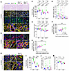 Figure 1
Pkd1 deletion drives cilia-dependent distal tubule distension independently of cell proliferation. (A and B) Periodic acid Schiff (PAS) staining and labeling of Ki67 (which stains proliferating cells), DNA, wheat germ agglutinin (WGA; which stains the brush border of PTs and all BMs), and aquaporin 2 (AQP2; a CD marker) (A) or calbindin (a marker of DCTs) (B) of kidneys from 8-week-old control, Pkd1Δtub, Pkd1Δtub Kif3aΔtub, and Pkd1Δtub Ift20Δtub male mice. Arrows: examples of internuclear distance measurements. Scale bars: 10 μm. (C–E) Quantification of mean PT, CD, and DCT cross-sectional area (C); proliferation index (percentage of Ki67+ cells; D); and internuclear distance (E) in PTs, CDs, and DCTs of kidneys from 8-week-old control, Pkd1Δtub, Pkd1Δtub Kif3aΔtub, and Pkd1Δtub Ift20Δtub mice. (F and G) Linear regression of tubule cross-sectional area and internuclear distance for CDs (F) and DCTs (G) in the same mice groups at 8 weeks. (H) Labeling of Ki67, DNA, WGA, and AQP2 of kidneys from 12-week-old control, Pkd1Δtub, and Pkd1Δtub Kif3aΔtub mice. Arrows: examples of internuclear distance measurements. Scale bars: 10 μm. (I–K) Quantification of mean tubule cross-sectional area (I), proliferation index (Ki67+ cells) (J), and internuclear distance (K) in PTs and CDs of kidneys from 12-week-old control, Pkd1Δtub, and Pkd1Δtub Kif3aΔtub mice. Each dot represents 1 male mouse. One-way ANOVA followed by Tukey-Kramer test: *P < 0.05, **P < 0.01, ***P < 0.001, ****P < 0.0001, or the indicated P value. Pkd1fl/fl and Pkd1fl/fl Ift20fl/fl littermates lacking cre or rtTA transgene were used as control. Confirmatory analyses on distal collecting tubules were only performed in the first cohort of mice analyzed.
Preferential distal tubule distension correlates with specific changes of the TBM. As tubule shape is determined by TBM geometry (16), we reasoned that CD distension could be linked to specific TBM properties. TBM stiffness depends on its thickness and composition: laminin and cross-linked collagen IV provide rigidity, while the sugar moieties of HS and chondroitin sulfate (CS) confer flexibility by increasing BM hydration (18). Studying these parameters in basal conditions, we noticed that CDs had thinner TBMs compared with PTs with higher HS content, suggesting an increased compliance (Figure 2, A and B), as historically suggested by pressure-volume measurements on isolated rabbit tubules (16). We further observed that distal nephron distension in Pkd1Δtub mice was associated with prominent TBM thinning (Figure 2C). TBM thinning also occurred in PTs but to a lesser extent than in CDs or DCTs (Figure 2C). A careful inspection of multiple tubule sections via transmission electron microscopy failed to detect any perforation or tubular cell protrusion through the TBM (Figure 2C and Supplemental Figure 3). Cilia ablation abolished the ability of PC1-deficient tubular cells to induce TBM thinning (Figure 2C). We further observed a specific increase in HS immunoreactivity of the TBM of PC1-deficient tubules undergoing distension, which was prevented by cilia disruption, without notable changes in collagen IV and laminin labeling (Figure 2, D–G). These results indicate that Pkd1 loss is associated with a cilia-dependent remodeling of the TBM. They also suggest that intrinsic properties of distal nephron TBMs may render this segment permissive to distension. Detailed transmission electron micrographs of TBMs from male mice are presented in Supplemental Figure 3.
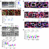 Figure 2
Pkd1 deletion induces cilia-dependent BM remodeling. (A) Transmission electron microscopy and quantification of PT and CD TBM thickness in 8-week-old control mice. Dashed lines underline TBM. Each dot represents 1 tubule (mean of 3–21 measurements per tubule; n = 3 mice). Scale bar: 0.5 μm. Student’s t test: ****P < 0.0001. (B) Labeling of PT brush border (WGA), CD (AQP2), and HS (left panel); collagen IV (COL4; middle panel); or laminin (right panel) and quantification of HS, COL4, and laminin signal intensity in 8-week-old control mice. Each dot represents 1 male mouse. Scale bars: 10 μm. Student’s t test: ***P < 0.001. (C) Transmission electron microscopy and quantification of TBM thickness of PTs, CDs, and DCTs from 8-week-old control, Pkd1Δtub, Pkd1Δtub Kif3aΔtub, and Pkd1Δtub Ift20Δtub mice (i.e., 2 weeks after the end of doxycycline treatment). Each dot represents 1 tubule (mean of 8–20 measurements per tubule; n = 4–5 mice per genotype). Scale bars: 0.5 μm. One-way ANOVA followed by Tukey-Kramer test: *P < 0.05, ****P < 0.0001, or the indicated P value. (D–G) Staining and quantification of HS (D and G), COL4 (E and G), and laminin (F and G) in CDs labeled with AQP2 from 8-week-old control, Pkd1Δtub, Pkd1Δtub Kif3aΔtub, and Pkd1Δtub Ift20Δtub mice. Each dot represents 1 male mouse. Scale bars: 10 μm. One-way ANOVA followed by Tukey-Kramer test: *P < 0.05, **P < 0.01, or the indicated P value. Data from Pkd1Δtub Kif3aΔtub and Pkd1Δtub Ift20Δtub mice were pooled for this statistical analysis. Pkd1fl/fl and Pkd1fl/fl Kif3afl/fl littermates lacking cre or rtTA transgene were used as control.
We further examined tubular remodeling in kidneys from Pkd1Δtub female mice, which display slower disease progression, at successive time points to better define the temporal relationship between TBM thinning and tubule distension (Supplemental Figure 4). TBM thickness was already decreased in Pkd1-deficient CDs at 8 weeks of age (Supplemental Figure 4, A–E), whereas an increase in internuclear distance and HS staining was only observed later at 12 weeks (Supplemental Figure 4, F–J). TBM thinning persisted at 18 weeks in Pkd1-deficient kidneys (Supplemental Figure 4, K–M), although multilayering of the TBM was occasionally observed (Supplemental Figure 4N). These results identify TBM thinning as an early event that precedes tubule distension and HS enrichment and that persists in overtly cystic kidneys.
TBM remodeling and tubule distension are more prominent features of cystogenesis driven by Pkd1 loss than by cilia ablation. Single cilia-deficient mouse models typically develop cystic kidney disease resembling the ADPKD phenotype (24, 25). Cyst formation occurs rapidly when cilia are ablated during kidney development (24), but the cystic phenotype is drastically delayed when cilia are ablated at later time points (25). With the strategy that we used for cilia ablation, cysts have only been detected in 9-month-old male mice (26). Indeed, contrary to single Pkd1 mutant mice and similarly to double Pkd1/cilia mutant mice, single Kif3a mutant mice did not show any spontaneous tubular distension or TBM thinning at 8 weeks. Proliferative index, internuclear distance, and TBM thickness remained unchanged after Kif3a inactivation in both proximal and distal tubules (Figure 3, A–F). We next conducted the same analyses in the context of neonatal tubular inactivation of Pkd1 or Kif3a, as cilia ablation in the developing kidney leads to rapid cyst formation (Figure 4, A–D). Lactating mothers at birth were fed doxycycline, which induced explosive and diffuse cystogenesis in 2-week-old Pkd1Δtub neonates, with increased tubule distension and marked HS accumulation in the TBM (Figure 4, A–D). In contrast, in single cilia-deleted neonates (Kif3aΔtub), the same treatment led to only a modest dilation of outer medullary tubules, with unchanged internuclear distance (Figure 4, A–C). Kif3a-deficient dilated tubules showed a mild, yet nonsignificant, increase in HS labeling compared with the outer medullary tubules of control littermates (Figure 4, A and D). Collectively, these findings indicate that cyst formation after cilia ablation is not initially associated with tubule distension or marked HS enrichment of the TBM, suggesting that the mechanisms of cyst formation after PC1 or cilia ablation do not fully overlap.
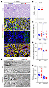 Figure 3
Kif3a inactivation alone does not lead to tubule distension. (A) PAS staining and labeling of Ki67 (which stains proliferating cells; arrowheads), DNA, WGA (which stains the brush border of PTs and all BMs), and AQP2 (a CD marker) of kidneys from 8-week-old control or Kif3aΔtub mice. Arrows: examples of internuclear distance measurements. Scale bars: 10 μm. (B–D) Quantification of KW/BW (B), proliferation index (percentage of Ki67+ cells; C), and internuclear distance (D) in PTs and CDs of kidneys from 8-week-old control or Kif3aΔtub mice. (E and F) Transmission electron microscopy (E) and quantification (F) of TBM thickness of PTs and CDs from 8-week-old control and Kif3aΔtub littermate mice (i.e., 2 weeks after the end of doxycycline treatment). Each dot represents 1 tubule (mean of 8–20 measurements per tubule; n = 3 mice per genotype). Scale bars: 200 nm. P values are from 1-way ANOVA followed by Tukey-Kramer test.
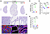 Figure 4
Comparison between neonatal cilia and Pkd1 loss reveals distinct modes of tubule dilation. (A) PAS staining and labeling of HS, DNA, WGA, and AQP2 of kidneys from 2-week-old control, Pkd1Δtub, and Kif3aΔtub mice. White arrows: examples of internuclear distance measurements. Scale bars: 1 mm (top), 50 μm (middle), 10 μm (bottom). (B–D) Quantification of KW/BW (B), internuclear distance (C), and HS staining (D) in the TBM of PTs, CDs, Pkd1Δtub cysts, and outer medullary tubules (OMTs) in kidneys from the same groups of mice, as indicated. Each dot represents 1 female (closed circle) or male (open circle) mouse. One-way ANOVA followed by Tukey-Kramer test: ****P < 0.0001, or the indicated P value. Pkd1fl/fl or Kif3afl/fl littermates lacking cre or rtTA transgene were used as control.
TBM remodeling and CD distension coincide with a specific transcriptional program. Our results suggest that cilia-dependent TBM remodeling promotes cyst formation in ADPKD and is decoupled from cell proliferation. Three nonmutually exclusive mechanisms can cause TBM distension without cell proliferation: (a) ECM digestion by proteases, which allows rapid BM remodeling during embryogenesis (27); (b) incorporation of BM softeners such as hydrating HS/CS, which allows rapid BM remodeling during Caenorhabditis elegans development (28); and (c) deformation of the BM by traction forces exerted by cells through cytoskeleton motor proteins, as documented during cancer invasion (29, 30). To gain insights into the effectors involved, we profiled the transcriptome of PTs and CDs microdissected from 8-week-old control and Pkd1Δtub mice (Figure 5, A–C, and Supplemental Figure 5A). PT- and CD-specific gene expression profiles confirmed dissection quality (Supplemental Figure 5B). Pdgfrb and Itgam expressions were undetectable in CD, indicating the absence of contamination of the extracts by fibroblasts or macrophages, respectively. Overall, Pkd1 inactivation induced a significant dysregulation in the expression of 2,151 genes in CDs but only 1,198 in PTs (Supplemental Figure 5C). Enrichment analyses showed an enrichment in gene sets related to cell proliferation in PC1-deficient PTs, but not in CDs (Figure 5B), a finding consistent with our immunolabeling experiments. More importantly, we identified a panel of 26 genes, which are upregulated in PC1-deficient CDs but not in PTs and are involved in the 3 aspects of BM remodeling that we prespecified, despite similar reduction in Pkd1 mRNA with secondary increases in Pkd2 and Glis2 expression (Figure 5C) (14). The panel includes 4 genes encoding ECM proteases, 3 genes allowing the production of BM softeners (e.g., enzymes involved in HS/CS biogenesis; Hs3st3a1, Chsy1), 6 additional ECM components, and 13 genes involved in cell–BM interactions or cytoskeleton dynamics. Further reanalysis of single-nucleus RNA-seq (snRNA-seq) datasets comparing human ADPKD and healthy kidneys (31) showed that the BM remodeling signature that we identified in distending CDs from Pkd1Δtub mice was significantly enriched in ADPKD human tubular cells (Figure 5, D–F). Of note, in these datasets of terminal human ADPKD kidneys, TBM remodeling gene induction was not restricted to distal segments, suggesting a generalization of the process at late stages.
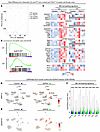 Figure 5
Transcriptomic profiling of microdissected PTs and CDs identifies a BM remodeling signature associated with tubule distension. (A) Principal component analysis of the transcriptome of PTs and CDs microdissected from control and Pkd1Δtub 8-week-old female mice (i.e., 2 weeks after the end of doxycycline treatment; n = 5 mice per genotype; each dot represents a tubule segment from 1 mouse). (B) GSEA comparing proliferation-related gene sets between control and mutant mice in PTs or CDs. (C) Heatmaps showing the relative expression for TBM remodeling candidate genes in CDs and PTs microdissected from control and Pkd1Δtub 8-week-old female mice (n = 5 mice per genotype). Gray in the heatmap of PT indicates undetectable expression. (D and E) Expression of MMP14 (D) and ADAMTS1 (E) in snRNA-seq data of control and ADPKD human kidneys. PEC, parietal epithelial cell; TAL, thick ascending limb of Henle’s loop; CNT-PC, connecting tubule and principal cell; ICA, type A intercalated cell; ICB, type B intercalated cell; PODO, podocyte; ENDO, endothelial cell; FIB, fibroblast; LEUK, leukocyte; URO, urothelium. (F) BM remodeling signature expression score in the indicated tubular cell population from control or ADPKD human kidneys. Bonferroni’s test: ****P < 0.0001. Pkd1fl/fl littermates lacking cre or rtTA transgene were used as control.
Primary cilia participate in the induction of BM remodeling genes. To gain insight into the mechanisms by which cilia elicit tubule distension, we first took advantage of the translatome dataset derived from ciliated and deciliated Pkd1–/– precystic tubules (14). Although BM remodeling genes were mostly induced in the distal nephron, which modestly contributes to the RNA pulled down in this dataset, we were able to replicate our findings for a set of genes, a fraction of which (e.g., Adamts1, Adamtsl4, Chsy1, Map6, Fgd3) showed cilia-dependent regulation (Supplemental Figure 6).
To complement this approach, we performed RNA-seq on PTs and CDs microdissected from Pkd1Δtub Ift20Δtub mice and control littermates (Figure 6A). Integration of these data with our previous RNA-seq results revealed that cilia ablation counteracted the majority of the transcriptional dysregulation triggered by Pkd1 ablation in CDs and PTs (Figure 6B and Supplemental Figure 7). Specifically, approximately half of the BM remodeling genes upregulated upon Pkd1 ablation were reduced in deciliated CD (Figure 6C), suggesting that cilia promote tubule distension through transcriptional regulation.
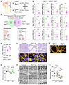 Figure 6
The primary cilium is instrumental in the induction of TBM remodeling genes downstream of PC1 loss and promotes tubule distension through GLIS2. (A) Schematic of the pipeline used to analyze cilia-dependent mediators of TBM remodeling: bulk RNA-seq was performed on CDs and PTs microdissected from control and Pkd1Δtub Ift20Δtub mice. Data were integrated with those obtained for Pkd1Δtub (Figure 5C). (B) Venn diagram showing the number of genes with significant variation between Pkd1Δtub and Pkd1Δtub Ift20Δtub mice and their corresponding littermate controls in CDs. The cilia-dependent and -independent genes of the BM remodeling signature are indicated below. (C) Dot plot showing the relative expression (fold increase compared with the relative controls) of the BM remodeling genes in ciliated and deciliated Pkd1-deficient CDs; only significant genes are shown. Each dot represents a tubule segment from 1 mouse. Student’s t test: *P < 0.05, **P < 0.01, ***P < 0.001. (D) Dot plot of Glis2 mRNA expression in CDs or PTs from ciliated or deciliated Pkd1-deficient tubules. Each dot represents a tubule segment from 1 mouse. Two-way ANOVA followed by Šidák’s test: ***P < 0.001, ****P < 0.0001 (P value for interaction between genotype and segments = 0.0041). (E) PAS staining and labeling of PTs (WGA+) and CDs (AQP2+) of kidneys from 8-week-old control, Pkd1Δtub, and Pkd1Δtub Glis2Δtub mice. Arrows: examples of internuclear distance measurements. Scale bars: 50 μm (top), 10 μm (bottom). (F) Immunolabeling of calbindin (a marker of DCTs and early CDs) of kidneys from 8-week-old Pkd1Δtub Glis2Δtub and Pkd1Δtub Kif3aΔtub mice. Scale bar: 10 μm. (G) Quantification of mean internuclear distance in PTs and CDs of kidneys from 8-week-old control, Pkd1Δtub, and Pkd1Δtub Glis2Δtub mice. Each dot represents 1 male (open circle) or female (closed circle) mouse. (H and I) Transmission electron microscopy (H) and quantification of TBM thickness (I) in PTs, CDs, and DCTs in kidneys from the same groups of mice. Each dot represents 1 tubule (mean of 8–20 measurements per tubule; n = 3 mice per genotype). Scale bars: 200 nm. One-way ANOVA followed by Tukey-Kramer test: *P < 0.05, **P < 0.01, ****P < 0.0001, or the indicated P value. Pkd1fl/fl and Pkd1fl/fl Glis2fl/fl littermates lacking cre or rtTA transgene were used as control.
The transcriptional regulator GLIS2 participates in TBM remodeling and Pkd1-deficient tubule distension, downstream of cilia. The transcriptional regulator GLIS2 was recently identified as an important mediator of CDCA (14) and was correspondingly downregulated in deciliated Pkd1-deficient PTs and CDs (Figure 6D). To assess the role of GLIS2 in tubule distension, we studied Pkd1Δtub and Pkd1Δtub Glis2Δtub mice. As previously described, Glis2 inactivation suppressed the cystic enlargement of Pkd1-deficient kidneys (14). Although Glis2 ablation drastically prevented tubule dilation, blinded examination of kidneys consistently revealed slight focal tubular dilation in the cortex of Pkd1Δtub Glis2Δtub kidneys that were not observed in cilia-deficient Pkd1Δtub animals (Figure 6E). These tubules were generally heterogeneously labeled by a calbindin-specific antibody, suggesting a DCT or early CD origin (Figure 6F). Overall, Glis2 ablation completely restored CD internuclear distance (Figure 6G) but only partially mitigated TBM thinning (Figure 6, H and I). Of note, in these mice housed in a different animal facility, there was only a trend toward proximal TBM thinning at this time point. Taken together, these results suggest that cilia regulate TBM remodeling through both GLIS2-dependent and -independent mechanisms.
Loss of Pkd1 is associated with altered tubule mechanics. We asked whether remodeling of the TBM alters the physical properties of tubules. We isolated CDs and PTs from 8-week-old Pkd1Δtub mice (with intact TBMs) and measured their diameter variations in response to increasing luminal pressure using a dedicated isolated tubule perfusion setup (Figure 7A) with physiologic pressure ranges (32). We observed that PC1-deficient CDs, which have undergone distension but not proliferation (i.e., dissected from 8-week-old mice; Figure 1, A–E), showed a steep increase in tubule diameter at low pressure (i.e., from 0 to 10 cmH2O; Figure 7, B–D). In contrast, PC1-deficient PTs, which have not yet undergone distension at this time point but are nonetheless larger than in control mice because of cell proliferation (Figure 1, A–E), showed similar pressure-diameter curves as control PTs (Figure 7, E and F). Repeating these experiments with PTs isolated from 12-week-old animals, a time point where distension has occurred in this segment (Figure 1, H and K), we found a similar steep increase in tubule diameter as in distended PC1-deficient CDs, although for a higher pressure threshold (i.e., from 21 to 31 cmH2O; Figure 7, G–I). Cilia ablation rescued the excessive dilation of PC1-deficient CDs (Figure 7, B–D) and PTs (Figure 7, G–I) by transmural pressure. Notably, decreasing pressure in distended PC1-deficient tubules resulted in a decrease of their diameter, indicating elastic deformation (Supplemental Figure 8). Together, these results demonstrate that tubule distension and TBM remodeling in ADPKD are associated with an alteration of tubule mechanics, characterized by a steep increase in tubule diameter with segment-specific pressure thresholds, which also appears as a cilia-dependent mechanism.
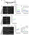 Figure 7
Pkd1 deletion alters tubule biomechanics. (A) Scheme of isolated intact tubule perfusion setting. Adapted from Journal of Clinical Investigation (16). (B and C) Representative images (B) and quantification of the variation in outer tubule diameter induced by a progressive increase in transmural pressure (TM; C) in CDs isolated from 8-week-old control, Pkd1Δtub, and Pkd1Δtub Kif3aΔtub male mice. Each dot represents 1 tubule (mean of 10 measurements per tubule) for a pressure increment. (D) Quantification of the slope of the pressure-diameter curves for each pressure increment in CDs isolated from the same groups of mice. Each dot represents 1 tubule. Student’s t test: *P < 0.05, **P < 0.01. (E and F) Representative images (E) and quantification of the variation in outer tubule diameter induced by incremental increase in TM pressure (F) in PTs isolated from 8-week-old control and Pkd1Δtub mice. Each dot represents 1 tubule (mean of 10 measurements per tubule) for a pressure increment. (G and H) Representative images (G) and quantification of the variation in outer tubule diameter induced by incremental increase in TM pressure (H) in PTs isolated from 12-week-old control, Pkd1Δtub, and Pkd1Δtub Kif3aΔtub mice. Each triangle represents 1 tubule (mean of 10 measurements per tubule) for a pressure increment. (I) Quantification of the slope of the pressure-diameter curves for each pressure increment in PTs isolated from 8- (circles) and 12-week-old (triangles) control, Pkd1Δtub, and Pkd1Δtub Kif3aΔtub mice. PTs from control animals at 8 (blue circle) and 12 weeks (blue triangle) were pooled in analyses. Each symbol represents 1 tubule. Scale bars: 50 μm. One-way ANOVA followed by Tukey-Kramer test: **P < 0.01. Pkd1fl/fl littermates lacking cre or rtTA transgene were used as control.
Changes in TBM mechanical properties result in permissive conditions for cystogenesis. Our results regarding the early modifications of TBM in ADPKD suggest that increased ECM deformability favors cystogenesis. To investigate this question in vitro, we used an established tubule-on-chip model, whereby immortalized tubular cells form ciliated tubules of physiological dimensions in a deformable ECM scaffold (33). Using edited ciliated clonal lines derived from mouse collecting ducts (mIMCD3) (34), we observed only minor differences between control and Pkd1–/– clones in the ability to dilate ECM scaffolds with a collagen content of 9.5 g/L (Figure 8A). However, by lowering collagen content, thereby reducing stiffness, we observed that Pkd1–/– clones dilated the ECM scaffold to a larger extent than the parental line (Figure 8, B and C). This increased dilation was not caused by increased cell proliferation but by an increase in internuclear distance (Figure 8, D–F). These in vitro results demonstrate that ECM mechanics affect the ability of Pkd1–/– cells to deform tubules, so that ECM deformability facilitates dilation. Ciliation of Pkd1-deficient cells was unaffected by collagen content (Figure 8, G and H), indicating that ECM mechanics do not regulate Pkd1–/– tubule dilation by affecting ciliogenesis.
 Figure 8
ECM mechanics modulate Pkd1–/– tubule-on-chip dilation in vitro. (A and B) Representative images of tubules-on-chip formed by parental or Pkd1–/– mIMCD3 clones in scaffolds containing 9.5 (A) or 6 (B) g/L collagen I, 1 and 11 days after confluency. Arrows indicate tubule diameter. Scale bars: 100 μm. (C) Quantification of the normalized tubule deformation in the corresponding tubules-on-chip at 11 days. Each dot represents 1 tubule from n = 8 for parental cells and n = 8, 5, and 4 chips for Pkd1–/– clones 1, 2, and 3, respectively. (D–F) Labeling (D) and quantification of Ki67 (E) and internuclear distance (F) in parental and Pkd1–/– tubules-on-chip. Each dot represents 1 tubule (parental cells: N = 3 chips/n = 9–11 tubules; Pkd1–/– cells: N = 3 chips/n = 9–12 tubules). Mann-Whitney test: **P < 0.01. Scale bar: 100 μm. The box-and-whisker plots depict the minimum and maximum values (whiskers), the upper and lower quartiles, and the median. (G) Representative orthogonal sections of ciliated Pkd1–/– tubules-on-chip embedded in 6 or 9.5 g/L collagen I and maintained under static conditions for 5 days. Cilia were identified by immunostaining of acetylated α-tubulin (green) and γ-tubulin (red). Scale bar: 100 μm. (H) Corresponding quantification of percentage of ciliated cells per tubule. Each dot represents 1 tubule (n = 3 tubules per condition). The reported P value was calculated using a Student’s t test. The data shown in this figure are derived from at least 2 different independent cell seeding experiments.
To test this concept in vivo, we inactivated Pxdn, which encodes peroxidasin, a collagen IV–specific cross-linking enzyme (in which germinal ablation induces a 20% decrease in TBM elastic modulus) (19), in Pkd1Δtub mice. Pxdn–/– single mutant mice have slightly smaller kidneys than control at baseline and do not spontaneously develop cysts, even in aging (Figure 9, A and B). The modest alteration of TBM mechanics caused by Pxdn inactivation translated into increased cyst burden in Pkd1Δtub mice (Figure 9, C–E). At the studied time point, cystogenesis was more pronounced in males compared with females (P value for sex by 2-way ANOVA < 0.001). However, the impact of Pxdn inactivation on cyst area was not affected by sex (P value for interaction between sex and genotype by 2-way ANOVA = 0.8). These results are consistent with a model where reduced TBM stiffness facilitates tubule deformation and cystogenesis. While cyst formation was increased in Pkd1Δtub Pxdn–/– mice compared with Pkd1Δtub mice, this did not translate into an increase in kidney weight/body weight ratio (KW/BW; Figure 9D). This discrepancy prompted us to assess tubular cell proliferation, which was overall reduced in the context of Pxdn inactivation (Figure 9, F and G), in line with the observation that stiffer substrates usually promote cell proliferation (35). The fact that reducing BM stiffness (via Pxdn inactivation) promoted tubule distension while reducing cell proliferation, further supports the view that, besides cell proliferation, BM mechanics and tubule distension are involved in cystogenesis.
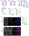 Figure 9
Decrease in TBM stiffness promotes cyst formation in vivo. (A) PAS staining and quantification of KW/BW of kidneys from 12-week-old control or Pxdn–/– female (closed circle) and male (open circle) mice. (B) PAS staining of kidney sections from aged Pxdn–/– mice showing no cyst development. (C–E) PAS staining (C), quantification of KW/BW (D), and tubular dilations (E) of kidneys from 12-week-old Pkd1Δtub female (closed circle) and male (open circle) mice with or without concomitant Pxdn gene inactivation. Scale bars: 0.5 mm (A–C). For A, D, and E, each dot represents 1 mouse. P value indicates the Pxdn genotype effect in 2-way ANOVA followed by Tukey-Kramer test (sex and Pxdn genotype; P value for interaction between sex and genotype = 0.3 and 0.8 in D and E, respectively). *P < 0.05, **P < 0.01. (F and G) Labeling (F) and quantification of Ki67+ cells (G) in CDs and PTs in kidneys from the same groups of mice. Each dot represents 1 mouse. Scale bars: 40 μm. P values indicate the Pxdn genotype effect in 2-way ANOVA (sex and Pxdn genotype; P value for interaction between sex and genotype = 0.5 and 0.1 in PTs and CDs, respectively; *P < 0.05).
Tubule obstruction exacerbates PC1-deficient tubule distension and precipitates cystogenesis. Tubular obstruction from enlarging cysts has been proposed to contribute to further cyst formation, and micropuncture and microdissection studies have shown that cysts frequently arise from obstructed tubules (36). We tested in vivo how alterations in TBM biomechanics affect tubule response to obstruction with respect to PC1 and cilia. After a single day of unilateral ureteral obstruction (UUO), Pkd1Δtub mice showed a disproportional increase in kidney weight compared with controls. This was not the case in kidneys with concomitant cilia ablation (Figure 10, A and B, and Supplemental Figure 9A). The kidney weight increase correlated with enlargement of the CDs and DCTs, but not PTs, and was associated with a marked increase in internuclear distance, indicating tubule distension (Figure 10, C–H, and Supplemental Figure 9, B–D). BrdU labeling in Pkd1-deficient CDs did not significantly change following obstruction (Supplemental Figure 9, E and F), suggesting that early cyst expansion after UUO occurs largely independently of cell proliferation, though the limited sample size of this analysis warrants caution in interpretation.
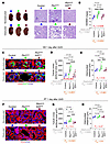 Figure 10
Acute ureteral obstruction accelerates Pkd1Δtub distal tubule dilation in a cilia-dependent manner. (A and B) Kidney images and PAS staining (A) and quantification (B) of KW/BW of obstructed and nonobstructed kidneys from 8-week-old control, Pkd1Δtub, and Pkd1Δtub Kif3aΔtub 1 day after UUO. Scale bars: 1 mm (left), 0.1 mm (right). (C–H) Labeling of DNA, WGA (which stains the brush border of PTs and all BMs), and AQP2 (a CD marker; C and F) and quantification of mean tubule cross-sectional area (D and G) and internuclear distance (E and H) of CDs (C–E) and PTs (F–H) of kidneys from the same animals. Each pair of linked symbols (circle and square) represents the obstructed (square) and nonobstructed (circle) kidneys of an individual female mouse. Scale bars: 10 μm. Two-way ANOVA, P value for obstruction (gray; Pobs), genotype (brown; Pgen), and their interaction (red; Pint), followed by Tukey-Kramer test: *P < 0.05, ***P < 0.001, ****P < 0.0001, or the indicated P value. Pkd1fl/fl or Pkd1fl/fl Kif3afl/fl littermates lacking cre or rtTA transgene were used as control.
Two-way ANOVA confirmed that a significant interaction between genotype and obstruction governs distal nephron distension (Figure 10, D and E, and Supplemental Figure 9, B–D). Of note, this first set of experiments was conducted on 8-week-old females, which at this age already display a thinner TBM in CDs but, contrary to males, have negligible tubule dilation at baseline. Similar results were obtained with male mice (Supplemental Figure 10).
Prolonging obstruction resulted in cystic transformation of PC1-deficient kidneys, with a 2- and 3-fold increase in KW/BW 4 and 14 days after the surgery, respectively (Figure 11, A–D, and Supplemental Figure 11, A and B). Cilia ablation reduced obstructed PC1-deficient kidney enlargement to the mild level observed in cilia-deficient mice with intact Pkd1 (i.e., Kif3aΔtub; Figure 11, B–D). Cystic transformation of Pkd1-deficient kidneys was mainly the consequence of an increase in DCT and CD cross-sectional area, while tubule internuclear distance remained higher than in control or PC1-deficient DCTs and CDs lacking cilia (Figure 12, A–C, and Supplemental Figure 11, C–H). We then asked if the cystic transformation of PC1-deficient tubules could be caused by an excessive proliferative response to obstruction. As expected, labeling cycling cells with PCNA or Ki67 revealed that, with time, obstruction increased the fraction of cycling cells in CDs and DCTs. However, the magnitude of this increase was similar across genotypes (Figure 12, D–F, and Supplemental Figure 12, A–D). Consistent with our baseline findings, nonobstructed PTs lacking PC1 displayed a higher fraction of cycling cells than control or deciliated PTs. Obstruction further increased the cycling cell fraction in all genotypes (Supplemental Figure 12, C–E). The proliferation rate did not correlate with PT deformation: obstruction reduced PT cross-sectional area in control animals, indicating atrophy. This compaction of PTs was similarly prevented by Pkd1 or cilia ablation (Figure 12, G and H).
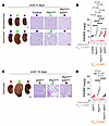 Figure 11
Ureteral obstruction triggers explosive cystogenesis in Pkd1Δtub distal tubules in a cilia-dependent manner. (A and B) Kidney images and PAS staining (A) and quantification (B) of KW/BW of obstructed and nonobstructed kidneys from control, Pkd1Δtub, Pkd1Δtub Kif3aΔtub, and Kif3aΔtub mice (in red) 4 days after UUO performed at 8 weeks of age. (C and D) Kidney images and PAS staining (C) and quantification (D) of KW/BW of kidneys from Kif3aΔtub and Pkd1Δtub Kif3aΔtub 14 days after UUO performed at 8 weeks of age. Single Kif3a mutant mice (Kif3aΔtub mice) are presented to show apparent epistasis of Kif3a over Pkd1 inactivation. Scale bars: 1 mm (kidney images), 0.1 mm (PAS). Each pair of linked symbols (circle and square) represents the obstructed (square) and nonobstructed (circle) kidneys of an individual female mouse. Two-way ANOVA, P value for obstruction (gray; Pobs), genotype (brown; Pgen), and their interaction (red; Pint), followed by Tukey-Kramer test: **P < 0.01, ***P < 0.001, ****P < 0.0001, or the indicated P value. Pkd1fl/fl or Pkd1fl/fl Kif3afl/fl littermates lacking cre or rtTA transgene were used as control.
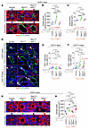 Figure 12
Accelerated cystogenesis after obstruction correlates with tubule distension and not proliferation. (A–C) Fluorescent labeling of DNA, WGA (which stains the brush border of PTs and all BMs), and AQP2 (a CD marker) (A) and quantification of CD mean tubule cross-sectional area (B) and mean internuclear distance (C) in kidneys from control, Pkd1Δtub, and Pkd1Δtub Kif3aΔtub 4 days after UUO performed at 8 weeks of age. (D–F) Fluorescent labeling of DNA, Ki67, and AQP2 (D) and quantification (E and F) of Ki67 staining (arrowheads; # indicates nonspecific red blood cell autofluorescence) in CDs of kidneys from control, Pkd1Δtub, and Pkd1Δtub Kif3aΔtub 4 (E) and 14 (F) days after UUO. (G and H) Representative images (G) and quantification of the PT cross-sectional area (H) in kidneys from control, Pkd1Δtub, Pkd1Δtub Kif3aΔtub, and Kif3aΔtub (in red) 4 days after UUO. Dashed lines in images indicate tubule perimeters. Scale bars: 10 μm. Each pair of linked symbols (circle and square) represents the obstructed (square) and nonobstructed (circle) kidneys of an individual female mouse. Two-way ANOVA, P value for obstruction (gray; Pobs), genotype (brown; Pgen), and their interaction (red; Pint), followed by Tukey-Kramer test: *P < 0.05, **P < 0.01, ***P < 0.001, ****P < 0.0001, or the indicated P value. Pkd1fl/fl or Pkd1fl/fl Kif3afl/fl littermates lacking cre or rtTA transgene were used as control.
In summary, the cystogenic response to obstruction corresponded with the observed effect of PC1 and cilia on TBM structure and mechanics: PC1-deficient CDs and DCTs have a thinner TBM and higher distensibility than PTs and correspondingly dilate more strongly, precipitating cystogenesis.
Transient obstruction is sufficient to precipitate irreversible cystogenesis. Previous reports showed that only a minority of PKD cysts are obstructed at any given time (22, 36), suggesting that permanent tubule obstruction is not a frequent driver of cystogenesis. To study the effect of a transient obstruction on cyst formation, we used a reversible UUO (R-UUO) model: Pkd1Δtub mice underwent ligation of the distal ureter, followed 4 days later by either surgical release of the obstruction or a sham operation, and the kidneys were analyzed at 14 days (i.e., 10 days after UUO reversion; Figure 13A). Notably, release of the obstruction after 4 days failed to reduce cyst growth at 14 days (Figure 13, B–D), indicating that transient obstruction is sufficient to commit tubules to an irreversible cystogenic program.
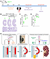 Figure 13
Transient obstruction triggers irreversible cystogenesis in Pkd1Δtub mice. (A) Scheme of the protocol of R-UUO. Left kidney of 8-week-old Pkd1Δtub mice was submitted to UUO, followed by a release of the obstruction (Reverse UUO) or a Sham operation (Sham Reverse UUO) 4 days after UUO, and mice were euthanized 10 days after the second surgery. (B–D) PAS staining (B) and quantification of KW/BW (C) and tubule dilations (D) of the kidneys from control or Pkd1Δtub mice submitted to R-UUO or sham R-UUO. Scale bars: 2 mm (top), 0.1 mm (bottom). Each symbol represents an individual female (closed symbol) or male (open symbol) mouse kidney. One-way ANOVA followed by Tukey-Kramer test: ****P < 0.0001 or the indicated P value. (E) Proposed “tire bulge” model for cyst formation in ADPKD: PC1 loss triggers cilia-dependent remodeling of BM. BM remodeling in turn promotes tubule distension, which is exacerbated by transmural hydrostatic pressure (P). Transient tubule obstruction increases luminal pressure, tubule dilation, and tubular cell stretch, precipitating cystogenesis.
Altogether, these results establish a TBM-centered model of cystogenesis in which Pkd1 loss drives cilia-dependent remodeling of TBM shape and mechanics, which, in conjunction with increased luminal pressure, produces tubule dilation in a manner resembling the formation of a tire bulge (Figure 13E). In this condition of increased tension, obstruction precipitates cystogenesis independent of cell proliferation.


Copyright © 2026 American Society for Clinical Investigation
ISSN: 0021-9738 (print), 1558-8238 (online)