
Advertisement
Research ArticleGastroenterologyInflammation
Open Access |  10.1172/JCI192804
Meflin confers antifibrotic properties to intestinal fibroblasts in inflammatory bowel disease
Jingxi Mu,1 Keiko Maeda,1 Tadashi Iida,1 Shinji Mii,2 Nobutoshi Esaki,2 Yukihiro Shiraki,2 Yasuyuki Mizutani,1 Masanao Nakamura,3 Takeshi Yamamura,1 Tsunaki Sawada,3 Eri Ishikawa,1 Kentaro Murate,1 Takashi Hirose,1 Kazuhiro Furukawa,1 Akina Oishi,1 Haruhiko Suzuki,4 Takayoshi Kishida,5 Goro Nakayama,5 Mitsuhiro Fujishiro,6 Hiroki Kawashima,1 and Atsushi Enomoto2,7
1Department of Gastroenterology and Hepatology,
2Department of Pathology,
3Department of Endoscopy,
4Department of Immunology, and
5Department of Gastroenterological Surgery, Nagoya University Graduate School of Medicine, Nagoya, Japan.
6Department of Gastroenterology, Graduate School of Medicine, The University of Tokyo, Tokyo, Japan.
7Center for One Medicine Innovative Translational Research, Gifu University Institute for Advanced Study, Gifu, Japan.
Address correspondence to: Atsushi Enomoto, Department of Pathology, Nagoya University Graduate School of Medicine, 65 Tsurumai-cho, Showa-ku, Nagoya 466-8550, Japan. Center for One Medicine Innovative Translational Research, Gifu University Institute for Advanced Study, Gifu, Japan. Phone: 81.52.744.2092; Email: enomoto.atsushi.d3@f.mail.nagoya-u.ac.jp. Or to: Keiko Maeda, Department of Gastroenterology and Hepatology, Nagoya University Graduate School of Medicine, 65 Tsurumai-cho, Showa-ku, Nagoya 466-8550, Japan. Phone: 81.52.744.2172; Email: maeda.keiko.v5@f.mail.nagoya-u.ac.jp.
Find articles by Mu, J. in: PubMed | Google Scholar
1Department of Gastroenterology and Hepatology,
2Department of Pathology,
3Department of Endoscopy,
4Department of Immunology, and
5Department of Gastroenterological Surgery, Nagoya University Graduate School of Medicine, Nagoya, Japan.
6Department of Gastroenterology, Graduate School of Medicine, The University of Tokyo, Tokyo, Japan.
7Center for One Medicine Innovative Translational Research, Gifu University Institute for Advanced Study, Gifu, Japan.
Address correspondence to: Atsushi Enomoto, Department of Pathology, Nagoya University Graduate School of Medicine, 65 Tsurumai-cho, Showa-ku, Nagoya 466-8550, Japan. Center for One Medicine Innovative Translational Research, Gifu University Institute for Advanced Study, Gifu, Japan. Phone: 81.52.744.2092; Email: enomoto.atsushi.d3@f.mail.nagoya-u.ac.jp. Or to: Keiko Maeda, Department of Gastroenterology and Hepatology, Nagoya University Graduate School of Medicine, 65 Tsurumai-cho, Showa-ku, Nagoya 466-8550, Japan. Phone: 81.52.744.2172; Email: maeda.keiko.v5@f.mail.nagoya-u.ac.jp.
Find articles by Maeda, K. in: PubMed | Google Scholar
1Department of Gastroenterology and Hepatology,
2Department of Pathology,
3Department of Endoscopy,
4Department of Immunology, and
5Department of Gastroenterological Surgery, Nagoya University Graduate School of Medicine, Nagoya, Japan.
6Department of Gastroenterology, Graduate School of Medicine, The University of Tokyo, Tokyo, Japan.
7Center for One Medicine Innovative Translational Research, Gifu University Institute for Advanced Study, Gifu, Japan.
Address correspondence to: Atsushi Enomoto, Department of Pathology, Nagoya University Graduate School of Medicine, 65 Tsurumai-cho, Showa-ku, Nagoya 466-8550, Japan. Center for One Medicine Innovative Translational Research, Gifu University Institute for Advanced Study, Gifu, Japan. Phone: 81.52.744.2092; Email: enomoto.atsushi.d3@f.mail.nagoya-u.ac.jp. Or to: Keiko Maeda, Department of Gastroenterology and Hepatology, Nagoya University Graduate School of Medicine, 65 Tsurumai-cho, Showa-ku, Nagoya 466-8550, Japan. Phone: 81.52.744.2172; Email: maeda.keiko.v5@f.mail.nagoya-u.ac.jp.
Find articles by
Iida, T.
in:
PubMed
|
Google Scholar
|

1Department of Gastroenterology and Hepatology,
2Department of Pathology,
3Department of Endoscopy,
4Department of Immunology, and
5Department of Gastroenterological Surgery, Nagoya University Graduate School of Medicine, Nagoya, Japan.
6Department of Gastroenterology, Graduate School of Medicine, The University of Tokyo, Tokyo, Japan.
7Center for One Medicine Innovative Translational Research, Gifu University Institute for Advanced Study, Gifu, Japan.
Address correspondence to: Atsushi Enomoto, Department of Pathology, Nagoya University Graduate School of Medicine, 65 Tsurumai-cho, Showa-ku, Nagoya 466-8550, Japan. Center for One Medicine Innovative Translational Research, Gifu University Institute for Advanced Study, Gifu, Japan. Phone: 81.52.744.2092; Email: enomoto.atsushi.d3@f.mail.nagoya-u.ac.jp. Or to: Keiko Maeda, Department of Gastroenterology and Hepatology, Nagoya University Graduate School of Medicine, 65 Tsurumai-cho, Showa-ku, Nagoya 466-8550, Japan. Phone: 81.52.744.2172; Email: maeda.keiko.v5@f.mail.nagoya-u.ac.jp.
Find articles by Mii, S. in: PubMed | Google Scholar
1Department of Gastroenterology and Hepatology,
2Department of Pathology,
3Department of Endoscopy,
4Department of Immunology, and
5Department of Gastroenterological Surgery, Nagoya University Graduate School of Medicine, Nagoya, Japan.
6Department of Gastroenterology, Graduate School of Medicine, The University of Tokyo, Tokyo, Japan.
7Center for One Medicine Innovative Translational Research, Gifu University Institute for Advanced Study, Gifu, Japan.
Address correspondence to: Atsushi Enomoto, Department of Pathology, Nagoya University Graduate School of Medicine, 65 Tsurumai-cho, Showa-ku, Nagoya 466-8550, Japan. Center for One Medicine Innovative Translational Research, Gifu University Institute for Advanced Study, Gifu, Japan. Phone: 81.52.744.2092; Email: enomoto.atsushi.d3@f.mail.nagoya-u.ac.jp. Or to: Keiko Maeda, Department of Gastroenterology and Hepatology, Nagoya University Graduate School of Medicine, 65 Tsurumai-cho, Showa-ku, Nagoya 466-8550, Japan. Phone: 81.52.744.2172; Email: maeda.keiko.v5@f.mail.nagoya-u.ac.jp.
Find articles by Esaki, N. in: PubMed | Google Scholar
1Department of Gastroenterology and Hepatology,
2Department of Pathology,
3Department of Endoscopy,
4Department of Immunology, and
5Department of Gastroenterological Surgery, Nagoya University Graduate School of Medicine, Nagoya, Japan.
6Department of Gastroenterology, Graduate School of Medicine, The University of Tokyo, Tokyo, Japan.
7Center for One Medicine Innovative Translational Research, Gifu University Institute for Advanced Study, Gifu, Japan.
Address correspondence to: Atsushi Enomoto, Department of Pathology, Nagoya University Graduate School of Medicine, 65 Tsurumai-cho, Showa-ku, Nagoya 466-8550, Japan. Center for One Medicine Innovative Translational Research, Gifu University Institute for Advanced Study, Gifu, Japan. Phone: 81.52.744.2092; Email: enomoto.atsushi.d3@f.mail.nagoya-u.ac.jp. Or to: Keiko Maeda, Department of Gastroenterology and Hepatology, Nagoya University Graduate School of Medicine, 65 Tsurumai-cho, Showa-ku, Nagoya 466-8550, Japan. Phone: 81.52.744.2172; Email: maeda.keiko.v5@f.mail.nagoya-u.ac.jp.
Find articles by Shiraki, Y. in: PubMed | Google Scholar
1Department of Gastroenterology and Hepatology,
2Department of Pathology,
3Department of Endoscopy,
4Department of Immunology, and
5Department of Gastroenterological Surgery, Nagoya University Graduate School of Medicine, Nagoya, Japan.
6Department of Gastroenterology, Graduate School of Medicine, The University of Tokyo, Tokyo, Japan.
7Center for One Medicine Innovative Translational Research, Gifu University Institute for Advanced Study, Gifu, Japan.
Address correspondence to: Atsushi Enomoto, Department of Pathology, Nagoya University Graduate School of Medicine, 65 Tsurumai-cho, Showa-ku, Nagoya 466-8550, Japan. Center for One Medicine Innovative Translational Research, Gifu University Institute for Advanced Study, Gifu, Japan. Phone: 81.52.744.2092; Email: enomoto.atsushi.d3@f.mail.nagoya-u.ac.jp. Or to: Keiko Maeda, Department of Gastroenterology and Hepatology, Nagoya University Graduate School of Medicine, 65 Tsurumai-cho, Showa-ku, Nagoya 466-8550, Japan. Phone: 81.52.744.2172; Email: maeda.keiko.v5@f.mail.nagoya-u.ac.jp.
Find articles by Mizutani, Y. in: PubMed | Google Scholar
1Department of Gastroenterology and Hepatology,
2Department of Pathology,
3Department of Endoscopy,
4Department of Immunology, and
5Department of Gastroenterological Surgery, Nagoya University Graduate School of Medicine, Nagoya, Japan.
6Department of Gastroenterology, Graduate School of Medicine, The University of Tokyo, Tokyo, Japan.
7Center for One Medicine Innovative Translational Research, Gifu University Institute for Advanced Study, Gifu, Japan.
Address correspondence to: Atsushi Enomoto, Department of Pathology, Nagoya University Graduate School of Medicine, 65 Tsurumai-cho, Showa-ku, Nagoya 466-8550, Japan. Center for One Medicine Innovative Translational Research, Gifu University Institute for Advanced Study, Gifu, Japan. Phone: 81.52.744.2092; Email: enomoto.atsushi.d3@f.mail.nagoya-u.ac.jp. Or to: Keiko Maeda, Department of Gastroenterology and Hepatology, Nagoya University Graduate School of Medicine, 65 Tsurumai-cho, Showa-ku, Nagoya 466-8550, Japan. Phone: 81.52.744.2172; Email: maeda.keiko.v5@f.mail.nagoya-u.ac.jp.
Find articles by Nakamura, M. in: PubMed | Google Scholar
1Department of Gastroenterology and Hepatology,
2Department of Pathology,
3Department of Endoscopy,
4Department of Immunology, and
5Department of Gastroenterological Surgery, Nagoya University Graduate School of Medicine, Nagoya, Japan.
6Department of Gastroenterology, Graduate School of Medicine, The University of Tokyo, Tokyo, Japan.
7Center for One Medicine Innovative Translational Research, Gifu University Institute for Advanced Study, Gifu, Japan.
Address correspondence to: Atsushi Enomoto, Department of Pathology, Nagoya University Graduate School of Medicine, 65 Tsurumai-cho, Showa-ku, Nagoya 466-8550, Japan. Center for One Medicine Innovative Translational Research, Gifu University Institute for Advanced Study, Gifu, Japan. Phone: 81.52.744.2092; Email: enomoto.atsushi.d3@f.mail.nagoya-u.ac.jp. Or to: Keiko Maeda, Department of Gastroenterology and Hepatology, Nagoya University Graduate School of Medicine, 65 Tsurumai-cho, Showa-ku, Nagoya 466-8550, Japan. Phone: 81.52.744.2172; Email: maeda.keiko.v5@f.mail.nagoya-u.ac.jp.
Find articles by Yamamura, T. in: PubMed | Google Scholar
1Department of Gastroenterology and Hepatology,
2Department of Pathology,
3Department of Endoscopy,
4Department of Immunology, and
5Department of Gastroenterological Surgery, Nagoya University Graduate School of Medicine, Nagoya, Japan.
6Department of Gastroenterology, Graduate School of Medicine, The University of Tokyo, Tokyo, Japan.
7Center for One Medicine Innovative Translational Research, Gifu University Institute for Advanced Study, Gifu, Japan.
Address correspondence to: Atsushi Enomoto, Department of Pathology, Nagoya University Graduate School of Medicine, 65 Tsurumai-cho, Showa-ku, Nagoya 466-8550, Japan. Center for One Medicine Innovative Translational Research, Gifu University Institute for Advanced Study, Gifu, Japan. Phone: 81.52.744.2092; Email: enomoto.atsushi.d3@f.mail.nagoya-u.ac.jp. Or to: Keiko Maeda, Department of Gastroenterology and Hepatology, Nagoya University Graduate School of Medicine, 65 Tsurumai-cho, Showa-ku, Nagoya 466-8550, Japan. Phone: 81.52.744.2172; Email: maeda.keiko.v5@f.mail.nagoya-u.ac.jp.
Find articles by Sawada, T. in: PubMed | Google Scholar
1Department of Gastroenterology and Hepatology,
2Department of Pathology,
3Department of Endoscopy,
4Department of Immunology, and
5Department of Gastroenterological Surgery, Nagoya University Graduate School of Medicine, Nagoya, Japan.
6Department of Gastroenterology, Graduate School of Medicine, The University of Tokyo, Tokyo, Japan.
7Center for One Medicine Innovative Translational Research, Gifu University Institute for Advanced Study, Gifu, Japan.
Address correspondence to: Atsushi Enomoto, Department of Pathology, Nagoya University Graduate School of Medicine, 65 Tsurumai-cho, Showa-ku, Nagoya 466-8550, Japan. Center for One Medicine Innovative Translational Research, Gifu University Institute for Advanced Study, Gifu, Japan. Phone: 81.52.744.2092; Email: enomoto.atsushi.d3@f.mail.nagoya-u.ac.jp. Or to: Keiko Maeda, Department of Gastroenterology and Hepatology, Nagoya University Graduate School of Medicine, 65 Tsurumai-cho, Showa-ku, Nagoya 466-8550, Japan. Phone: 81.52.744.2172; Email: maeda.keiko.v5@f.mail.nagoya-u.ac.jp.
Find articles by Ishikawa, E. in: PubMed | Google Scholar
1Department of Gastroenterology and Hepatology,
2Department of Pathology,
3Department of Endoscopy,
4Department of Immunology, and
5Department of Gastroenterological Surgery, Nagoya University Graduate School of Medicine, Nagoya, Japan.
6Department of Gastroenterology, Graduate School of Medicine, The University of Tokyo, Tokyo, Japan.
7Center for One Medicine Innovative Translational Research, Gifu University Institute for Advanced Study, Gifu, Japan.
Address correspondence to: Atsushi Enomoto, Department of Pathology, Nagoya University Graduate School of Medicine, 65 Tsurumai-cho, Showa-ku, Nagoya 466-8550, Japan. Center for One Medicine Innovative Translational Research, Gifu University Institute for Advanced Study, Gifu, Japan. Phone: 81.52.744.2092; Email: enomoto.atsushi.d3@f.mail.nagoya-u.ac.jp. Or to: Keiko Maeda, Department of Gastroenterology and Hepatology, Nagoya University Graduate School of Medicine, 65 Tsurumai-cho, Showa-ku, Nagoya 466-8550, Japan. Phone: 81.52.744.2172; Email: maeda.keiko.v5@f.mail.nagoya-u.ac.jp.
Find articles by Murate, K. in: PubMed | Google Scholar
1Department of Gastroenterology and Hepatology,
2Department of Pathology,
3Department of Endoscopy,
4Department of Immunology, and
5Department of Gastroenterological Surgery, Nagoya University Graduate School of Medicine, Nagoya, Japan.
6Department of Gastroenterology, Graduate School of Medicine, The University of Tokyo, Tokyo, Japan.
7Center for One Medicine Innovative Translational Research, Gifu University Institute for Advanced Study, Gifu, Japan.
Address correspondence to: Atsushi Enomoto, Department of Pathology, Nagoya University Graduate School of Medicine, 65 Tsurumai-cho, Showa-ku, Nagoya 466-8550, Japan. Center for One Medicine Innovative Translational Research, Gifu University Institute for Advanced Study, Gifu, Japan. Phone: 81.52.744.2092; Email: enomoto.atsushi.d3@f.mail.nagoya-u.ac.jp. Or to: Keiko Maeda, Department of Gastroenterology and Hepatology, Nagoya University Graduate School of Medicine, 65 Tsurumai-cho, Showa-ku, Nagoya 466-8550, Japan. Phone: 81.52.744.2172; Email: maeda.keiko.v5@f.mail.nagoya-u.ac.jp.
Find articles by Hirose, T. in: PubMed | Google Scholar
1Department of Gastroenterology and Hepatology,
2Department of Pathology,
3Department of Endoscopy,
4Department of Immunology, and
5Department of Gastroenterological Surgery, Nagoya University Graduate School of Medicine, Nagoya, Japan.
6Department of Gastroenterology, Graduate School of Medicine, The University of Tokyo, Tokyo, Japan.
7Center for One Medicine Innovative Translational Research, Gifu University Institute for Advanced Study, Gifu, Japan.
Address correspondence to: Atsushi Enomoto, Department of Pathology, Nagoya University Graduate School of Medicine, 65 Tsurumai-cho, Showa-ku, Nagoya 466-8550, Japan. Center for One Medicine Innovative Translational Research, Gifu University Institute for Advanced Study, Gifu, Japan. Phone: 81.52.744.2092; Email: enomoto.atsushi.d3@f.mail.nagoya-u.ac.jp. Or to: Keiko Maeda, Department of Gastroenterology and Hepatology, Nagoya University Graduate School of Medicine, 65 Tsurumai-cho, Showa-ku, Nagoya 466-8550, Japan. Phone: 81.52.744.2172; Email: maeda.keiko.v5@f.mail.nagoya-u.ac.jp.
Find articles by Furukawa, K. in: PubMed | Google Scholar
1Department of Gastroenterology and Hepatology,
2Department of Pathology,
3Department of Endoscopy,
4Department of Immunology, and
5Department of Gastroenterological Surgery, Nagoya University Graduate School of Medicine, Nagoya, Japan.
6Department of Gastroenterology, Graduate School of Medicine, The University of Tokyo, Tokyo, Japan.
7Center for One Medicine Innovative Translational Research, Gifu University Institute for Advanced Study, Gifu, Japan.
Address correspondence to: Atsushi Enomoto, Department of Pathology, Nagoya University Graduate School of Medicine, 65 Tsurumai-cho, Showa-ku, Nagoya 466-8550, Japan. Center for One Medicine Innovative Translational Research, Gifu University Institute for Advanced Study, Gifu, Japan. Phone: 81.52.744.2092; Email: enomoto.atsushi.d3@f.mail.nagoya-u.ac.jp. Or to: Keiko Maeda, Department of Gastroenterology and Hepatology, Nagoya University Graduate School of Medicine, 65 Tsurumai-cho, Showa-ku, Nagoya 466-8550, Japan. Phone: 81.52.744.2172; Email: maeda.keiko.v5@f.mail.nagoya-u.ac.jp.
Find articles by Oishi, A. in: PubMed | Google Scholar
1Department of Gastroenterology and Hepatology,
2Department of Pathology,
3Department of Endoscopy,
4Department of Immunology, and
5Department of Gastroenterological Surgery, Nagoya University Graduate School of Medicine, Nagoya, Japan.
6Department of Gastroenterology, Graduate School of Medicine, The University of Tokyo, Tokyo, Japan.
7Center for One Medicine Innovative Translational Research, Gifu University Institute for Advanced Study, Gifu, Japan.
Address correspondence to: Atsushi Enomoto, Department of Pathology, Nagoya University Graduate School of Medicine, 65 Tsurumai-cho, Showa-ku, Nagoya 466-8550, Japan. Center for One Medicine Innovative Translational Research, Gifu University Institute for Advanced Study, Gifu, Japan. Phone: 81.52.744.2092; Email: enomoto.atsushi.d3@f.mail.nagoya-u.ac.jp. Or to: Keiko Maeda, Department of Gastroenterology and Hepatology, Nagoya University Graduate School of Medicine, 65 Tsurumai-cho, Showa-ku, Nagoya 466-8550, Japan. Phone: 81.52.744.2172; Email: maeda.keiko.v5@f.mail.nagoya-u.ac.jp.
Find articles by Suzuki, H. in: PubMed | Google Scholar
1Department of Gastroenterology and Hepatology,
2Department of Pathology,
3Department of Endoscopy,
4Department of Immunology, and
5Department of Gastroenterological Surgery, Nagoya University Graduate School of Medicine, Nagoya, Japan.
6Department of Gastroenterology, Graduate School of Medicine, The University of Tokyo, Tokyo, Japan.
7Center for One Medicine Innovative Translational Research, Gifu University Institute for Advanced Study, Gifu, Japan.
Address correspondence to: Atsushi Enomoto, Department of Pathology, Nagoya University Graduate School of Medicine, 65 Tsurumai-cho, Showa-ku, Nagoya 466-8550, Japan. Center for One Medicine Innovative Translational Research, Gifu University Institute for Advanced Study, Gifu, Japan. Phone: 81.52.744.2092; Email: enomoto.atsushi.d3@f.mail.nagoya-u.ac.jp. Or to: Keiko Maeda, Department of Gastroenterology and Hepatology, Nagoya University Graduate School of Medicine, 65 Tsurumai-cho, Showa-ku, Nagoya 466-8550, Japan. Phone: 81.52.744.2172; Email: maeda.keiko.v5@f.mail.nagoya-u.ac.jp.
Find articles by Kishida, T. in: PubMed | Google Scholar
1Department of Gastroenterology and Hepatology,
2Department of Pathology,
3Department of Endoscopy,
4Department of Immunology, and
5Department of Gastroenterological Surgery, Nagoya University Graduate School of Medicine, Nagoya, Japan.
6Department of Gastroenterology, Graduate School of Medicine, The University of Tokyo, Tokyo, Japan.
7Center for One Medicine Innovative Translational Research, Gifu University Institute for Advanced Study, Gifu, Japan.
Address correspondence to: Atsushi Enomoto, Department of Pathology, Nagoya University Graduate School of Medicine, 65 Tsurumai-cho, Showa-ku, Nagoya 466-8550, Japan. Center for One Medicine Innovative Translational Research, Gifu University Institute for Advanced Study, Gifu, Japan. Phone: 81.52.744.2092; Email: enomoto.atsushi.d3@f.mail.nagoya-u.ac.jp. Or to: Keiko Maeda, Department of Gastroenterology and Hepatology, Nagoya University Graduate School of Medicine, 65 Tsurumai-cho, Showa-ku, Nagoya 466-8550, Japan. Phone: 81.52.744.2172; Email: maeda.keiko.v5@f.mail.nagoya-u.ac.jp.
Find articles by Nakayama, G. in: PubMed | Google Scholar
1Department of Gastroenterology and Hepatology,
2Department of Pathology,
3Department of Endoscopy,
4Department of Immunology, and
5Department of Gastroenterological Surgery, Nagoya University Graduate School of Medicine, Nagoya, Japan.
6Department of Gastroenterology, Graduate School of Medicine, The University of Tokyo, Tokyo, Japan.
7Center for One Medicine Innovative Translational Research, Gifu University Institute for Advanced Study, Gifu, Japan.
Address correspondence to: Atsushi Enomoto, Department of Pathology, Nagoya University Graduate School of Medicine, 65 Tsurumai-cho, Showa-ku, Nagoya 466-8550, Japan. Center for One Medicine Innovative Translational Research, Gifu University Institute for Advanced Study, Gifu, Japan. Phone: 81.52.744.2092; Email: enomoto.atsushi.d3@f.mail.nagoya-u.ac.jp. Or to: Keiko Maeda, Department of Gastroenterology and Hepatology, Nagoya University Graduate School of Medicine, 65 Tsurumai-cho, Showa-ku, Nagoya 466-8550, Japan. Phone: 81.52.744.2172; Email: maeda.keiko.v5@f.mail.nagoya-u.ac.jp.
Find articles by
Fujishiro, M.
in:
PubMed
|
Google Scholar
|
1Department of Gastroenterology and Hepatology,
2Department of Pathology,
3Department of Endoscopy,
4Department of Immunology, and
5Department of Gastroenterological Surgery, Nagoya University Graduate School of Medicine, Nagoya, Japan.
6Department of Gastroenterology, Graduate School of Medicine, The University of Tokyo, Tokyo, Japan.
7Center for One Medicine Innovative Translational Research, Gifu University Institute for Advanced Study, Gifu, Japan.
Address correspondence to: Atsushi Enomoto, Department of Pathology, Nagoya University Graduate School of Medicine, 65 Tsurumai-cho, Showa-ku, Nagoya 466-8550, Japan. Center for One Medicine Innovative Translational Research, Gifu University Institute for Advanced Study, Gifu, Japan. Phone: 81.52.744.2092; Email: enomoto.atsushi.d3@f.mail.nagoya-u.ac.jp. Or to: Keiko Maeda, Department of Gastroenterology and Hepatology, Nagoya University Graduate School of Medicine, 65 Tsurumai-cho, Showa-ku, Nagoya 466-8550, Japan. Phone: 81.52.744.2172; Email: maeda.keiko.v5@f.mail.nagoya-u.ac.jp.
Find articles by Kawashima, H. in: PubMed | Google Scholar
1Department of Gastroenterology and Hepatology,
2Department of Pathology,
3Department of Endoscopy,
4Department of Immunology, and
5Department of Gastroenterological Surgery, Nagoya University Graduate School of Medicine, Nagoya, Japan.
6Department of Gastroenterology, Graduate School of Medicine, The University of Tokyo, Tokyo, Japan.
7Center for One Medicine Innovative Translational Research, Gifu University Institute for Advanced Study, Gifu, Japan.
Address correspondence to: Atsushi Enomoto, Department of Pathology, Nagoya University Graduate School of Medicine, 65 Tsurumai-cho, Showa-ku, Nagoya 466-8550, Japan. Center for One Medicine Innovative Translational Research, Gifu University Institute for Advanced Study, Gifu, Japan. Phone: 81.52.744.2092; Email: enomoto.atsushi.d3@f.mail.nagoya-u.ac.jp. Or to: Keiko Maeda, Department of Gastroenterology and Hepatology, Nagoya University Graduate School of Medicine, 65 Tsurumai-cho, Showa-ku, Nagoya 466-8550, Japan. Phone: 81.52.744.2172; Email: maeda.keiko.v5@f.mail.nagoya-u.ac.jp.
Find articles by
Enomoto, A.
in:
PubMed
|
Google Scholar
|
Published May 19, 2026 - More info
J Clin Invest. 2026;136(13):e192804. https://doi.org/10.1172/JCI192804.
© 2026 Mu et al. This work is licensed under the Creative Commons Attribution 4.0 International License. To view a copy of this license, visit http://creativecommons.org/licenses/by/4.0/.
Received: February 25, 2025; Accepted: May 14, 2026


-
Results
Meflin expression in intestinal stromal cells of fibrotic tissues in patients with CD. We analyzed NCBI GEO, a publicly available database (GSE165512, GSE83687) (17, 18) using DESeq2 and found significant upregulation of Meflin in tissue biopsy samples taken from the inflamed area of the colon of patients with CD compared with people who were in the non-IBD control group (Figure 1A). Analysis of a publicly available scRNA-seq dataset available from the Broad Institute Single Cell Portal (SCP1884) obtained from cells isolated from both the ileum and colon of patients with CD (19), revealed that Meflin expression was limited to the intestinal stromal cells, with no expression in epithelial or immune cell populations (Figure 1B and Supplemental Figure 1; supplemental material available online with this article; https://doi.org/10.1172/JCI192804DS1). Uniform manifold approximation and projection (UMAP) clustering analysis revealed that Meflin expression was enriched in stromal cells positive for PDGFRA and PDPN, canonical fibroblast marker genes encoding platelet-derived growth factor receptor α (PDGFRα) and podoplanin, respectively. In contrast, the expression of ACTA2, which encodes the myofibroblast marker α-SMA, was enriched in different stromal clusters that may also comprise pericytes and smooth muscle cells (Figure 1, B and C). Subsequent fluorescence in situ hybridization (FISH) analysis confirmed that most Meflin+ stromal cells coexpressed PDGFRA but showed limited ACTA2 expression in the submucosal layer of CD-affected intestine (Figure 1D). Based on these findings and given the central role of intestinal stromal cells in fibrosis, we hypothesized that Meflin may play a crucial role in fibrosis in patients with CD.
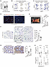 Figure 1
Downregulation of Meflin expression in fibroblasts in the intestinal stricture regions. (A) Normalized Meflin (ISLR) mRNA expression in colon tissue biopsies obtained from non-IBD healthy individuals (n = 80) and patients with CD (n = 48). (B) UMAP feature plots show Meflin (ISLR) expression in stromal cell clusters but not in epithelial and immune cells. (C) UMAP feature plots showing normalized PDGFRA, PDPN, and ACTA2, and expressions in the stromal cell cluster. The arrows indicate cell clusters that coexpress Meflin, PDGFRA, and PDPN. The solid arrowhead indicates the cluster in the top right, which shows high ACTA2 expression but lacks Meflin expression. (D) FISH analysis for Meflin (red) and either PDGFRA (green, left panel) or ACTA2 (green, right panel) in the submucosal layer of the CD-affected intestine. Boxed areas are magnified in insets. Arrows denote double-positive cells. (E) Representative macroscopic image of the ileum surgically resected from a patient with CD. Stricture and nonstricture areas are indicated with arrowheads (left panel) and quantitative real-time polymerase chain reaction (qPCR) analysis for Meflin (right panel, n = 10 pairs). (F) Representative images of Meflin ISH in intestinal tissue sections from non-IBD and nonstricture and stricture areas of patients with CD (Case 1 and 2) and their quantification. Arrows denote Meflin+ cells. (G) Intestinal tissue sections of patients with CD were analyzed by IHC for α-SMA, followed by quantification of α-SMA-positive areas. (H) qPCR analysis of the intestines from patients with CD (n = 10 pairs). (F and G) 5 high-power fields (HPFs) per area were quantified for each patient. Small gray dots indicate individual HPFs, and black triangles indicate patient-level means used for statistical analysis. Paired samples from 10 patients were analyzed. Scale bars: 40 μm. 2-tailed t tests. *P < 0.05; **P < 0.01; ***P < 0.001.
Analysis of a publicly available transcriptomic dataset from NCBI Gene Expression Omnibus (GEO; GSE158952) of intestinal biopsy samples from patients with CD (20) showed that Meflin expression was lower in the ileum, which is more prone to stricture formation, than the rectum (Supplemental Figure 2A). Real-time quantitative polymerase chain reaction (RT-qPCR) analysis of tissues from surgically resected intestines of patients with CD showed that Meflin expression was significantly higher in nonstricture areas than in stricture areas with fibrosis (Figure 1E). Meflin expression was increased in the nonstricture areas of patients with CD compared with tissues from people in the non-IBD control group both at the mRNA and protein levels, whereas it was significantly downregulated in the stricture areas that were strongly positive for α-SMA (Figure 1, F and G and Supplemental Figure 2, B and C). This was further corroborated by analysis of the previously published RNA-seq dataset from Mukherjee et al. (8), which showed that Meflin expression was downregulated in the inflamed and stricture areas (Supplemental Figure 2D). Differences in the expression of fibroinflammatory markers such as ACTA2, COL1A1, TGFB1, and IL-10 between nonstricture and stricture areas were also confirmed by RT-qPCR (Figure 1H).
To investigate the mechanism underlying the regulation of Meflin expression, we treated human intestinal fibroblasts isolated from patients with CD with TGF-β, which resulted in an upregulation of ACTA2 and significant downregulation Meflin expression (Supplemental Figure 2, E and F). The Pearson correlation analysis of the gene expression profiles of balloon-assisted enteroscopic tissue biopsy samples taken from nonstricture ileal areas of patients with CD (n = 24, Supplemental Table 1) showed significant correlations between Meflin and TGFB1 and COL1A1 (Supplemental Figure 2G). Taken together, these findings suggest the involvement of Meflin downregulation in fibroblasts in the progression of intestinal fibrosis in patients with CD.
Meflin deficiency aggravates intestinal fibrosis in the DSS-induced chronic colitis model. Similar to the higher expression in biopsy samples taken from the rectum than in the ileum in patients with CD in a publicly available dataset from NCBI Gene Expression Omnibus (GEO; GSE158952) (20), Meflin expression was more abundant in the mouse colon than in the small intestine (Supplemental Figure 3A). In the colon, Meflin+ fibroblasts were located in the subepithelial region and around the crypts within the lamina propria, as well as in the submucosal layer and muscularis propria (Figure 2A).
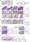 Figure 2
Meflin deficiency aggravates intestinal fibrosis in the DSS-induced colitis model. (A) ISH for Meflin (Islr) in the mouse colon. Arrowheads denote Meflin+ cells. SM, submucosa; MP, musclularis propria. (B) Schematic diagram of DSS-induced chronic colitis and fibrosis model. (C and D) Colonic tissue sections from WT mice at different time points after DSS administration are examined by H&E staining, IHC for α-SMA, Sirius Red staining (C), and ISH for Meflin (D). Note that muscularis mucosa thickness is increased over time (arrowheads). Quantification (n = 3–5/group). Arrows denote Meflin+ cells. 1-way ANOVA. (E) Representative images of H&E-stained colon sections from WT and Meflin-KO mice after 9 weeks of DSS administration (n = 7 and 4 for WT and Meflin-KO mice, respectively), followed by the quantification of submucosa thickness. (F and G) Representative images of IHC for α-SMA (F) and Sirius Red staining (G) on colon sections from WT and Meflin-KO mice after DSS administration, followed by the quantifications of α-SMA–positive areas, collagen layer thickness, Sirius Red+ areas, and fibrosis score. (C–G) Five HPFs per area were quantified for each mouse. Small gray dots indicate individual HPFs, and black triangles indicate mouse-level means used for statistical analysis. Scale bars: 40 μm. 2-tailed Student’s t tests unless otherwise indicated. *P < 0.05; **P < 0.01; ****P < 0.0001.
We first compared the littermates of WT and Meflin-knockout (KO) mice of the same sex and age, which we previously generated (9, 12–14) and found that there were no preexisting differences at baseline in either overall morphology or fibrotic indicators, including the expression of profibrotic and inflammatory genes such as Tgfb1, Acta2, Col3a1, and Mmp9 (Supplemental Figure 3, B and C). To clarify the involvement of Meflin in inflammation-induced intestinal fibrosis, we used the chronic dextran sodium sulfate–induced (DSS-induced) colitis model, which is widely recognized and used in modeling fibrogenesis of IBD (21–23) (Figure 2B). Progressive intestinal inflammation and fibrosis were confirmed by histological evaluation, immunohistochemistry (IHC) for α-SMA, and Sirius Red staining (Figure 2C). The in situ hybridization (ISH) assay revealed that Meflin expression was initially upregulated, peaking at the early stage of fibrosis approximately 6 weeks after starting DSS administration, but was significantly downregulated once intestinal fibrosis was established at week 9, as evidenced by extensive collagen deposition and α-SMA expression (Figure 2, C and D).
To investigate the potential involvement of Meflin in fibrosis progression, we subjected Meflin-KO mice to the DSS-induced colitis model. Histological analysis at 9 weeks after starting DSS administration revealed that intestinal inflammation in Meflin-KO mice was more prominent than that in WT mice, as assessed by the inflammatory histological scoring system (24) and quantification of the minimum and maximum submucosal thickness (Figure 2E). Correspondingly, intestinal fibrosis was significantly more severe in Meflin-KO mice than in WT mice (Figure 2, F and G). These results suggested that Meflin plays a protective role against the development of inflammation and fibrosis.
Meflin deficiency accelerates intestinal fibrosis in a spontaneous colitis model induced by IL-10 deficiency. We then used IL-10–KO mice deficient in the expression of the antiinflammatory cytokine IL-10, which is known to spontaneously develop small intestinal and colonic intestinal colitis and fibrosis without DSS or any other chemical administration (Supplemental Figure 3D) (25, 26), and confirmed the progressive development of intestinal inflammation and fibrosis over time (Figure 3A). ISH analysis of Meflin expression at different time points showed that there were a large number of Meflin+ fibroblasts at the early stages of fibrosis development from 10–12 weeks; however, their numbers significantly decreased at later stages from 14–16 weeks (Figure 3B).
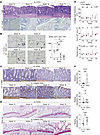 Figure 3
Meflin deficiency accelerates intestinal fibrosis in the IL-10–KO spontaneous colitis model. (A and B) Colon tissue sections from IL-10–KO mice at weeks 10, 12, 14, and 16 are subjected to H&E staining (upper panels), ISH for Acta2 (lower panels) (A), and ISH for Meflin (B), followed by quantification (n = 3–5/group). Arrows indicate Meflin-positive cells. (C–E) Colon tissue sections from IL-10–KO and IL-10/Meflin–DKO mice at weeks 10, 12, 14, and 16 are subjected to IHC for α-SMA (C) and Sirius Red staining (D), followed by collagen layer thickness, quantification of Sirius Red+ areas, and fibrosis score (E) (n = 3–5/ group). (F) qPCR analysis for Acta2, Il6, and Tgfb1 mRNA in total RNAs isolated from all the colons of IL-10–KO and IL-10/Meflin–DKO mice (n = 14 and 16, respectively). Each dot represents an individual sample. 2-tailed Student’s t tests. (B and E). 5 HPFs per area were quantified for each mouse. Small gray dots indicate individual HPFs, and black triangles indicate mouse-level means used for statistical analysis. Scale bars: 40 μm. 1-way ANOVA for B and 2-tailed Student’s t tests for E. *P < 0.05; ** P < 0.01; *** P < 0.001; ****P < 0.0001.
We generated IL-10/Meflin double–knockout (DKO) mice (Supplemental Figure 3, D and E) and compared the extent of fibrosis between IL-10–KO and IL-10/Meflin–DKO mice at different time points. A more accelerated fibrogenesis was observed in IL-10/Meflin-DKO mice compared with IL-10–KO mice (Figure 3, C–E, and Supplemental Figure 3F). Consistent with this finding, the expressions of profibrotic and inflammatory genes, such as Acta2, Il6, and Tgfb1, were more dominant in the fibrotic intestine of IL-10/Meflin DKO mice than in IL-10–KO mice (Figure 3F). Our results demonstrate that Meflin deficiency in fibroblasts aggravates the intestinal fibroinflammatory reaction induced by IL-10 deficiency in mice.
Antifibrotic effect of pharmacological induction of Meflin expression in fibroblasts on intestinal fibrosis. We previously screened a chemical library and identified Am80 (tamibarotene), a synthetic retinoid that has been clinically used as a therapeutic agent for acute promyelocytic leukemia (27), as a reagent that effectively induces Meflin expression in fibroblasts (14, 15, 28). The functional doses and potential cytotoxic effects were evaluated previously (14). Since Meflin expression peaked at the early stage of fibrogenesis, approximately 6 weeks after DSS induction (Figure 2D), we first investigated the effect of the pharmacological induction of Meflin expression by orally administering Am80 to mice subjected to DSS for 7–9 weeks (days 43–63) (Figure 4A). Am80-mediated significant induction of Meflin expression was confirmed by ISH (Figure 4B). Unexpectedly, mice treated with Am80 exhibited a continuous decrease in body weight (Figure 4C). Additionally, gross observation of the colon and histopathological evaluation with H&E staining revealed that the Am80 group exhibited more severe intestinal inflammation, suggesting that Am80 administration exacerbated intestinal inflammation (Figure 4, D and E). However, intestinal fibrosis was significantly ameliorated by Am80 administration, as shown by decreases in the number of α-SMA+ myofibroblasts and collagen deposition (Figure 4, F and G).
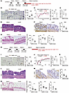 Figure 4
Antifibrotic effect of Am80 administration on intestinal fibrosis in the DSS mouse model. (A) Schematic diagram showing the experimental setup. Oral administration of Am80 (3 mg/kg/day) or DMSO every day was initiated simultaneously with the third dose of DSS administration from days 43–63. (B) Colon tissue sections from WT mice after Am80 or DMSO administration for 9 weeks are analyzed for Meflin expression using ISH. Boxed areas are magnified in insets. Arrows denote Meflin+ cells. (C and D) Body weight changes over time and colon length at Day 63 are measured. (E–G) Colon tissue sections after Am80 administration are stained with H&E (E), IHC for α-SMA (F), and Sirius Red (G), followed by collagen layer thickness, quantification of Sirius Red+ areas, and fibrosis score. (F and G) (n = 6 and 7 for the control and Am80 groups, respectively). (H) Oral administration of Am80 (3 mg/kg/day) or DMSO every day to WT mice from weeks 10 to 13 after the 3 cycles of DSS administration. (I) Colon tissue sections are analyzed for Meflin expression using ISH. Boxed areas are magnified in insets. Arrows denote Meflin+ cells. (n = 4 and 5 for the control and Am80 groups, respectively). (J and K) Body weight changes over time and colon length at Day 92 are evaluated. (L–N) Representative images for H&E, IHC for α-SMA, and Sirius Red staining performed in colon tissue sections from WT mice after DSS and subsequent Am80 administrations (n = 7 and 5 for the control and Am80 groups, respectively). (B, F, G, I, M, and N) 5 HPFs per area were quantified for each mouse. Small gray dots indicate individual HPFs, and black triangles indicate mouse-level means used for statistical analysis. Scale bars: 40 μm. 2-tailed Student’s t tests. *P < 0.05; ** P < 0.01; *** P < 0.001; ****P < 0.0001.
To explore the optimal timing of Am80 administration to maximize its beneficial therapeutic effect, we next administered Am80 during weeks 10–13, corresponding with the phase in which acute inflammation resolves in the DSS-induced colitis model (Figure 4H) (24). As with the previous dosing regimen (Figure 4A), significant upregulation of Meflin expression was achieved using this regimen (Figure 4I). However, no significant differences in inflammatory indicators, including body weight changes, colon length, or histopathological assessments, were observed between Am80-treated and control groups (Figure 4, J–L). Importantly, the Am80-treated group showed significant mitigation of intestinal fibrosis, as indicated by decreases in the number of α-SMA+ myofibroblasts, submucosal collagen deposition, and fibrosis scoring (Figure 4, M and N). These histological findings were further supported by significant downregulation of fibrotic marker genes, including Acta2, Col1a1, Col3a1, and Fn1, in Am80-treated mice (Supplemental Figure 4, A and B).
We next administered Am80 to the DSS-induced colitis model through the early to fibrotic phase (days 1–42) to test its preventive effect on disease progression (Supplemental Figure 5A). No significant differences in the inflammatory indicators, including body weight changes, colon length, and histopathological assessments, were observed between the Am80-treated and control groups (Supplemental Figure 5, B–D). Interestingly, Am80 administration from the start of DSS induction significantly attenuated the development of intestinal fibrosis, as demonstrated by reduced fibrotic area and collagen deposition at a later time point (week 9) (Supplemental Figure 5, E–G). These data suggest that there are specific therapeutic windows and dosing regimens in which Am80 can be used to suppress, resolve, or even prevent intestinal fibrosis in a DSS-induced colitis model.
Given that the established therapy for patients with IBD has been based on the antiinflammatory effect of the antitumor necrosis factor α (TNF-α) antibody, we next evaluated the synergistic effect of the combination of Am80 and anti–TNF-α antibody on the disease state of the DSS model and examined whether the aggravative effect of Am80 observed in the earlier experiment, shown in Figure 4, A–G, was rescued by the anti–TNF-α antibody (Supplemental Figure 6A). Coadministration of the anti–TNF-α antibody with Am80 did not ameliorate the excessive inflammation induced by Am80, as indicated by comparable body weights, colon length, and histological inflammation (Supplemental Figure 6, B-D). However, the fibrotic reaction was significantly reduced by Am80 administration, regardless of its combination with the anti–TNF-α antibody. Meanwhile, anti–TNF-α monotherapy failed to suppress myofibroblast activation or collagen deposition (Supplemental Figure 6, E–H). The expression levels of inflammatory cytokines, including Il6, Tnf, and Il10, were not significantly different between the Am80 monotherapy and the Am80/anti–TNF-α antibody combination groups (Supplemental Figure 6I).
Next, we evaluated the therapeutic potential of combining Am80 with an anti–IL-12/23 p40 antibody, which is widely used in clinical practice, particularly for patients with CD who are refractory to anti–TNF-α therapy (29, 30). We coadministered an anti–IL-12 p40 antibody with Am80 in the DSS-induced colitis model (Figure 5A). Interestingly, the coadministration of an anti–IL-12 p40 antibody with Am80 ameliorated the excessive inflammation induced by Am80, as indicated by improved body weight loss, restored colon length, and reduced histological inflammation when compared with mice treated with Am80 alone (Figure 5, B–D). Moreover, the fibrotic reaction was significantly reduced by Am80 administration, regardless of its combination with the anti–IL-12 p40 antibody (Figure 5, E and F). Collectively, these findings indicate that the anti–IL-12 p40 antibody effectively mitigates Am80-associated intestinal inflammation while preserving Am80’s antifibrotic effects, thereby broadening the therapeutic window of Am80 when used in combination with clinically relevant antiinflammatory therapy.
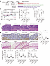 Figure 5
Combined administration of Am80 and anti–IL-12 p40 antibodies attenuates intestinal fibrosis and suppresses Am80-associated intestinal inflammation. (A) Schematic diagram showing the experimental setup. Oral administration of Am80 (3 mg/kg/day, every day), anti–IL-12 p40 antibodies (every other day), or the combination of Am80 and anti–IL-12 p40 antibody administration was initiated simultaneously with the 3rd dose of DSS administration from days 43–63. (B and C) Body weight changes over time and colon length at Day 63 are measured, followed by quantification. (D–F) Colon tissue sections of mice of the indicated groups are stained with H&E (D), examined for α-SMA by IHC (E), and stained by Sirius Red (F), followed by the quantification of collagen layer thickness, Sirius Red+ areas, and fibrosis score (n = 4 for control and n = 5 for Am80, anti-IL-12 p40 Ab, and Am80/anti–IL-12 combination groups). (B right panel, C) Each dot represents an individual sample. (E and F) 5 HPFs per area were quantified for each mouse. Small gray dots indicate individual HPFs, and black triangles indicate mouse-level means used for statistical analysis. Scale bars: 40 μm. One-way ANOVA. *P < 0.05; **P < 0.01; ***P < 0.001; ****P < 0.0001.
Am80-induced reversal of intestinal fibrosis is mediated by Meflin expression. Given that Am80 is a synthetic retinoid, it may exert many effects on several types of cells besides the induction of Meflin expression in fibroblasts. We next subjected Meflin-KO mice to the DSS-induced colitis model, followed by treatment with Am80 using the same protocols for WT mice (Figure 6A). Meflin-KO mice administered Am80 from days 43–63 exhibited comparable body weight, colon length, and intestinal inflammation to those of the control groups (Figure 6, B–D). No significant difference in fibrotic response was observed between the Am80-treated and control groups (Figure 6, E and F). These results suggest that both the hyperactivated intestinal inflammation and reduced fibrosis induced by Am80 are dependent on Meflin expression.
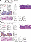 Figure 6
Am80-induced reversion of established intestinal fibrosis is mediated by Meflin expression. (A) Schematic diagram showing the experimental setup. (B and C) Body weight changes over time, and colon length at Day 63 is measured, followed by quantification. (D–F) Colon tissue sections from Meflin-KO mice after Am80 administration are stained with H&E (D), IHC for α-SMA (E), and Sirius Red (F), followed by collagen layer thickness, quantification of Sirius Red+ areas, and fibrosis score. (E and F) (n = 5 and 4 for the control and Am80 groups, respectively). (G) Am80 (3 mg/kg/day) or DMSO is orally administered every day to Meflin-KO mice from weeks 10–13 after the 3 cycles of DSS administration. (H and I) Body weight changes over time, and colon length at Day 92 is evaluated. (J–L) Representative images for H&E, IHC, for α-SMA, and Sirius Red staining performed in colon tissues from Meflin-KO mice after DSS and subsequent Am80 administrations (n = 4 and 6 for the control and Am80 groups, respectively), followed by quantification. (B right panel, C, H right panel, I) Each dot represents an individual sample. (E, F, K, L) 5 HPFs per area were quantified for each mouse. Small gray dots indicate individual HPFs, and black triangles indicate mouse-level means used for statistical analysis. Scale bars: 40 μm. Statistical significance was determined using 2-tailed Student’s t tests.
We then examined whether the efficacy of Am80 in mitigating intestinal fibrosis after the resolution of acute inflammation also depends on the expression of Meflin. Meflin-KO mice subjected to DSS were orally administered Am80 after fibrosis was established for 10–13 weeks (Figure 6G). We did not observe any significant differences in body weight, colon length, inflammatory reaction, number of α-SMA+ myofibroblasts, collagen deposition, or fibrosis scores between Am80-treated and control mice (Figure 6, H–L). Moreover, no significant difference was found in fibrotic and inflammatory gene expression in RT-qPCR analysis (Supplemental Figure 7, A and B). Our results indicate that the efficacy of Am80 in suppressing the fibrotic response and reversing established intestinal fibrosis is mediated by Meflin expression induced by Am80 in the DSS colitis model.
Am80 reverses fibrotic response in cultured fibroblasts and tissue pieces harvested from intestinal stenotic regions of patients with CD. To test whether Am80 could be used to treat patients with CD, we isolated intestinal fibroblasts from both nonstricture and the stricture areas of surgically resected intestines of patients with CD and cultured them in the presence or absence of Am80, followed by gene expression analysis (Figure 7A). Immunofluorescence (IF) staining showed more prominent expression of α-SMA in intestinal fibroblasts isolated from stricture areas than in those isolated from nonstricture areas (Figure 7B). We also confirmed that Meflin expression was significantly downregulated in intestinal fibroblasts from stricture areas compared with those from nonstricture areas (Figure 7C). The treatment of fibroblasts isolated from the stricture area with Am80 for 48 hours resulted in a significant decrease in α-SMA expression (Figure 7D), which was accompanied by the upregulation of expressions of Meflin and IL-10 and downregulation of collagen type I (COL1A1) expression (Figure 7E).
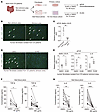 Figure 7
Am80 administration reverses fibrotic response of fibroblasts and tissue pieces isolated from patients with CD. (A) Intestinal fibroblasts and ileal tissue pieces are isolated from the intestinal nonstenotic and stenotic areas surgically resected from patients with CD, followed by culture on plastic. These samples are treated with Am80 (50, 100, 300 μg/mL) or DMSO for 48 or 72 hours, followed by the indicated experiments. (B) Primary cultured intestinal fibroblasts from the nonstenotic (left) and stenotic (right) areas are fixed and stained for α-SMA using IF. Arrows denote fibroblasts that are strongly positive for α-SMA. Scale bars: 20 μm. (C) qPCR analysis for Meflin mRNA expression in the primary cultured intestinal fibroblasts (n = 4 and 5 for the nonstenotic and stenotic groups, respectively). (D) Intestinal fibroblasts cultured in the absence (left) or presence (right) of Am80 are fixed and subjected to IF for α-SMA. Arrows denote fibroblasts that are strongly positive for α-SMA. Scale bars: 20 μm. (E) qPCR analysis for the expression of the indicated genes in the intestinal fibroblasts cultured in the absence (left) or presence (right) of Am80 (n = 5 for each group). (F and G) Ileal tissue pieces obtained from the stenotic regions of intestine are incubated with different concentrations of Am80 (50 and 300 μg/mL) or DSMO for 24 hours, followed by the measurement of TGF-β (F) and IL-6 (G) in the culture supernatants using enzyme-linked immunosorbent assays (n = 7 to 12 for each group). (C, E, F, G) Statistical significance was determined using 2-tailed Student’s t tests. *P < 0.05; **P < 0.01; ****P < 0.0001.
To further test the antifibrotic effect of Am80 in a model that recapitulates human intestinal fibrosis, we prepared ileal tissue pieces from surgically resected specimens of 7 patients with CD and put them in ex vivo tissue culture for 72 hours either in the presence or absence of different doses of Am80 (50–300 μg/mL) in the medium (Figure 7A). The measurement of the secretion of profibrotic cytokines using ELISA showed that the amounts of active and latent form of TGF-β and IL-6 were significantly reduced by Am80 treatment (Figure 7, F and G), suggesting that Am80 may be potent in inhibiting the secretion of profibrotic cytokines and ameliorating established intestinal fibrosis in patients with CD.
Meflin deficiency leads to the upregulation of the noncanonical Wnt ligand WNT5A and its receptor ROR2. Given the role of Meflin in intestinal fibrogenesis, we sought to elucidate the underlying mechanisms. We isolated colonic fibroblasts from both WT and Meflin-KO mice after DSS administration (Supplemental Figure 8A) and transduced those fibroblasts with mouse Meflin (mMeflin) cDNA using a lentiviral expression system (Figure 8A). Notably, the elevated expression of fibrotic markers, including Fn1, Col3a1, and Col6a1, observed in Meflin-KO fibroblasts was significantly reversed by mMeflin restoration (Figure 8B). These results confirmed that Meflin may confer an antifibrotic role to fibroblasts. To further characterize Meflin+ fibroblasts, we analyzed a publicly available single-cell RNA-seq dataset from NCBI Gene Expression Omnibus (GEO; GSE211275) for mouse and human colonic mesenchymal cells (8). The analysis revealed that Meflin and various BMP signaling components are highly coexpressed within several intestinal mesenchymal cell populations across the species (Supplemental Figure 8, B–F, and Supplemental Tables 2 and 3), consistent with our previous studies, which showed that Meflin augments BMP7 signaling in fibroblasts (12, 16). However, Meflin deficiency or exogenous mMeflin transduction had little-to-no effect on BMP7-mediated SMAD1/5/9 phosphorylation, cell proliferation, or the expression of collagen genes (Col1a1 and Col3a1) (Supplemental Figure 8, G–I). Basal Smad1/5/9 phosphorylation and the proliferation rate of Meflin-KO fibroblasts were lower and higher, respectively, compared with WT fibroblasts, and these differences were negated by BMP7 stimulation, which is of unknown significance at present (Supplemental Figure 8, G and H). These data showed the limited functional involvement of Meflin in BMP7/SMAD signaling at least in intestinal fibroblasts.
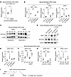 Figure 8
Meflin may inhibit intestinal fibrosis by suppressing the expression of WNT5A and its receptor ROR2. (A) Colonic fibroblasts isolated from WT mice after 9 weeks of DSS administration were transduced with control or mouse Meflin (mMeflin) cDNA using a lentiviral expression system, followed by qPCR analysis (n = 3 per group). CTL, control. 2-tailed Student’s t tests. (B) qPCR analysis for the expression of Fn1, Col3a1, and Col6a1 in the indicated fibroblasts (n = 3–6 per group). (C) Colonic fibroblasts isolated from WT and Meflin-KO mice after 9 weeks of DSS administration were cultured with or without recombinant BMP7 (rBMP7, 100 ng/mL) for 1 hour, followed by Western blot analysis for WNT5A/B and β-actin. The data from 2 independent experimental batches are shown. kDa, kilodaltons. (D) Colonic fibroblasts isolated from nonstricture and stricture areas of patients with CD were treated with Am80 (1 μM) for 48 hours, followed by Western blot analysis for Meflin, WNT5A/B, and GAPDH. (E) qPCR analysis for Ror2 mRNA expression in colonic fibroblasts isolated from WT and Meflin-KO mice. Restoring Meflin expression (mMeflin) in Meflin-KO fibroblasts significantly suppressed Ror2 expression (n = 6 per group). (F) Colonic fibroblasts isolated from WT and Meflin-KO mice were treated with rBMP7 (20 ng/ml) for 48 hours, followed by qPCR analysis for Ror2 mRNA expression (n = 9 per group). (G and H) Colonic fibroblasts were transfected with control siRNA (siControl) or siRor2, followed by qPCR analysis for Ror2 (G) in Col1a1 (H) mRNA levels (n = 3 per group). (I) Summary of the findings obtained by colonic fibroblasts from WT and Meflin-KO mice and Ror2 knockdown. (B, E, F) Each dot represents an individual sample. Scale bars: 40 μm. 1-way ANOVA unless otherwise indicated. *P < 0.05; **P < 0.01; ***P < 0.001; ****P < 0.0001.
We next sought to identify other changes in gene expression associated with Meflin. Gene ontology enrichment analysis of high–Meflin-expressing cell clusters identified from scRNA-seq datasets of intestinal tissues from patients with CD (GSE134809) and from the DSS-induced colitis model (GSE172261) showed an enrichment of genes associated with Wnt signaling (Supplemental Figure 9A). Further analysis of both datasets revealed that Meflin is coexpressed with the noncanonical Wnt ligand Wnt5a in some intestinal stromal populations (Supplemental Figure 9, B-E). Our analysis also demonstrated that Meflin+ cells are distinct from CDH11+ cells, which have been reported to promote intestinal fibrosis (8) (Supplemental Figure 9F). Considering a recent report that fibroblasts positive for WNT5A are highly enriched in the strictured lesions to play a central role in fibrogenesis in patients with CD (8), we hypothesized that Meflin suppresses the fibrogenesis by regulating WNT5A expression.
We observed an upregulation of WNT5A expression in Meflin-KO fibroblasts and human intestinal fibroblasts isolated from the stricture area of the ileum of patients with CD, and this was modestly promoted by recombinant BMP7 (rBMP7) stimulation and suppressed by Am80 treatment, consistent with previous reports demonstrating that retinoic acid signaling counteracts Wnt signaling (31, 32) (Figure 8, C and D). Meflin KO fibroblasts exhibited an upregulation of the WNT5A receptor Ror2 (33, 34), which was negated by mMeflin restoration but independent of rBMP7 stimulation (Figure 8, E and F). Importantly, siRNA-mediated knockdown of Ror2 in Meflin-KO fibroblasts significantly reduced Col1a1 expression (Figure 8, G and H). These data suggested that Meflin deficiency in fibroblasts is associated with increases in the expression of WNT5A and its receptor ROR2 that induce fibrogenesis, which could be modulated by BMP7 and Am80 (Figure 8I).
Meflin deficiency is associated with Wnt signaling dysregulation in the DSS-induced colitis model. Finally, we examined the dysregulation of WNT5A and ROR2 expression in tissue samples of the DSS-induced colitis model. ISH analysis showed that the expression of Wnt5a in Meflin+ fibroblasts was more evident in mice undergoing DSS-induced chronic inflammation than in control mice (Figure 9A). The number of Wnt5a+ fibroblasts was higher in Meflin-KO mice treated with DSS than in WT mice (Figure 9B). In contrast, the number of Meflin+ fibroblasts positive for Wnt2b, a canonical Wnt ligand (35), was reduced in the DSS model (Supplemental Figure 10A), and Wnt2b expression in fibroblasts was significantly downregulated in Meflin-KO mice subjected to the DSS model (Supplemental Figure 10B). Consistent with the results that showed the high expression of Ror2 in intestinal fibroblasts isolated from Meflin KO mice (Figure 8, E and F), ISH analysis confirmed the upregulation of Ror2 in the intestinal fibroblasts of Meflin-KO mice after DSS treatment, suggesting activation of the noncanonical Wnt pathway in Meflin-KO mice (Figure 9C). Conversely, the expression of the canonical Wnt target genes Axin2 and R-spondin 3 (Rspo3) in fibroblasts was attenuated in Meflin-KO mice compared with that in WT mice (Figure 9, D and E). We also found the compensatory upregulation of Wntless (encoded by the Wls gene), which plays an indispensable role in the transportation and secretion of Wnt ligands (36), in fibroblasts of Meflin-KO mice treated with DSS (Figure 9F, Supplemental Figure 10C).
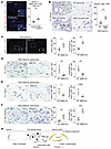 Figure 9
Dysregulated Wnt signaling in Meflin-deficient mice. (A) Colon tissue sections prepared from intestines of WT mice treated with DSS are stained for Meflin (Islr, red) and Wnt5a (green) mRNA by FISH (left), followed by quantification of the positivity of Wnt5a in Meflin+ fibroblasts (arrows) (right). Boxed areas are magnified in insets (n = 3 per group). (B–F) Colon tissue sections prepared from the intestines of WT and Meflin-KO mice treated with DSS are stained for Wnt5a (B), Ror2 (C), Axin2 (D), Rspo3 (E), and Wls (F) using ISH or FISH (left panels), followed by quantification of the numbers of positive cells and percentage of those cells per all stromal cells with a fibroblast-like morphology (right panels). (n = 3–4 for each group). Arrows indicate the positive cells. (G) Schematic illustration showing the hypothesis proposed by the present study. (A–F) 5 HPFs per area were quantified for each mouse. Small gray dots indicate individual HPFs, and black triangles indicate mouse-level means used for statistical analysis. Scale bars: 40 μm. Statistical significance was determined using 2-tailed Student’s t tests. *P < 0.05; **P < 0.01; ***P < 0.001.
Collectively, these data suggest that Meflin deficiency is associated with the upregulated production of noncanonical Wnt ligands and their activation, thus contributing to the activation of intestinal fibrogenesis.


Copyright © 2026 American Society for Clinical Investigation
ISSN: 0021-9738 (print), 1558-8238 (online)