
Advertisement
Research ArticleCardiologyImmunology
Open Access |  10.1172/JCI189684
Fate-mapping infiltrating monocytes following experimental myocardial infarction reveals differentiation trajectories in the infarcted heart
Andrew L. Koenig,1 Farid F. Kadyrov,1 Junedh M. Amrute,1 Steven Yang,1 Carla J. Weinheimer,1 Jessica M. Nigro,1 Attila Kovacs,1 Wenjun Li,2 Gabriella B. Smith,1 Lance Yeh,1 Daniel Kreisel,2,3 and Kory J. Lavine1,3,4
1Center for Cardiovascular Research, Division of Cardiology, Department of Medicine,
2Department of Surgery,
3Department of Pathology and Immunology, and
4Department of Developmental Biology, Washington University School of Medicine, Saint Louis, Missouri, USA.
Address correspondence to: Kory J. Lavine, Division of Cardiology, Department of Medicine, Washington University School of Medicine, 660 S Euclid, Campus Box 8086, St. Louis, Missouri 63110, USA. Phone: 314.362.1171; Email: klavine@wustl.edu.
Authorship note: ALK and KJL are co-senior authors.
Find articles by Koenig, A. in: PubMed | Google Scholar
1Center for Cardiovascular Research, Division of Cardiology, Department of Medicine,
2Department of Surgery,
3Department of Pathology and Immunology, and
4Department of Developmental Biology, Washington University School of Medicine, Saint Louis, Missouri, USA.
Address correspondence to: Kory J. Lavine, Division of Cardiology, Department of Medicine, Washington University School of Medicine, 660 S Euclid, Campus Box 8086, St. Louis, Missouri 63110, USA. Phone: 314.362.1171; Email: klavine@wustl.edu.
Authorship note: ALK and KJL are co-senior authors.
Find articles by Kadyrov, F. in: PubMed | Google Scholar
1Center for Cardiovascular Research, Division of Cardiology, Department of Medicine,
2Department of Surgery,
3Department of Pathology and Immunology, and
4Department of Developmental Biology, Washington University School of Medicine, Saint Louis, Missouri, USA.
Address correspondence to: Kory J. Lavine, Division of Cardiology, Department of Medicine, Washington University School of Medicine, 660 S Euclid, Campus Box 8086, St. Louis, Missouri 63110, USA. Phone: 314.362.1171; Email: klavine@wustl.edu.
Authorship note: ALK and KJL are co-senior authors.
Find articles by Amrute, J. in: PubMed | Google Scholar
1Center for Cardiovascular Research, Division of Cardiology, Department of Medicine,
2Department of Surgery,
3Department of Pathology and Immunology, and
4Department of Developmental Biology, Washington University School of Medicine, Saint Louis, Missouri, USA.
Address correspondence to: Kory J. Lavine, Division of Cardiology, Department of Medicine, Washington University School of Medicine, 660 S Euclid, Campus Box 8086, St. Louis, Missouri 63110, USA. Phone: 314.362.1171; Email: klavine@wustl.edu.
Authorship note: ALK and KJL are co-senior authors.
Find articles by Yang, S. in: PubMed | Google Scholar
1Center for Cardiovascular Research, Division of Cardiology, Department of Medicine,
2Department of Surgery,
3Department of Pathology and Immunology, and
4Department of Developmental Biology, Washington University School of Medicine, Saint Louis, Missouri, USA.
Address correspondence to: Kory J. Lavine, Division of Cardiology, Department of Medicine, Washington University School of Medicine, 660 S Euclid, Campus Box 8086, St. Louis, Missouri 63110, USA. Phone: 314.362.1171; Email: klavine@wustl.edu.
Authorship note: ALK and KJL are co-senior authors.
Find articles by Weinheimer, C. in: PubMed | Google Scholar
1Center for Cardiovascular Research, Division of Cardiology, Department of Medicine,
2Department of Surgery,
3Department of Pathology and Immunology, and
4Department of Developmental Biology, Washington University School of Medicine, Saint Louis, Missouri, USA.
Address correspondence to: Kory J. Lavine, Division of Cardiology, Department of Medicine, Washington University School of Medicine, 660 S Euclid, Campus Box 8086, St. Louis, Missouri 63110, USA. Phone: 314.362.1171; Email: klavine@wustl.edu.
Authorship note: ALK and KJL are co-senior authors.
Find articles by Nigro, J. in: PubMed | Google Scholar
1Center for Cardiovascular Research, Division of Cardiology, Department of Medicine,
2Department of Surgery,
3Department of Pathology and Immunology, and
4Department of Developmental Biology, Washington University School of Medicine, Saint Louis, Missouri, USA.
Address correspondence to: Kory J. Lavine, Division of Cardiology, Department of Medicine, Washington University School of Medicine, 660 S Euclid, Campus Box 8086, St. Louis, Missouri 63110, USA. Phone: 314.362.1171; Email: klavine@wustl.edu.
Authorship note: ALK and KJL are co-senior authors.
Find articles by Kovacs, A. in: PubMed | Google Scholar
1Center for Cardiovascular Research, Division of Cardiology, Department of Medicine,
2Department of Surgery,
3Department of Pathology and Immunology, and
4Department of Developmental Biology, Washington University School of Medicine, Saint Louis, Missouri, USA.
Address correspondence to: Kory J. Lavine, Division of Cardiology, Department of Medicine, Washington University School of Medicine, 660 S Euclid, Campus Box 8086, St. Louis, Missouri 63110, USA. Phone: 314.362.1171; Email: klavine@wustl.edu.
Authorship note: ALK and KJL are co-senior authors.
Find articles by Li, W. in: PubMed | Google Scholar
1Center for Cardiovascular Research, Division of Cardiology, Department of Medicine,
2Department of Surgery,
3Department of Pathology and Immunology, and
4Department of Developmental Biology, Washington University School of Medicine, Saint Louis, Missouri, USA.
Address correspondence to: Kory J. Lavine, Division of Cardiology, Department of Medicine, Washington University School of Medicine, 660 S Euclid, Campus Box 8086, St. Louis, Missouri 63110, USA. Phone: 314.362.1171; Email: klavine@wustl.edu.
Authorship note: ALK and KJL are co-senior authors.
Find articles by Smith, G. in: PubMed | Google Scholar
1Center for Cardiovascular Research, Division of Cardiology, Department of Medicine,
2Department of Surgery,
3Department of Pathology and Immunology, and
4Department of Developmental Biology, Washington University School of Medicine, Saint Louis, Missouri, USA.
Address correspondence to: Kory J. Lavine, Division of Cardiology, Department of Medicine, Washington University School of Medicine, 660 S Euclid, Campus Box 8086, St. Louis, Missouri 63110, USA. Phone: 314.362.1171; Email: klavine@wustl.edu.
Authorship note: ALK and KJL are co-senior authors.
Find articles by Yeh, L. in: PubMed | Google Scholar
1Center for Cardiovascular Research, Division of Cardiology, Department of Medicine,
2Department of Surgery,
3Department of Pathology and Immunology, and
4Department of Developmental Biology, Washington University School of Medicine, Saint Louis, Missouri, USA.
Address correspondence to: Kory J. Lavine, Division of Cardiology, Department of Medicine, Washington University School of Medicine, 660 S Euclid, Campus Box 8086, St. Louis, Missouri 63110, USA. Phone: 314.362.1171; Email: klavine@wustl.edu.
Authorship note: ALK and KJL are co-senior authors.
Find articles by Kreisel, D. in: PubMed | Google Scholar
1Center for Cardiovascular Research, Division of Cardiology, Department of Medicine,
2Department of Surgery,
3Department of Pathology and Immunology, and
4Department of Developmental Biology, Washington University School of Medicine, Saint Louis, Missouri, USA.
Address correspondence to: Kory J. Lavine, Division of Cardiology, Department of Medicine, Washington University School of Medicine, 660 S Euclid, Campus Box 8086, St. Louis, Missouri 63110, USA. Phone: 314.362.1171; Email: klavine@wustl.edu.
Authorship note: ALK and KJL are co-senior authors.
Find articles by Lavine, K. in: PubMed | Google Scholar
Published July 7, 2026 - More info
J Clin Invest. 2026;136(17):e189684. https://doi.org/10.1172/JCI189684.
© 2026 Koenig et al. This work is licensed under the Creative Commons Attribution 4.0 International License. To view a copy of this license, visit http://creativecommons.org/licenses/by/4.0/.
Received: December 3, 2024; Accepted: June 24, 2026

Related article:
Abstract
Monocytes and macrophages promote tissue repair following myocardial infarction, but the mechanisms tuning their effector functions remain elusive. While macrophages are essential in clearing debris and resolving inflammation, they can also contribute to uncontrolled inflammation and provoke additional damage. Thus, factors that influence macrophage differentiation trajectories and phenotypes play an important role in cardiac repair outcomes. By combining genetic lineage tracing with cell-specific targeting, the study from Koenig et al. sheds light on key signaling events that shape monocyte fate decisions in the injured myocardium, establishing a differentiation hierarchy among monocyte-macrophage subsets. The findings also reveal that macrophages with an IFN response signature give rise to MHCIIhi macrophages, which, in turn, contribute to regulatory T cell generation and cardioprotection. This work underscores the importance of understanding cardiac macrophage phenotypic plasticity within a broader framework of lineage relationships.
Authors
Ecem Tugba Sakalli, Giuseppe Rizzo, Alma Zernecke, Gustavo Campos Ramos
-
Results
Monocytes are recruited to the heart early after MI and their progeny chronically persist within the myocardium. To trace the fates of monocytes and their progeny after MI, we subjected Ccr2CreERT2Rosa26LSL–tdTomato mice to closed-chest ischemia reperfusion injury to model reperfused MI (23–25). Ccr2CreERT2 mice harbor a tamoxifen-inducible CRE recombinase inserted into the 3’ UTR of the Ccr2 locus. We optimized a tamoxifen induction strategy to exclusively label circulating monocytes and not cardiac macrophages. A single injection of tamoxifen (60 mg/kg, IP) into naive mice achieved selective tdTomato labeling of LY6Chi monocytes in the blood at greater than 80% efficiency 24 hours after injection. Cardiac macrophages did not express tdTomato at day 1 or day 7 after injection (Figure 1A, Supplemental Figure 1; supplemental material available online with this article; https://doi.org/10.1172/JCI189684DS1). We also did not detect substantial tdTomato labeling of neutrophils or lymphocytes in the blood or their progenitors within the bone marrow compartment (Supplemental Figure 2). As such, this strategy allows lineage tracing of infiltrating monocytes and their progeny independent of resident macrophages.
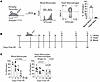 Figure 1
Recruited monocyte-derived populations persist in the heart after injury. (A) Representative flow cytometry plots demonstrating that a single dose of tamoxifen administered to Ccr2CreERT2Rosa26tdTomato mice results in 80% labeling efficiency in monocytes in the blood, while resident cardiac macrophages are not labeled. (B) Schematic of experimental strategy for monocyte lineage tracing after MI. (C) Bar graphs demonstrating tdTomato labeling of blood monocytes (left) and cardiac monocyte and macrophages. Single inj., single dose labeling; cont, continuous dose labeling. Brown-Forsythe 1-way ANOVA.
We then applied this labeling strategy to mice subjected to closed-chest ischemia reperfusion injury. Tamoxifen was injected 1 day before injury. Blood was collected from mice at days 1, 3, 5, 7, 13, and 28 after injury and hearts were collected at days 3, 7, 13, and 28 after injury (Figure 1B). Flow cytometry analysis demonstrated greater than 80% tdTomato labeling efficiency in LY6Chi monocytes in the blood 1 day after MI. The percent of LY6Chi monocytes labeled progressively decreased throughout the first 7 days after MI and was absent thereafter. Intriguingly, the progeny of monocytes that entered the heart during this labeling window persisted for at least 28 days after MI. Continuous labeling of monocytes by tamoxifen injection (60 mg/kg IP, every 3 days) did not demonstrate additional labeling of cardiac macrophages (Figure 1C). Labeling of macrophages within the heart was validated at each time point by visualization of tdTomato and immunostaining for CD68 (Figure 2, A and B). These findings indicate that monocytes enter the heart predominately within the first 7 days after MI and their progeny, including monocyte-derived macrophages, persist within the myocardium for at least 28 days after MI.
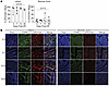 Figure 2
Recruited monocyte-derived populations are concentrated within the infarcted area. (A) Quantification of tdTomato+ macrophages relative to total CD68-positive macrophages measured by IHC. (B) Representative IHC images. Magnification ×20. Each data point represents an individual animal. Brown-Forsythe 1-way ANOVA.
Macrophage states dynamically shift over time after injury. To delineate the temporal dynamics of recruited monocytes and their progeny, we performed single-cell RNA-seq of monocytes and macrophages sorted from Ccr2CreERT2Rosa26LSL–tdTomato hearts collected at days 3, 7, 13, and 28 after MI (Figure 1B and Supplemental Figure 3A). Libraries were constructed for each time point, sequenced, aligned using Cell Ranger, and then integrated, clustered, and analyzed using Seurat (26, 27). We identified 10 transcriptionally distinct cell clusters: monocytes, dendritic cells, and 8 macrophage states (Figure 3A). We performed differential expression analysis and constructed z-score signatures to support that the observed clusters were transcriptionally distinct (Figure 3, B and C, Supplemental Figure 4). When cells were compared across time points, it was evident that macrophage states markedly shifted over time. At 3 days after MI, the predominant populations within the heart consisted of monocytes, proliferating macrophages, Arg1+ macrophages, type-I interferon (IFN) activated macrophages, and Trem2+ macrophages. Through days 7 and 13 after MI, we observed decreases in frequency of monocytes, proliferating cells, Arg1+ macrophages, and type I IFN–activated macrophages. Trem2+ macrophages and Gdf15+ macrophages expanded during this time interval. By day 28 after MI, macrophages had transitioned to a predominantly MHCIIhi state (Figure 4A).
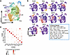 Figure 3
Recruited monocyte-derived populations are transcriptionally diverse. (A) Clustered UMAP projection of combined monocyte lineage tracing single-cell libraries from each time point. (B) Feature plots of calculated z-scores for each cluster. Blue box, list of genes in each z-score. (C) Dot plot of z-scores by cluster.
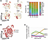 Figure 4
Monocyte-derived populations dynamically shift as a function of time after MI. (A) Gaussian kernel plots indicating cell density at each time point after MI (left) and proportion of cells in each cluster split by time point (right). (B) UMAP plot of Tomato+ cells. (C) Line graph of proportion of tdTomato+ cells in each cluster across time points.
We additionally performed custom alignment of the libraries to the tdTomato coding sequence and observed that all identified clusters were labeled by expression of tdTomato (Figure 4B). Interestingly, a proportion of macrophages within the resident cluster were also labeled by tdTomato, indicating that recruited macrophages have the potential to adopt a resident-like fate consistent with our prior findings (28). When comparing differential expression of tdTomato– and tdTomato+ resident macrophages, we identified that monocyte-derived resident-like (tdTomato+) macrophages demonstrate reduced expression of the tissue resident macrophage markers Folr2 and Lyve1 and enriched expression of Ccl2, Ccl7, and Ccl12 compared with cardiac resident (tdTomato–) macrophages (Supplemental Figure 4).
We next examined the dynamics of monocyte-derived cells as a function of time and found that tdTomato+ cells were substantially distributed across cell states at day 3 after MI. At days 7 and 14 after MI, the number of tdTomato+ monocytes, Arg1+ macrophages, type I IFN–activated macrophages, and proliferating macrophages declined, and Gdf15+ and Trem2+ macrophages progressively increased. At day 28 after MI, the majority of tdTomato+ cells were MHCIIhi macrophages (Figure 4C). Together, these results demonstrate that monocyte recruitment occurs within the first week after MI, their progeny persist within the heart at chronic stages, and monocytes display substantial plasticity differentiating into multiple transcriptionally distinct subsets that dynamically shift over time.
Monocyte-derived macrophage and dendritic cell–like states exist outside of the cardiac resident macrophage compartment. We utilized Cx3cr1CreERT2Rosa26LSL–tdTomato mice to evaluate the contribution of resident macrophages to the diverse subsets of macrophage states that emerge following MI. Utilizing a pulse-chase strategy, we injected mice with tamoxifen (60 mg/kg daily X5 days) 2 weeks before ischemia reperfusion injury (Figure 3A). This strategy specifically labels long-lived resident macrophages with tdTomato. Monocytes are not labeled, as they are replenished by unlabeled monocytes during the chase period (13). Macrophages and monocytes were sorted via FACS into tdTomato+ (resident) and tdTomato– (recruited) libraries for single-cell RNA-seq (Figure 5A and Supplemental Figure 3B). We observed that cardiac resident macrophages clustered independently of recruited macrophages and displayed distinct transcriptional profiles. Differential gene expression analysis demonstrated that resident and recruited macrophages express distinct transcriptional signatures. Many of the genes enriched in monocyte-derived macrophages were markers of the diverse populations seen in Ccr2CreERT2 lineage tracing. Conversely, classical markers of tissue resident macrophages were enriched within the resident cardiac macrophage library (Cd163, Mrc1, Cd207, Vsig4, Folr2, Cbr2). The resident macrophage library is also enriched for components of the complement pathway (C1qa, C1qc, C4b). This is consistent with the role of resident macrophages initiating the inflammatory cascade after injury and the role of the complement pathway in recruiting monocytes (29, 30). At this earlier time point after injury, expression of MHCII genes appear to be enriched primarily in the resident macrophage library. In interpreting this finding, it should be noted that very few recruited MHCIIhi macrophages exist within the heart at this early time point. Thus, these data are most consistent with studies demonstrating existence of MHCIIhi and MHCIIlo resident macrophages (Figure 5, B and C) (31). To validate that the resident macrophage lineage tracing labeling was efficient, we aligned the single cell libraries to the tdTomato coding sequence and found that tdTomato was exclusively expressed in cells found within the resident macrophage library (Figure 5, D and E). These data indicate that resident macrophages do not contribute to the diverse macrophage subsets marked by Ccr2-CreERT2 lineage tracing, consistent with their origin from infiltrating monocytes.
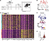 Figure 5
Monocyte-derived populations exist independent of cardiac resident macrophages. (A) Schematic of experimental strategy for resident macrophage lineage tracing (left) and FACS gating strategy for constructing tdTomato+ (resident) and tdTomato– (recruited) libraries. (B) Combined UMAP plot of resident and recruited libraries. Red indicates cells derived from cardiac resident macrophages. (C) Heatmap of genes differentially expressed in cells derived from either cardiac resident macrophages or recruited monocytes. (D) Feature plot of tdTomato expression split by library. (E) Violin plot of tdTomato expression split by library.
Macrophage states identified after MI in mice are conserved in human heart failure. We utilized human single-cell RNA-seq data obtained from a prior study to determine if the observed monocyte, macrophage, and dendritic cell populations are conserved in the human heart. Transcription signatures for each mouse cluster were converted to their corresponding human homologues. Z-scores for these marker genes were calculated across human cardiac monocytes, macrophages, and dendritic cell clusters (Figure 6, A–C) (32). This analysis demonstrated that transcriptional signatures enriched in each mouse monocyte, macrophage, and dendritic cell cluster were conserved in corresponding populations of monocytes, macrophages, and dendritic cells found in the human heart, indicating that mice may serve as an appropriate model to investigate human monocyte differentiation.
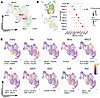 Figure 6
Monocyte-derived populations are conserved between mice and humans. (A) UMAP plot of monocytes, macrophages, and dendritic cells from human donor (n = 2) and heart failure samples (n = 5). (B)Schematic of analysis strategy (left) and dot plot of denoting the expression of genes enriched in each mouse monocyte, macrophage, and dendritic cell population (displayed as a z-score) in corresponding human monocyte, macrophage, and dendritic cell populations. Genes with mouse and human homologues are included in this analysis. (C) UMAP feature plots of each mouse z-score signature in the human object.
Arg1+ and IFN-activated macrophages are predicted as early transient macrophage states. We utilized scVelo and Velocyto.R to perform RNA Velocity lineage tracing analysis (33, 34). We presented the data using t-distributed Stochastic Neighbor Embedding (t-SNE) projections split by time point after MI (Figure 7, A and B). RNA Velocity suggested that monocytes differentiate into Arg1+ and IFN-activated macrophage populations as early intermediates, which subsequently differentiate to terminal Trem2+ and MHCIIhi macrophages, respectively (Figure 7A). Splitting the t-SNE plot by time point after MI revealed 2 distinct populations of Trem2+ macrophages, one arising at day 3 and a second evident beginning at day 7. The early Trem2+ macrophage population expressed a distinct transcriptional signature that includes Arg1 and Spp1. The later Trem2+ macrophage population displayed enriched expression of Ninj1, Creg1, and Gstm1 (Figure 7A and Supplemental Figure 5A). Each of these Trem2+ macrophage populations was also observed in an independent trajectory analysis using Palantir (35). Palantir predicted that cDC2s, Trem2+ macrophages, and MHCIIhi macrophages were terminal cell states, and that type I IFN–activated macrophages and Arg1+ macrophages represented intermediate states of differentiation based on entropy and pseudotime scores (Figure 7C). Plotting z-scores of each population also demonstrated increasing expression of terminal state markers further down the differentiation trajectories (Figure 7D). Consistent with this framework, monocytes, Arg1+ macrophages, and type I interferon activated macrophages were enriched at early time points post-MI and positioned directly adjacent to monocytes in pseudotime vs. entropy waterfall plots (Supplemental Figure 5B).
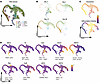 Figure 7
Monocyte-derived populations are temporally distinct. (A and B) Palantir tSNE projection of combined time points labeled by cluster with superimposed RNA velocity information (A) and split by time point (B). (C) Palantir tSNE projection labeled by pseudotime and differentiation potential. (D) Feature plots of monocyte, macrophage, and dependent cell populations z-scores overlayed on the tSNE projection.
To assess whether these diverse macrophage populations exist in different regions of the heart, we utilized a published spatial transcriptomic dataset of mouse MI (36). We plotted z-scores of the individual macrophage states onto transverse heart sections at 7 days after MI and observed that individual monocyte, conventional dendritic cells (cDC), and macrophage states were enriched in different regions of the infarct. The infarcted area can be visualized by loss of cardiomyocyte z-score expression. Arg1+, Trem2+, Gdf15+, and Ccl8+ macrophages were enriched near the ischemic core of the infarct, distal to the border zone. Type I IFN–activated macrophages, MHCIIhi macrophages, and cDC2s were enriched at the periphery of the infarct nearer to the border zone. Resident macrophages were predominantly found outside of the infarct in the remote zone of the heart, while proliferating macrophages are distributed within the infarct and remote zones (Figure 8). Taken together, these data predict that monocytes differentiate into spatially restricted macrophage subsets and that type I IFN–activated macrophages and Arg1+ macrophages represent distinct intermediate macrophage populations.
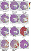 Figure 8
Monocyte-derived populations are spatially distinct. Spatial transcriptomic expression plots of monocyte, macrophage and dependent cell populations, and cardiomyocyte z-scores at day 7 after MI. Images were acquired with a 10X objective.
Recruited macrophages begin to acquire transcriptional identity during extravasation. To investigate where fate decisions are initially specified during the process of monocyte recruitment and extravasation into the heart, we performed closed chest ischemia-reperfusion injury on WT mice and harvested animals at 2 days after MI. Mice were administered BV421 conjugated anti-CD45 antibodies retro-orbitally 5 minutes before harvest to label leukocytes in continuity with the circulation (intravascular compartment) (Figure 9A). Upon harvest, hearts were perfused to flush blood cells which had not adhered to the endothelium. Macrophages and monocytes were then sorted by FACS into intravascular (BV421+) and extravascular (BV421–) libraries for single-cell RNA-seq.
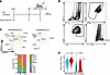 Figure 9
Monocyte fate specifications is initiated prior to myocardial infiltration. (A) Experimental strategy for single-cell RNA-seq of intravascular (anti-CD45 BV421+) and extravascular (anti-CD45 BV421–) compartments. Anti-CD45 BV421 was administered via retro-orbital injection 5 minutes prior to collection. (B) Representative FACS plots and gating strategy demonstrating anti-CD45 BV421 antibody staining in cardiac monocytes and macrophages. (C) Clustered UMAP plots of intravascular (BV421+) and extravascular (BV421–) libraries and stacked bar chart showing distribution of monocyte, macrophage, and dendritic cell populations in each compartment. (D) Violin plot of monocyte signature gene expression (expressed as a z-score) in monocytes and macrophages located within the intravascular and extravascular compartments. 2 tailed t test with Welch’s correction.
Approximately 2.5% of all macrophages and monocytes collected from the heart were BV421+, indicating that these cells had either adhered to the endothelium or were in the process of extravasating from the vasculature into the myocardium (Figure 9B and Supplemental Figure 6). Libraries were sequenced and cell clusters annotated by reference mapping to the Ccr2CreERT2Rosa26LSL–tdTomato monocyte lineage tracing object. As expected, the majority of intravascular cells mapped to the monocyte cluster. Interestingly, we also observed mapping of intravascular cells to each monocyte-derived macrophage populations, suggesting that macrophage fate determination may begin during or prior to monocyte extravasation (Figure 9C). Combined pseudotime analysis using the monocyte lineage tracing and IV anti-CD45 antibody–labeled libraries also demonstrated distribution of intravascular cells throughout all clusters (Supplemental Figure 5, C and D). Consistent with reference mapping, marker genes enriched in each of the monocyte, macrophage, and dendritic cell clusters were expressed within corresponding cells in both the intravascular and extravascular compartments (Supplemental Figure 6). Monocytes and macrophages present within the intravascular compartment displayed transcriptional signatures of monocytes (monocyte z-score), suggesting that they recently differentiated from monocytes (Figure 9D).
To corroborate observations from our sequencing data, we performed ischemia reperfusion injury on Mx1GFP mice (type I IFN reporter) and harvested hearts 2 days after MI. Mice were injected with BV421-conjugated anti-CD45 antibodies 5 minutes before collection. FACS analysis revealed that 4% of monocytes and macrophages within the heart were BV421+, indicating that they were localized within the intravascular compartment. GFP-positive cells were evident within the intravascular and extravascular monocyte and macrophage pool. Very few GFP-expressing cells were detected in the peripheral blood. These findings confirm that type I IFN activation within monocytes and macrophages is initiated during extravasation (Figure 10, A and B). To further validate acquisition of fate during extravasation, we performed a syngeneic cervical heterotopic heart transplantation of a WT heart into Ccr2CreERT2Rosa25LSL–tdTomatoMx1GFP mice (37). Hearts were subjected to 1 hour of cold ischemia before implantation. After 24 hours, intravital imaging was performed and we observed Mx1GFP positive macrophages adhered to the vascular endothelium of coronary veins (lumen marked by Q-dots) and extravasated cells within the myocardial interstitial space (Figure 10, C and D, and Supplemental Video 1). Taken together, these results demonstrate that cardiac infiltrating monocytes begin to acquire cell state specific transcriptional identity during extravasation and differentiation into macrophages.
 Figure 10
IFN is activated in recruited monocytes during extravasation. (A and B) Representative FACS plots and quantification of GFP+ monocytes and macrophages within the heart and blood of Mx1GFP mice after MI. Intravascular and extravascular cardiac monocytes and macrophages expressing GFP is shown. Two tailed t-test with Welch’s correction. (C and D) Experimental strategy for heterotopic heart transplantation and representative images demonstrating Mx1GFP-positive recruited macrophages (arrowheads) in surface vessels of the WT donor heart. *P < 0.05, **P < 0.01. Scale bars 40 μM (D, left); 10 μm (D, right).
Type I IFN signaling in monocytes and monocyte-derived macrophages protects from adverse remodeling after MI. To decipher the functional implications of these diverse macrophage populations, we focused on the IFN-activated macrophage subset given their proximal position within the predicted differentiation trajectory and suggested clinical relevance (38). Utilizing Ccr2CreERT2Ifnar1Fl mice, we selectively deleted Ifnar1 in recruited monocytes and their progeny using a single dose of tamoxifen, injected IP, referred to as “Mono KO” (Figure 11A) (39). Control and Ccr2CreERT2Ifnar1Fl mice were subjected to MI and echocardiography performed at day 28 after injury. IFNAR1 deletion was validated using flow cytometry of induced peritoneal macrophages (Supplemental Figure 7A). Ccr2CreERT2Ifnar1Fl hearts demonstrated significantly decreased left ventricular ejection fraction, increased end systolic and diastolic left ventricular volumes, and increased akinetic area compared with controls (Figure 11, B and C). Analysis of cardiomyocyte size via wheat-germ agglutinin staining revealed increased cardiomyocyte hypertrophy in the border and remote zones of the left ventricle along with increased fibrosis measured by trichrome staining. We observed consistent findings in Ccr2CreERT2Ifnar1Fl mice subjected to continuous tamoxifen treatment, which will delete Ifnar1 in resident CCR2+ macrophages, monocytes, and their progeny, referred to as “All CCR2+ KO”. (Figure 11, D–F). These data are consistent with the conclusions that type I IFN primarily functions within monocytes and their progeny recruited to the heart early after injury and that type I IFN signaling within infiltrating monocytes and their progeny confers a protective effect on MI remodeling. Intriguingly, these findings are in contrast to studies demonstrating protective effects in global Ifnar1–/– mice, suggesting that type I IFN signaling has cell type–specific effects following MI (38). We next utilized spatial transcriptomics to delineate different type I IFN signaling pathways. We observed that Irf3 expression was observed throughout the infarct, while Irf7 expression was enriched in the border zone Figure 11G). IRF3 was reported to regulate IFN activation in response to cytoplasmic DNA sensing (38, 40, 41). In contrast, IRF7 functions downstream TLRs and is amplified in response to type I IFN signaling. The expression of IRF7 is consistent with the most robust IFN signal in the border zone, which is the location of type I IFN–activated macrophages.
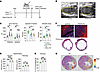 Figure 11
Type 1 IFN signaling in monocytes attenuate post-MI remodeling. (A) Experimental strategy for deletion of Ifnar in recruited monocytes and their progeny. (B) Representative long-axis echocardiographic images at end diastole showing enhanced LV remodeling in Ccr2ERT2CreIfnarFl mice compared to controls. (C) Quantification of echocardiography measurements of LV end systolic volume, ejection fraction, and akinetic area 4 weeks after MI. Two tailed t-test with Welch’s correction. (D) Measurement of cardiomyocyte size by WGA staining 4 weeks after MI. Two tailed t-test with Welch’s correction. (E) Measurement of infarct size assessed by fibrosis as a percentage of LV diameter 4 weeks after MI. Two tailed t-test with Welch’s correction. (F) Representative images of WGA staining at the border zone and trichrome staining in control and Ccr2ERT2CreIfnarFl hearts. Scale bar: 100 μM. (G) Spatial transcriptomic expression plots of Irf7 and Irf3.
Type I IFN signaling is essential for the differentiation of MHCIIhi macrophages. We next sought to determine the effect of monocyte Ifnar1 deletion on the diversity of recruited monocytes and macrophages. We performed ischemia reperfusion injury on control and Ccr2CreERT2Ifnar1Fl mice given a single IP injection of tamoxifen 1 day prior to injury. Hearts were collected 7 days after injury and processed for single-cell RNA-seq (Figure 12A). Libraries were sequenced and clusters annotated by reference mapping to the Ccr2CreERT2Rosa26LSL–tdTomato object. As expected, the proportion of IFN-activated macrophages was reduced. Interestingly, the proportion of MHCIIhi macrophages was also dramatically reduced in Ccr2CreERT2Ifnar1Fl mice compared with litter mate controls (Figure 12B and Supplemental Figure 7B). Combined pseudotime analysis using monocyte lineage tracing, IV labeling, and Ccr2CreERT2Ifnar1Fl libraries demonstrated a reduction in terminal MHCIIhi macrophages (Supplemental Figure 7C). Immunostaining confirmed that MHCIIhi macrophages were significantly reduced in Ccr2CreERT2Ifnar1Fl hearts compared with controls at 28 days after injury (Figure 12C).
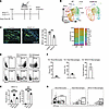 Figure 12
Type 1 IFN–activated macrophages give rise to MHCIIhi macrophages. (A) Experimental strategy for deletion of Ifnar in recruited monocytes and their progeny. (B) UMAP projection and stacked bar chart comparing distribution of cells in control and Ccr2ERT2CreIfnarFl hearts. (C) Representative images and bar chart quantifying MHCII+ macrophages per 20 × field in the infarct of control and Ccr2ERT2CreIfnarFl hearts. 2 tailed t test with Welch’s correction. Scale bar: 100 μM. (D and E) FACS plots of elicited peritoneal macrophages isolated from Mx1GFP mice stimulated with either PBS or IFN-β showing GFP and MHC-II expression (D). Quantification of the percent GFP+ and MHCII+ macrophages (E). 2 tailed t test with Welch’s correction. (F and G) Quantification and representative FACs plots demonstrating Mx1ERT2CreRosa26LSL–tdTomato labeling in circulating monocytes, macrophages in the heart, and percent of tdTomato+ macrophages which were MHCIIhi.
To assess whether type I IFN signaling to monocytes directly influences MHCIIhi macrophage differentiation, we utilized thioglycolate-induced peritoneal macrophages (pMacs) from Mx1GFP mice. pMacs were cultured in complete media with 25 nM IFN-β for 3 days followed by 4 days without IFN-β (IFN Withdrawal) or for 7 days with IFN-β (IFN Continuous). After 7 days of culture, pMacs were collected and flow cytometry was performed to identify percentages of GFP+ and MHCIIhi macrophages. Continuous treatment with IFN-β resulted in GFP expression in 98.7% of pMacs compared with less than 0.25% of controls. Continuous treatment also significantly increased the proportion of MHCIIhi pMacs from 3.7% to 10.4%. Withdrawal of IFN-β reduced the percentage of MHCII high macrophages showing a reversible effect (Figure 12, D and E). These results indicate that type I IFN signaling directly regulates the acquisition of the MHCIIhi macrophage fate.
We additionally generated an Mx1CreERT2Rosa26LSL–tdTomato reporter line to evaluate contribution of IFN-activated macrophages to other macrophage populations. Using an angiotensin II/phenylephrine infusion model of cardiac injury in conjunction with flow cytometry, we found that less than 5% of circulating monocytes were tdTomato+ and that 10% of all macrophages in the heart were tdTomato+. Importantly, 60% of tdTomato+ macrophages were MHCIIhi, indicating that IFN-activated macrophages give rise to MHCIIhi macrophages and represent a major contributor to this population (Figure 12, F and G).
Mechanisms by which type I IFN signaling in macrophages confers protection. To investigate mechanisms by which IFN-activated macrophages are protective in myocardial infarction, we (a) assessed the role of MHC-II expression (regulated by type I IFN signaling) in monocyte-derived macrophages and (b) explored cell types that may interact with type I IFN–activated macrophages. We first investigated the functional role of MHCII in monocyte-derived macrophages following myocardial injury. Utilizing Ccr2CreERT2MHCIIFl mice, we selectively deleted MHCII in recruited monocytes and their progeny using a single dose of tamoxifen, injected IP (Supplemental Figure 8A) (42). Control and Ccr2CreERT2MHCIIFl mice were subjected to reperfused MI and echocardiography performed at day 28 after injury. Ccr2CreERT2MHCIIFlx hearts demonstrated significantly decreased left ventricular ejection fraction, increased end systolic and diastolic left ventricular volumes, and increased akinetic area compared with controls (Supplemental Figure 8, B and C). Analysis of cardiomyocyte size via wheat-germ agglutinin staining revealed increased cardiomyocyte hypertrophy in the border and remote zones of the left ventricle; however, no change in fibrosis was observed (Supplemental Figure 8, D–F). These results demonstrate that MHC-II expression in monocyte-derived macrophages is protective in myocardial infarction and are consistent with prior reports indicating that antigen-specific CD4+ T cells may be protective in the heart (43).
We then utilized a single-cell RNA-seq dataset encompassing stromal cell types in the heart at 8 days after ischemia-reperfusion injury (Figure 13A). We referenced mapped macrophages from this dataset to derive annotations and performed cell-cell communication analysis using CellChat (Figure 13B) (44). We observed predicted communication between IFN-activated macrophage and multiple other cell types within the heart (Figure 13C). Recent studies have demonstrated the importance of regulatory T cells (Tregs) in dampening inflammation and remodeling in myocardial infarction (45). This led us to evaluate the PD-L1 signaling pathway, which is responsible for promoting of Treg specification. We identified that IFN-activated macrophages, along with dendritic cells, are the primary producers of PD-L1 among macrophage populations (Figure 13, D and E). To evaluate the impact of Ifnar1 deletion in monocyte-derived macrophages on Treg abundance, we performed immunostaining for FOXP3 (Treg marker) and observed that Ifnar1 deletion in monocyte-derived macrophages resulted in significantly reduced numbers of Tregs in the infarct at 28 days after injury (Figure 13F) (46). Similarly, MHC-II deletion results in reduced numbers of Tregs, consistent with evidence in cancer that MHC-IIhi macrophages can induce Treg development (Figure 13G) (47). To elucidate the source of type I IFN in ischemic injury, we utilized a publicly available human single nuclei RNA-seq dataset (48) This dataset demonstrated elevated levels of type I IFN ligands, (particularly IFNB1) across cardiac cell types in ischemic samples, indicating ubiquitous induction of type I IFN signaling in acute myocardial infarction (Supplemental Figure 9). These results indicate that IFN activated macrophages directly contribute to the development of Tregs through PD-L1 expression, providing a plausible explanation for the observed protective effects on inflammation and remodeling.
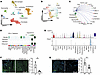 Figure 13
Type 1 IFN–activated macrophages promote Foxp3+ Treg development. (A) UMAP projection of single-cell data of all cell types obtained from hearts at D8 after MI. (B) UMAP projection of macrophages from experiment in panel A reference mapped to the monocyte lineage tracing dataset. (C) CellChat diagram demonstrating signaling from IFN-activated macrophages. (D) Heatmap of the PD-L1 signaling network. (E) Violin plot of Pd-L1 (Cd274) and Pd1 (Pdcd1). (F) Representative images and bar graph quantifying Foxp3+ cells per 20 × field in the infarct in control and Ccr2ERT2CreIfnarFl hearts. (G) Representative images and bar chart quantifying Foxp3+ cells per 20X field in the infarct in control and Ccr2ERT2CreAb1Fl hearts. *P < 0.05, **P < 0.01, ***P < 0.001 Two tailed t-test with Welch’s correction. Scale bars: 100 μm.


Copyright © 2026 American Society for Clinical Investigation
ISSN: 0021-9738 (print), 1558-8238 (online)