Abstract
GATA transcription factors play critical roles in restricting cell lineage differentiation during development. Here, we show that conditional inactivation of GATA-6 in VSMCs results in perinatal mortality from a spectrum of cardiovascular defects, including interrupted aortic arch and persistent truncus arteriosus. Inactivation of GATA-6 in neural crest recapitulates these abnormalities, demonstrating a cell-autonomous requirement for GATA-6 in neural crest–derived SMCs. Surprisingly, the observed defects do not result from impaired SMC differentiation but rather are associated with severely attenuated expression of semaphorin 3C, a signaling molecule critical for both neuronal and vascular patterning. Thus, the primary function of GATA-6 during cardiovascular development is to regulate morphogenetic patterning of the cardiac outflow tract and aortic arch. These findings provide new insights into the conserved functions of the GATA-4, -5, and -6 subfamily members and identify GATA-6 and GATA-6–regulated genes as candidates involved in the pathogenesis of congenital heart disease.
Authors
John J. Lepore, Patricia A. Mericko, Lan Cheng, Min Min Lu, Edward E. Morrisey, Michael S. Parmacek
Abstract
An α1-adrenergic receptor (α1-AR) antagonist increased heart failure in the Antihypertensive and Lipid-Lowering Treatment to Prevent Heart Attack Trial (ALLHAT), but it is unknown whether this adverse result was due to α1-AR inhibition or a nonspecific drug effect. We studied cardiac pressure overload in mice with double KO of the 2 main α1-AR subtypes in the heart, α1A (Adra1a) and α1B (Adra1b). At 2 weeks after transverse aortic constriction (TAC), KO mouse survival was only 60% of WT, and surviving KO mice had lower ejection fractions and larger end-diastolic volumes than WT mice. Mechanistically, final heart weight and myocyte cross-sectional area were the same after TAC in KO and WT mice. However, KO hearts after TAC had increased interstitial fibrosis, increased apoptosis, and failed induction of the fetal hypertrophic genes. Before TAC, isolated KO myocytes were more susceptible to apoptosis after oxidative and β-AR stimulation, and β-ARs were desensitized. Thus, α1-AR deletion worsens dilated cardiomyopathy after pressure overload, by multiple mechanisms, indicating that α1-signaling is required for cardiac adaptation. These results suggest that the adverse cardiac effects of α1-antagonists in clinical trials are due to loss of α1-signaling in myocytes, emphasizing concern about clinical use of α1-antagonists, and point to a revised perspective on sympathetic activation in heart failure.
Authors
Timothy D. O’Connell, Philip M. Swigart, M.C. Rodrigo, Shinji Ishizaka, Shuji Joho, Lynne Turnbull, Laurence H. Tecott, Anthony J. Baker, Elyse Foster, William Grossman, Paul C. Simpson
Abstract
Having identified renin in cardiac mast cells, we assessed whether its release leads to cardiac dysfunction. In Langendorff-perfused guinea pig hearts, mast cell degranulation with compound 48/80 released Ang I–forming activity. This activity was blocked by the selective renin inhibitor BILA2157, indicating that renin was responsible for Ang I formation. Local generation of cardiac Ang II from mast cell–derived renin also elicited norepinephrine release from isolated sympathetic nerve terminals. This action was mediated by Ang II-type 1 (AT1) receptors. In 2 models of ischemia/reperfusion using Langendorff-perfused guinea pig and mouse hearts, a significant coronary spillover of renin and norepinephrine was observed. In both models, this was accompanied by ventricular fibrillation. Mast cell stabilization with cromolyn or lodoxamide markedly reduced active renin overflow and attenuated both norepinephrine release and arrhythmias. Similar cardioprotection was observed in guinea pig hearts treated with BILA2157 or the AT1 receptor antagonist EXP3174. Renin overflow and arrhythmias in ischemia/reperfusion were much less prominent in hearts of mast cell–deficient mice than in control hearts. Thus, mast cell–derived renin is pivotal for activating a cardiac renin-angiotensin system leading to excessive norepinephrine release in ischemia/reperfusion. Mast cell–derived renin may be a useful therapeutic target for hyperadrenergic dysfunctions, such as arrhythmias, sudden cardiac death, myocardial ischemia, and congestive heart failure.
Authors
Christina J. Mackins, Seiichiro Kano, Nahid Seyedi, Ulrich Schäfer, Alicia C. Reid, Takuji Machida, Randi B. Silver, Roberto Levi
Abstract
Previous work showed that calmodulin (CaM) and Ca2+-CaM–dependent protein kinase II (CaMKII) are somehow involved in cardiac hypertrophic signaling, that inositol 1,4,5-trisphosphate receptors (InsP3Rs) in ventricular myocytes are mainly in the nuclear envelope, where they associate with CaMKII, and that class II histone deacetylases (e.g., HDAC5) suppress hypertrophic gene transcription. Furthermore, HDAC phosphorylation in response to neurohumoral stimuli that induce hypertrophy, such as endothelin-1 (ET-1), activates HDAC nuclear export, thereby regulating cardiac myocyte transcription. Here we demonstrate a detailed mechanistic convergence of these 3 issues in adult ventricular myocytes. We show that ET-1, which activates plasmalemmal G protein–coupled receptors and InsP3 production, elicits local nuclear envelope Ca2+ release via InsP3R. This local Ca2+ release activates nuclear CaMKII, which triggers HDAC5 phosphorylation and nuclear export (derepressing transcription). Remarkably, this Ca2+-dependent pathway cannot be activated by the global Ca2+ transients that cause contraction at each heartbeat. This novel local Ca2+ signaling in excitation-transcription coupling is analogous to but separate (and insulated) from that involved in excitation-contraction coupling. Thus, myocytes can distinguish simultaneous local and global Ca2+ signals involved in contractile activation from those targeting gene expression.
Authors
Xu Wu, Tong Zhang, Julie Bossuyt, Xiaodong Li, Timothy A. McKinsey, John R. Dedman, Eric N. Olson, Ju Chen, Joan Heller Brown, Donald M. Bers
Abstract
Thousands die each year from sudden infant death syndrome (SIDS). Neither the cause nor basis for varied prevalence in different populations is understood. While 2 cases have been associated with mutations in type Vα, cardiac voltage-gated sodium channels (SCN5A), the “Back to Sleep” campaign has decreased SIDS prevalence, consistent with a role for environmental influences in disease pathogenesis. Here we studied SCN5A in African Americans. Three of 133 SIDS cases were homozygous for the variant S1103Y. Among controls, 120 of 1,056 were carriers of the heterozygous genotype, which was previously associated with increased risk for arrhythmia in adults. This suggests that infants with 2 copies of S1103Y have a 24-fold increased risk for SIDS. Variant Y1103 channels were found to operate normally under baseline conditions in vitro. As risk factors for SIDS include apnea and respiratory acidosis, Y1103 and wild-type channels were subjected to lowered intracellular pH. Only Y1103 channels gained abnormal function, demonstrating late reopenings suppressible by the drug mexiletine. The variant appeared to confer susceptibility to acidosis-induced arrhythmia, a gene-environment interaction. Overall, homozygous and rare heterozygous SCN5A missense variants were found in approximately 5% of cases. If our findings are replicated, prospective genetic testing of SIDS cases and screening with counseling for at-risk families warrant consideration.
Authors
Leigh D. Plant, Peter N. Bowers, Qianyong Liu, Thomas Morgan, Tingting Zhang, Matthew W. State, Weidong Chen, Rick A. Kittles, Steve A.N. Goldstein
Abstract
Authors
Tomohisa Nagoshi, Takashi Matsui, Takuma Aoyama, Annarosa Leri, Piero Anversa, Ling Li, Wataru Ogawa, Federica del Monte, Judith K. Gwathmey, Luanda Grazette, Brian Hemmings, David A. Kass, Hunter C. Champion, Anthony Rosenzweig
Abstract
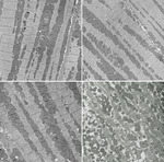
Focal adhesion kinase (FAK) is a cytoplasmic tyrosine kinase that plays a major role in integrin signaling pathways. Although cardiovascular defects were observed in FAK total KO mice, the embryonic lethality prevented investigation of FAK function in the hearts of adult animals. To circumvent these problems, we created mice in which FAK is selectively inactivated in cardiomyocytes (CFKO mice). We found that CFKO mice develop eccentric cardiac hypertrophy (normal LV wall thickness and increased left chamber dimension) upon stimulation with angiotensin II or pressure overload by transverse aortic constriction as measured by echocardiography. We also found increased heart/body weight ratios, elevated markers of cardiac hypertrophy, multifocal interstitial fibrosis, and increased collagen I and VI expression in CFKO mice compared with control littermates. Spontaneous cardiac chamber dilation and increased expression of hypertrophy markers were found in the older CFKO mice. Analysis of cardiomyocytes isolated from CFKO mice showed increased length but not width. The myocardium of CFKO mice exhibited disorganized myofibrils with increased nonmyofibrillar space filled with swelled mitochondria. Last, decreased tyrosine phosphorylation of FAK substrates p130Cas and paxillin were observed in CFKO mice compared with the control littermates. Together, these results provide strong evidence for a role of FAK in the regulation of heart hypertrophy in vivo.
Authors
Xu Peng, Marc S. Kraus, Huijun Wei, Tang-Long Shen, Romain Pariaut, Ana Alcaraz, Guangju Ji, Lihong Cheng, Qinglin Yang, Michael I. Kotlikoff, Ju Chen, Kenneth Chien, Hua Gu, Jun-Lin Guan
Abstract
We report that dietary modification from a soy-based diet to a casein-based diet radically improves disease indicators and cardiac function in a transgenic mouse model of hypertrophic cardiomyopathy. On a soy diet, males with a mutation in the α-myosin heavy chain gene progress to dilation and heart failure. However, males fed a casein diet no longer deteriorate to severe, dilated cardiomyopathy. Remarkably, their LV size and contractile function are preserved. Further, this diet prevents a number of pathologic indicators in males, including fibrosis, induction of β-myosin heavy chain, inactivation of glycogen synthase kinase 3β (GSK3β), and caspase-3 activation.
Authors
Brian L. Stauffer, John P. Konhilas, Elizabeth D. Luczak, Leslie A. Leinwand
Abstract
In the face of systemic risk factors, certain regions of the arterial vasculature remain relatively resistant to the development of atherosclerotic lesions. The biomechanically distinct environments in these arterial geometries exert a protective influence via certain key functions of the endothelial lining; however, the mechanisms underlying the coordinated regulation of specific mechano-activated transcriptional programs leading to distinct endothelial functional phenotypes have remained elusive. Here, we show that the transcription factor Kruppel-like factor 2 (KLF2) is selectively induced in endothelial cells exposed to a biomechanical stimulus characteristic of atheroprotected regions of the human carotid and that this flow-mediated increase in expression occurs via a MEK5/ERK5/MEF2 signaling pathway. Overexpression and silencing of KLF2 in the context of flow, combined with findings from genome-wide analyses of gene expression, demonstrate that the induction of KLF2 results in the orchestrated regulation of endothelial transcriptional programs controlling inflammation, thrombosis/hemostasis, vascular tone, and blood vessel development. Our data also indicate that KLF2 expression globally modulates IL-1β–mediated endothelial activation. KLF2 therefore serves as a mechano-activated transcription factor important in the integration of multiple endothelial functions associated with regions of the arterial vasculature that are relatively resistant to atherogenesis.
Authors
Kush M. Parmar, H. Benjamin Larman, Guohao Dai, Yuzhi Zhang, Eric T. Wang, Sripriya N. Moorthy, Johannes R. Kratz, Zhiyong Lin, Mukesh K. Jain, Michael A. Gimbrone Jr., Guillermo García-Cardeña
Abstract
The majority of acute clinical manifestations of atherosclerosis are due to the physical rupture of advanced atherosclerotic plaques. It has been hypothesized that macrophages play a key role in inducing plaque rupture by secreting proteases that destroy the extracellular matrix that provides physical strength to the fibrous cap. Despite reports detailing the expression of multiple proteases by macrophages in rupture-prone regions, there is no direct proof that macrophage-mediated matrix degradation can induce plaque rupture. We aimed to test this hypothesis by retrovirally overexpressing the candidate enzyme MMP-9 in macrophages of advanced atherosclerotic lesions of apoE–/– mice. Despite a greater than 10-fold increase in the expression of MMP-9 by macrophages, there was only a minor increase in the incidence of plaque fissuring. Subsequent analysis revealed that macrophages secrete MMP-9 predominantly as a proform, and this form is unable to degrade the matrix component elastin. Expression of an autoactivating form of MMP-9 in macrophages in vitro greatly enhances elastin degradation and induces significant plaque disruption when overexpressed by macrophages in advanced atherosclerotic lesions of apoE–/– mice in vivo. These data show that enhanced macrophage proteolytic activity can induce acute plaque disruption and highlight MMP-9 as a potential therapeutic target for stabilizing rupture-prone plaques.
Authors
Peter J. Gough, Ivan G. Gomez, Paul T. Wille, Elaine W. Raines


Copyright © 2025 American Society for Clinical Investigation
ISSN: 0021-9738 (print), 1558-8238 (online)


