
Advertisement
Commentary
Open Access |  10.1172/JCI196740
Stress, plasticity, and fibrosis: unfolding the role of the IRE1α/RIDD/Fgfr2 axis
SeungHye Han
Division of Pulmonary and Critical Care, Department of Medicine, Feinberg School of Medicine, Northwestern University, Chicago, Illinois, USA.
Address correspondence to: SeungHye Han, Division of Pulmonary and Critical Care Medicine, Department of Medicine, Northwestern University, 303 E Superior Street, Chicago, Illinois 60611. Email: shan@northwestern.edu.
Find articles by Han, S. in: PubMed | Google Scholar
Published October 15, 2025 - More info
J Clin Invest. 2025;135(20):e196740. https://doi.org/10.1172/JCI196740.
© 2025 Han This work is licensed under the Creative Commons Attribution 4.0 International License. To view a copy of this license, visit http://creativecommons.org/licenses/by/4.0/.

Related article:
Abstract
Stress-induced epithelial plasticity is central to lung regeneration, fibrosis, and malignancy, but how cellular stress leads to differentiation is incompletely understood. Here, we found a central role for IRE1α, a conserved mediator of the unfolded protein response (UPR), in stimulating the plasticity of alveolar type 2 (AT2) cells. In single-cell RNA-seq, IRE1α activity was associated with loss of AT2 identity and progression toward a damage-associated transitional state unique to fibrosis. AT2 plasticity required destructive regulated IRE1α-dependent decay (RIDD), which we demonstrated by deploying PAIR2, a kinase modulator that inhibits RIDD while preserving IRE1α’s adaptive XBP1 mRNA splicing activity. In vivo, selective inhibition of RIDD with PAIR2 reduced AT2 differentiation into profibrotic transitional cells and protected mice from bleomycin-induced pulmonary fibrosis. Mechanistically, we identified the Fgfr2 mRNA as a direct and regulated substrate for IRE1α’s RNase in primary AT2 cells and in a biochemically reconstituted cell-free system. Loss of Fgf signaling caused AT2 differentiation, while gain of signaling protected cells from IRE1α-induced differentiation. We propose that IRE1α downregulates Fgf signaling through RIDD, provoking loss of AT2 identity and differentiation towards a profibrotic phenotype. Thus, IRE1α’s RIDD activity emerges as a novel target for treatment of pulmonary fibrosis and potentially other diseases driven by aberrant epithelial cell plasticity.
Authors
Vincent C. Auyeung, Tavienne L. Steinberg, Alina Olivier, Luka Suzuki, Mary E. Moreno, Imran S. Khan, Michael S. Downey, Maike Thamsen, Lu Guo, Dustin J. Maly, Bradley J. Backes, Dean Sheppard, Feroz R. Papa
Lung fibrosis is a progressive and complex process characterized by the replacement of the alveolar space with extracellular matrix, leading to tissue stiffening and, ultimately, to respiratory failure. Therapeutic options remain limited, and the few currently approved medications do not reverse disease progression or prolong survival. In this issue of the JCI, Auyeung et al. (1) examined the molecular drivers of intermediate epithelial cell states observed in injured and fibrotic lungs, focusing on a damage-associated transient progenitor (DATP) population associated with aberrant epithelial plasticity and lung fibrosis. Their observations reveal an important link between a downstream effector of ER stress and pathologic epithelial differentiation in the context of lung fibrosis.
-
Cellular stress response pathways and lung disease
Cellular stress response pathways are evolutionarily conserved signaling networks in eukaryotes that detect disruptions, such as protein misfolding, oxidative stress, nutrient deprivation, and infection, and orchestrate programs that restore homeostasis or eliminate irreparably damaged cells. The ER unfolded protein response (UPR) and the integrated stress response (ISR) are two central, interconnected signaling networks activated by these cellular disruptions (Figure 1).
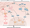 Figure 1
Cellular stress response pathways: the unfolded protein response and the integrated stress response. Cells respond to a variety of stressors by activating two major coordinated signaling networks to restore homeostasis: the unfolded protein response (UPR) and the integrated stress response (ISR). The UPR is activated when three ER sensors, ATF6, IRE1, and PERK, detect the accumulation of misfolded and unfolded proteins in the ER. Upon ER stress, ATF6 is transported to the Golgi apparatus, where it is cleaved. The released cytosolic fragment of ATF6 (ATF6f) then enters the nucleus and induces UPR gene expression. Upon activation, IRE1α dimerizes and autophosphorylates its kinase domain. The activated endoribonuclease domain excises a small intron from Xbp1 mRNA, producing XBP1s, a transcription factor that induces the UPR target genes. IRE1α also degrades select mRNAs (Regulated IRE1-dependent decay [RIDD]), ultimately reducing the protein-folding load. PERK activation leads to the phosphorylation of eukaryotic initiation factor 2 alpha (eIF2α), resulting in a global reduction in protein translation to prevent further ER burden, but paradoxically allowing selective translation of ATF4 that upregulates stress response genes. PERK is also a part of the ISR. The ISR integrates stress signals beyond the ER. In addition to PERK, three other kinases, PKR, HRI, and GCN2, can also phosphorylate eIF2α in response to viral infection, heme deficiency/mitochondrial dysfunction, and amino acid starvation, respectively. These inputs converge to modulate translation and reprogram gene expression, promoting cellular adaptation, or, if stress is severe, initiating programmed cell death. The study by Auyeung and colleagues (1) indicates that pathological cellular stress responses can impair alveolar epithelial differentiation.
The UPR activates when it senses accumulation of unfolded or misfolded proteins in the ER. Three sensors can initiate UPR: Inositol-requiring enzyme 1 alpha (IRE1α), protein kinase RNA-activated–like (PKR-like) ER kinase (PERK), and activating transcription factor 6 (ATF6) (6). UPR activation attenuates global protein translation to reduce ER-client protein load, upregulates molecular chaperones to enhance folding capacity, and enhances ER-associated degradation (ERAD) to clear misfolded polypeptides. Given the diversified outcomes of UPR activation, it follows that UPR sensors elicit crosstalk between multiple pathways. For instance, IRE1α activates the Xbp1s pathway, the JNK signaling cascade, and the regulated IRE1α-dependent decay (RIDD) pathway.
The ISR, on the other hand, is triggered by various cellular stressors, including viral infection, amino acid deprivation, mitochondrial dysfunction, and heme deficiency, as well as ER stress (7). Four kinases, PKR, GCN2, HRI, and PERK, function as stress sensors for the ISR; when activated, the ISR sensor phosphorylates eIF2α, reducing global protein translation yet paradoxically increasing translation of select transcription factors, such as ATF4, to induce stress response genes. If these stresses persist, apoptosis is initiated.
The UPR and ISR cellular stress response pathways have been implicated in the pathogenesis of pulmonary fibrosis, and targeting their activation has improved lung fibrosis in animal models (8–14). However, the precise molecular mechanisms underlying their effects on fibrosis remain unknown.


Copyright © 2026 American Society for Clinical Investigation
ISSN: 0021-9738 (print), 1558-8238 (online)