
Advertisement
Research ArticleGastroenterologyImmunologyOncology
Open Access |  10.1172/JCI196722
Targeting the N-acetyltransferase 10/DKK2 axis enhances CD8+ T cell antitumor activity in colorectal cancer models
Mengmeng Li,1,2 Xiaoya Zhao,2 Jun Wu,3 Shimeng Zhou,2 Yao Fu,4 Chen Chen,1 Zhuang Ma,2 Jiawen Xu,2 Yun Qian,2 Zhangding Wang,1 Bo Wang,1 Qiang Wang,1 Qingqing Ding,5 Changyu Chen,6 Honggang Wang,7 Xiaozhong Yang,7 Weijie Dai,7 Wenjie Zhang,1 and Shouyu Wang1,2
1Department of Hepatobiliary Surgery, The First Affiliated Hospital of Anhui Medical University, MOE Innovation Center for Basic Research in Tumor Immunotherapy, Anhui Province Key Laboratory of Tumor Immune Microenvironment and Immunotherapy, Hefei, Anhui Province, China.
2Medical School of Nanjing University, Nanjing, Jiangsu Province, China.
3Department of Thoracic Surgery, Northern Jiangsu People’s Hospital, Yangzhou, China.
4Department of Pathology, The First Affiliated Hospital of Anhui Medical University, Hefei, Anhui Province, China.
5Department of Geriatric Oncology, The First Affiliated Hospital of Nanjing Medical University, Nanjing, Jiangsu Province, China.
6Department of General Surgery, The First Affiliated Hospital of Anhui Medical University, Hefei, Anhui Province, China.
7Department of Gastroenterology, The Affiliated Huaian No. 1 People’s Hospital of Nanjing Medical University, Huai’an, Jiangsu Province, China.
Address correspondence to: Shouyu Wang or Wenjie Zhang, Department of Hepatobiliary Surgery, The First Affiliated Hospital of Anhui Medical University, Hefei 230022, Anhui Province, China. Email: shouyuwang@ahmu.edu.cn (SW); Email: drzhangwj@163.com (WZ). Or to: Weijie Dai, Department of Gastroenterology, The Affiliated Huaian No. 1 People’s Hospital of Nanjing Medical University, Huai’an 223300, Jiangsu Province, China. Email: daiweijie@njmu.edu.cn.
Authorship note: ML, XZ, JW, SZ, and YF contributed equally to this work.
Find articles by Li, M. in: PubMed | Google Scholar
1Department of Hepatobiliary Surgery, The First Affiliated Hospital of Anhui Medical University, MOE Innovation Center for Basic Research in Tumor Immunotherapy, Anhui Province Key Laboratory of Tumor Immune Microenvironment and Immunotherapy, Hefei, Anhui Province, China.
2Medical School of Nanjing University, Nanjing, Jiangsu Province, China.
3Department of Thoracic Surgery, Northern Jiangsu People’s Hospital, Yangzhou, China.
4Department of Pathology, The First Affiliated Hospital of Anhui Medical University, Hefei, Anhui Province, China.
5Department of Geriatric Oncology, The First Affiliated Hospital of Nanjing Medical University, Nanjing, Jiangsu Province, China.
6Department of General Surgery, The First Affiliated Hospital of Anhui Medical University, Hefei, Anhui Province, China.
7Department of Gastroenterology, The Affiliated Huaian No. 1 People’s Hospital of Nanjing Medical University, Huai’an, Jiangsu Province, China.
Address correspondence to: Shouyu Wang or Wenjie Zhang, Department of Hepatobiliary Surgery, The First Affiliated Hospital of Anhui Medical University, Hefei 230022, Anhui Province, China. Email: shouyuwang@ahmu.edu.cn (SW); Email: drzhangwj@163.com (WZ). Or to: Weijie Dai, Department of Gastroenterology, The Affiliated Huaian No. 1 People’s Hospital of Nanjing Medical University, Huai’an 223300, Jiangsu Province, China. Email: daiweijie@njmu.edu.cn.
Authorship note: ML, XZ, JW, SZ, and YF contributed equally to this work.
Find articles by Zhao, X. in: PubMed | Google Scholar
1Department of Hepatobiliary Surgery, The First Affiliated Hospital of Anhui Medical University, MOE Innovation Center for Basic Research in Tumor Immunotherapy, Anhui Province Key Laboratory of Tumor Immune Microenvironment and Immunotherapy, Hefei, Anhui Province, China.
2Medical School of Nanjing University, Nanjing, Jiangsu Province, China.
3Department of Thoracic Surgery, Northern Jiangsu People’s Hospital, Yangzhou, China.
4Department of Pathology, The First Affiliated Hospital of Anhui Medical University, Hefei, Anhui Province, China.
5Department of Geriatric Oncology, The First Affiliated Hospital of Nanjing Medical University, Nanjing, Jiangsu Province, China.
6Department of General Surgery, The First Affiliated Hospital of Anhui Medical University, Hefei, Anhui Province, China.
7Department of Gastroenterology, The Affiliated Huaian No. 1 People’s Hospital of Nanjing Medical University, Huai’an, Jiangsu Province, China.
Address correspondence to: Shouyu Wang or Wenjie Zhang, Department of Hepatobiliary Surgery, The First Affiliated Hospital of Anhui Medical University, Hefei 230022, Anhui Province, China. Email: shouyuwang@ahmu.edu.cn (SW); Email: drzhangwj@163.com (WZ). Or to: Weijie Dai, Department of Gastroenterology, The Affiliated Huaian No. 1 People’s Hospital of Nanjing Medical University, Huai’an 223300, Jiangsu Province, China. Email: daiweijie@njmu.edu.cn.
Authorship note: ML, XZ, JW, SZ, and YF contributed equally to this work.
Find articles by Wu, J. in: PubMed | Google Scholar
1Department of Hepatobiliary Surgery, The First Affiliated Hospital of Anhui Medical University, MOE Innovation Center for Basic Research in Tumor Immunotherapy, Anhui Province Key Laboratory of Tumor Immune Microenvironment and Immunotherapy, Hefei, Anhui Province, China.
2Medical School of Nanjing University, Nanjing, Jiangsu Province, China.
3Department of Thoracic Surgery, Northern Jiangsu People’s Hospital, Yangzhou, China.
4Department of Pathology, The First Affiliated Hospital of Anhui Medical University, Hefei, Anhui Province, China.
5Department of Geriatric Oncology, The First Affiliated Hospital of Nanjing Medical University, Nanjing, Jiangsu Province, China.
6Department of General Surgery, The First Affiliated Hospital of Anhui Medical University, Hefei, Anhui Province, China.
7Department of Gastroenterology, The Affiliated Huaian No. 1 People’s Hospital of Nanjing Medical University, Huai’an, Jiangsu Province, China.
Address correspondence to: Shouyu Wang or Wenjie Zhang, Department of Hepatobiliary Surgery, The First Affiliated Hospital of Anhui Medical University, Hefei 230022, Anhui Province, China. Email: shouyuwang@ahmu.edu.cn (SW); Email: drzhangwj@163.com (WZ). Or to: Weijie Dai, Department of Gastroenterology, The Affiliated Huaian No. 1 People’s Hospital of Nanjing Medical University, Huai’an 223300, Jiangsu Province, China. Email: daiweijie@njmu.edu.cn.
Authorship note: ML, XZ, JW, SZ, and YF contributed equally to this work.
Find articles by Zhou, S. in: PubMed | Google Scholar
1Department of Hepatobiliary Surgery, The First Affiliated Hospital of Anhui Medical University, MOE Innovation Center for Basic Research in Tumor Immunotherapy, Anhui Province Key Laboratory of Tumor Immune Microenvironment and Immunotherapy, Hefei, Anhui Province, China.
2Medical School of Nanjing University, Nanjing, Jiangsu Province, China.
3Department of Thoracic Surgery, Northern Jiangsu People’s Hospital, Yangzhou, China.
4Department of Pathology, The First Affiliated Hospital of Anhui Medical University, Hefei, Anhui Province, China.
5Department of Geriatric Oncology, The First Affiliated Hospital of Nanjing Medical University, Nanjing, Jiangsu Province, China.
6Department of General Surgery, The First Affiliated Hospital of Anhui Medical University, Hefei, Anhui Province, China.
7Department of Gastroenterology, The Affiliated Huaian No. 1 People’s Hospital of Nanjing Medical University, Huai’an, Jiangsu Province, China.
Address correspondence to: Shouyu Wang or Wenjie Zhang, Department of Hepatobiliary Surgery, The First Affiliated Hospital of Anhui Medical University, Hefei 230022, Anhui Province, China. Email: shouyuwang@ahmu.edu.cn (SW); Email: drzhangwj@163.com (WZ). Or to: Weijie Dai, Department of Gastroenterology, The Affiliated Huaian No. 1 People’s Hospital of Nanjing Medical University, Huai’an 223300, Jiangsu Province, China. Email: daiweijie@njmu.edu.cn.
Authorship note: ML, XZ, JW, SZ, and YF contributed equally to this work.
Find articles by Fu, Y. in: PubMed | Google Scholar
1Department of Hepatobiliary Surgery, The First Affiliated Hospital of Anhui Medical University, MOE Innovation Center for Basic Research in Tumor Immunotherapy, Anhui Province Key Laboratory of Tumor Immune Microenvironment and Immunotherapy, Hefei, Anhui Province, China.
2Medical School of Nanjing University, Nanjing, Jiangsu Province, China.
3Department of Thoracic Surgery, Northern Jiangsu People’s Hospital, Yangzhou, China.
4Department of Pathology, The First Affiliated Hospital of Anhui Medical University, Hefei, Anhui Province, China.
5Department of Geriatric Oncology, The First Affiliated Hospital of Nanjing Medical University, Nanjing, Jiangsu Province, China.
6Department of General Surgery, The First Affiliated Hospital of Anhui Medical University, Hefei, Anhui Province, China.
7Department of Gastroenterology, The Affiliated Huaian No. 1 People’s Hospital of Nanjing Medical University, Huai’an, Jiangsu Province, China.
Address correspondence to: Shouyu Wang or Wenjie Zhang, Department of Hepatobiliary Surgery, The First Affiliated Hospital of Anhui Medical University, Hefei 230022, Anhui Province, China. Email: shouyuwang@ahmu.edu.cn (SW); Email: drzhangwj@163.com (WZ). Or to: Weijie Dai, Department of Gastroenterology, The Affiliated Huaian No. 1 People’s Hospital of Nanjing Medical University, Huai’an 223300, Jiangsu Province, China. Email: daiweijie@njmu.edu.cn.
Authorship note: ML, XZ, JW, SZ, and YF contributed equally to this work.
Find articles by Chen, C. in: PubMed | Google Scholar
1Department of Hepatobiliary Surgery, The First Affiliated Hospital of Anhui Medical University, MOE Innovation Center for Basic Research in Tumor Immunotherapy, Anhui Province Key Laboratory of Tumor Immune Microenvironment and Immunotherapy, Hefei, Anhui Province, China.
2Medical School of Nanjing University, Nanjing, Jiangsu Province, China.
3Department of Thoracic Surgery, Northern Jiangsu People’s Hospital, Yangzhou, China.
4Department of Pathology, The First Affiliated Hospital of Anhui Medical University, Hefei, Anhui Province, China.
5Department of Geriatric Oncology, The First Affiliated Hospital of Nanjing Medical University, Nanjing, Jiangsu Province, China.
6Department of General Surgery, The First Affiliated Hospital of Anhui Medical University, Hefei, Anhui Province, China.
7Department of Gastroenterology, The Affiliated Huaian No. 1 People’s Hospital of Nanjing Medical University, Huai’an, Jiangsu Province, China.
Address correspondence to: Shouyu Wang or Wenjie Zhang, Department of Hepatobiliary Surgery, The First Affiliated Hospital of Anhui Medical University, Hefei 230022, Anhui Province, China. Email: shouyuwang@ahmu.edu.cn (SW); Email: drzhangwj@163.com (WZ). Or to: Weijie Dai, Department of Gastroenterology, The Affiliated Huaian No. 1 People’s Hospital of Nanjing Medical University, Huai’an 223300, Jiangsu Province, China. Email: daiweijie@njmu.edu.cn.
Authorship note: ML, XZ, JW, SZ, and YF contributed equally to this work.
Find articles by Ma, Z. in: PubMed | Google Scholar
1Department of Hepatobiliary Surgery, The First Affiliated Hospital of Anhui Medical University, MOE Innovation Center for Basic Research in Tumor Immunotherapy, Anhui Province Key Laboratory of Tumor Immune Microenvironment and Immunotherapy, Hefei, Anhui Province, China.
2Medical School of Nanjing University, Nanjing, Jiangsu Province, China.
3Department of Thoracic Surgery, Northern Jiangsu People’s Hospital, Yangzhou, China.
4Department of Pathology, The First Affiliated Hospital of Anhui Medical University, Hefei, Anhui Province, China.
5Department of Geriatric Oncology, The First Affiliated Hospital of Nanjing Medical University, Nanjing, Jiangsu Province, China.
6Department of General Surgery, The First Affiliated Hospital of Anhui Medical University, Hefei, Anhui Province, China.
7Department of Gastroenterology, The Affiliated Huaian No. 1 People’s Hospital of Nanjing Medical University, Huai’an, Jiangsu Province, China.
Address correspondence to: Shouyu Wang or Wenjie Zhang, Department of Hepatobiliary Surgery, The First Affiliated Hospital of Anhui Medical University, Hefei 230022, Anhui Province, China. Email: shouyuwang@ahmu.edu.cn (SW); Email: drzhangwj@163.com (WZ). Or to: Weijie Dai, Department of Gastroenterology, The Affiliated Huaian No. 1 People’s Hospital of Nanjing Medical University, Huai’an 223300, Jiangsu Province, China. Email: daiweijie@njmu.edu.cn.
Authorship note: ML, XZ, JW, SZ, and YF contributed equally to this work.
Find articles by Xu, J. in: PubMed | Google Scholar
1Department of Hepatobiliary Surgery, The First Affiliated Hospital of Anhui Medical University, MOE Innovation Center for Basic Research in Tumor Immunotherapy, Anhui Province Key Laboratory of Tumor Immune Microenvironment and Immunotherapy, Hefei, Anhui Province, China.
2Medical School of Nanjing University, Nanjing, Jiangsu Province, China.
3Department of Thoracic Surgery, Northern Jiangsu People’s Hospital, Yangzhou, China.
4Department of Pathology, The First Affiliated Hospital of Anhui Medical University, Hefei, Anhui Province, China.
5Department of Geriatric Oncology, The First Affiliated Hospital of Nanjing Medical University, Nanjing, Jiangsu Province, China.
6Department of General Surgery, The First Affiliated Hospital of Anhui Medical University, Hefei, Anhui Province, China.
7Department of Gastroenterology, The Affiliated Huaian No. 1 People’s Hospital of Nanjing Medical University, Huai’an, Jiangsu Province, China.
Address correspondence to: Shouyu Wang or Wenjie Zhang, Department of Hepatobiliary Surgery, The First Affiliated Hospital of Anhui Medical University, Hefei 230022, Anhui Province, China. Email: shouyuwang@ahmu.edu.cn (SW); Email: drzhangwj@163.com (WZ). Or to: Weijie Dai, Department of Gastroenterology, The Affiliated Huaian No. 1 People’s Hospital of Nanjing Medical University, Huai’an 223300, Jiangsu Province, China. Email: daiweijie@njmu.edu.cn.
Authorship note: ML, XZ, JW, SZ, and YF contributed equally to this work.
Find articles by Qian, Y. in: PubMed | Google Scholar
1Department of Hepatobiliary Surgery, The First Affiliated Hospital of Anhui Medical University, MOE Innovation Center for Basic Research in Tumor Immunotherapy, Anhui Province Key Laboratory of Tumor Immune Microenvironment and Immunotherapy, Hefei, Anhui Province, China.
2Medical School of Nanjing University, Nanjing, Jiangsu Province, China.
3Department of Thoracic Surgery, Northern Jiangsu People’s Hospital, Yangzhou, China.
4Department of Pathology, The First Affiliated Hospital of Anhui Medical University, Hefei, Anhui Province, China.
5Department of Geriatric Oncology, The First Affiliated Hospital of Nanjing Medical University, Nanjing, Jiangsu Province, China.
6Department of General Surgery, The First Affiliated Hospital of Anhui Medical University, Hefei, Anhui Province, China.
7Department of Gastroenterology, The Affiliated Huaian No. 1 People’s Hospital of Nanjing Medical University, Huai’an, Jiangsu Province, China.
Address correspondence to: Shouyu Wang or Wenjie Zhang, Department of Hepatobiliary Surgery, The First Affiliated Hospital of Anhui Medical University, Hefei 230022, Anhui Province, China. Email: shouyuwang@ahmu.edu.cn (SW); Email: drzhangwj@163.com (WZ). Or to: Weijie Dai, Department of Gastroenterology, The Affiliated Huaian No. 1 People’s Hospital of Nanjing Medical University, Huai’an 223300, Jiangsu Province, China. Email: daiweijie@njmu.edu.cn.
Authorship note: ML, XZ, JW, SZ, and YF contributed equally to this work.
Find articles by Wang, Z. in: PubMed | Google Scholar
1Department of Hepatobiliary Surgery, The First Affiliated Hospital of Anhui Medical University, MOE Innovation Center for Basic Research in Tumor Immunotherapy, Anhui Province Key Laboratory of Tumor Immune Microenvironment and Immunotherapy, Hefei, Anhui Province, China.
2Medical School of Nanjing University, Nanjing, Jiangsu Province, China.
3Department of Thoracic Surgery, Northern Jiangsu People’s Hospital, Yangzhou, China.
4Department of Pathology, The First Affiliated Hospital of Anhui Medical University, Hefei, Anhui Province, China.
5Department of Geriatric Oncology, The First Affiliated Hospital of Nanjing Medical University, Nanjing, Jiangsu Province, China.
6Department of General Surgery, The First Affiliated Hospital of Anhui Medical University, Hefei, Anhui Province, China.
7Department of Gastroenterology, The Affiliated Huaian No. 1 People’s Hospital of Nanjing Medical University, Huai’an, Jiangsu Province, China.
Address correspondence to: Shouyu Wang or Wenjie Zhang, Department of Hepatobiliary Surgery, The First Affiliated Hospital of Anhui Medical University, Hefei 230022, Anhui Province, China. Email: shouyuwang@ahmu.edu.cn (SW); Email: drzhangwj@163.com (WZ). Or to: Weijie Dai, Department of Gastroenterology, The Affiliated Huaian No. 1 People’s Hospital of Nanjing Medical University, Huai’an 223300, Jiangsu Province, China. Email: daiweijie@njmu.edu.cn.
Authorship note: ML, XZ, JW, SZ, and YF contributed equally to this work.
Find articles by Wang, B. in: PubMed | Google Scholar
1Department of Hepatobiliary Surgery, The First Affiliated Hospital of Anhui Medical University, MOE Innovation Center for Basic Research in Tumor Immunotherapy, Anhui Province Key Laboratory of Tumor Immune Microenvironment and Immunotherapy, Hefei, Anhui Province, China.
2Medical School of Nanjing University, Nanjing, Jiangsu Province, China.
3Department of Thoracic Surgery, Northern Jiangsu People’s Hospital, Yangzhou, China.
4Department of Pathology, The First Affiliated Hospital of Anhui Medical University, Hefei, Anhui Province, China.
5Department of Geriatric Oncology, The First Affiliated Hospital of Nanjing Medical University, Nanjing, Jiangsu Province, China.
6Department of General Surgery, The First Affiliated Hospital of Anhui Medical University, Hefei, Anhui Province, China.
7Department of Gastroenterology, The Affiliated Huaian No. 1 People’s Hospital of Nanjing Medical University, Huai’an, Jiangsu Province, China.
Address correspondence to: Shouyu Wang or Wenjie Zhang, Department of Hepatobiliary Surgery, The First Affiliated Hospital of Anhui Medical University, Hefei 230022, Anhui Province, China. Email: shouyuwang@ahmu.edu.cn (SW); Email: drzhangwj@163.com (WZ). Or to: Weijie Dai, Department of Gastroenterology, The Affiliated Huaian No. 1 People’s Hospital of Nanjing Medical University, Huai’an 223300, Jiangsu Province, China. Email: daiweijie@njmu.edu.cn.
Authorship note: ML, XZ, JW, SZ, and YF contributed equally to this work.
Find articles by Wang, Q. in: PubMed | Google Scholar
1Department of Hepatobiliary Surgery, The First Affiliated Hospital of Anhui Medical University, MOE Innovation Center for Basic Research in Tumor Immunotherapy, Anhui Province Key Laboratory of Tumor Immune Microenvironment and Immunotherapy, Hefei, Anhui Province, China.
2Medical School of Nanjing University, Nanjing, Jiangsu Province, China.
3Department of Thoracic Surgery, Northern Jiangsu People’s Hospital, Yangzhou, China.
4Department of Pathology, The First Affiliated Hospital of Anhui Medical University, Hefei, Anhui Province, China.
5Department of Geriatric Oncology, The First Affiliated Hospital of Nanjing Medical University, Nanjing, Jiangsu Province, China.
6Department of General Surgery, The First Affiliated Hospital of Anhui Medical University, Hefei, Anhui Province, China.
7Department of Gastroenterology, The Affiliated Huaian No. 1 People’s Hospital of Nanjing Medical University, Huai’an, Jiangsu Province, China.
Address correspondence to: Shouyu Wang or Wenjie Zhang, Department of Hepatobiliary Surgery, The First Affiliated Hospital of Anhui Medical University, Hefei 230022, Anhui Province, China. Email: shouyuwang@ahmu.edu.cn (SW); Email: drzhangwj@163.com (WZ). Or to: Weijie Dai, Department of Gastroenterology, The Affiliated Huaian No. 1 People’s Hospital of Nanjing Medical University, Huai’an 223300, Jiangsu Province, China. Email: daiweijie@njmu.edu.cn.
Authorship note: ML, XZ, JW, SZ, and YF contributed equally to this work.
Find articles by Ding, Q. in: PubMed | Google Scholar
1Department of Hepatobiliary Surgery, The First Affiliated Hospital of Anhui Medical University, MOE Innovation Center for Basic Research in Tumor Immunotherapy, Anhui Province Key Laboratory of Tumor Immune Microenvironment and Immunotherapy, Hefei, Anhui Province, China.
2Medical School of Nanjing University, Nanjing, Jiangsu Province, China.
3Department of Thoracic Surgery, Northern Jiangsu People’s Hospital, Yangzhou, China.
4Department of Pathology, The First Affiliated Hospital of Anhui Medical University, Hefei, Anhui Province, China.
5Department of Geriatric Oncology, The First Affiliated Hospital of Nanjing Medical University, Nanjing, Jiangsu Province, China.
6Department of General Surgery, The First Affiliated Hospital of Anhui Medical University, Hefei, Anhui Province, China.
7Department of Gastroenterology, The Affiliated Huaian No. 1 People’s Hospital of Nanjing Medical University, Huai’an, Jiangsu Province, China.
Address correspondence to: Shouyu Wang or Wenjie Zhang, Department of Hepatobiliary Surgery, The First Affiliated Hospital of Anhui Medical University, Hefei 230022, Anhui Province, China. Email: shouyuwang@ahmu.edu.cn (SW); Email: drzhangwj@163.com (WZ). Or to: Weijie Dai, Department of Gastroenterology, The Affiliated Huaian No. 1 People’s Hospital of Nanjing Medical University, Huai’an 223300, Jiangsu Province, China. Email: daiweijie@njmu.edu.cn.
Authorship note: ML, XZ, JW, SZ, and YF contributed equally to this work.
Find articles by Chen, C. in: PubMed | Google Scholar
1Department of Hepatobiliary Surgery, The First Affiliated Hospital of Anhui Medical University, MOE Innovation Center for Basic Research in Tumor Immunotherapy, Anhui Province Key Laboratory of Tumor Immune Microenvironment and Immunotherapy, Hefei, Anhui Province, China.
2Medical School of Nanjing University, Nanjing, Jiangsu Province, China.
3Department of Thoracic Surgery, Northern Jiangsu People’s Hospital, Yangzhou, China.
4Department of Pathology, The First Affiliated Hospital of Anhui Medical University, Hefei, Anhui Province, China.
5Department of Geriatric Oncology, The First Affiliated Hospital of Nanjing Medical University, Nanjing, Jiangsu Province, China.
6Department of General Surgery, The First Affiliated Hospital of Anhui Medical University, Hefei, Anhui Province, China.
7Department of Gastroenterology, The Affiliated Huaian No. 1 People’s Hospital of Nanjing Medical University, Huai’an, Jiangsu Province, China.
Address correspondence to: Shouyu Wang or Wenjie Zhang, Department of Hepatobiliary Surgery, The First Affiliated Hospital of Anhui Medical University, Hefei 230022, Anhui Province, China. Email: shouyuwang@ahmu.edu.cn (SW); Email: drzhangwj@163.com (WZ). Or to: Weijie Dai, Department of Gastroenterology, The Affiliated Huaian No. 1 People’s Hospital of Nanjing Medical University, Huai’an 223300, Jiangsu Province, China. Email: daiweijie@njmu.edu.cn.
Authorship note: ML, XZ, JW, SZ, and YF contributed equally to this work.
Find articles by Wang, H. in: PubMed | Google Scholar
1Department of Hepatobiliary Surgery, The First Affiliated Hospital of Anhui Medical University, MOE Innovation Center for Basic Research in Tumor Immunotherapy, Anhui Province Key Laboratory of Tumor Immune Microenvironment and Immunotherapy, Hefei, Anhui Province, China.
2Medical School of Nanjing University, Nanjing, Jiangsu Province, China.
3Department of Thoracic Surgery, Northern Jiangsu People’s Hospital, Yangzhou, China.
4Department of Pathology, The First Affiliated Hospital of Anhui Medical University, Hefei, Anhui Province, China.
5Department of Geriatric Oncology, The First Affiliated Hospital of Nanjing Medical University, Nanjing, Jiangsu Province, China.
6Department of General Surgery, The First Affiliated Hospital of Anhui Medical University, Hefei, Anhui Province, China.
7Department of Gastroenterology, The Affiliated Huaian No. 1 People’s Hospital of Nanjing Medical University, Huai’an, Jiangsu Province, China.
Address correspondence to: Shouyu Wang or Wenjie Zhang, Department of Hepatobiliary Surgery, The First Affiliated Hospital of Anhui Medical University, Hefei 230022, Anhui Province, China. Email: shouyuwang@ahmu.edu.cn (SW); Email: drzhangwj@163.com (WZ). Or to: Weijie Dai, Department of Gastroenterology, The Affiliated Huaian No. 1 People’s Hospital of Nanjing Medical University, Huai’an 223300, Jiangsu Province, China. Email: daiweijie@njmu.edu.cn.
Authorship note: ML, XZ, JW, SZ, and YF contributed equally to this work.
Find articles by Yang, X. in: PubMed | Google Scholar
1Department of Hepatobiliary Surgery, The First Affiliated Hospital of Anhui Medical University, MOE Innovation Center for Basic Research in Tumor Immunotherapy, Anhui Province Key Laboratory of Tumor Immune Microenvironment and Immunotherapy, Hefei, Anhui Province, China.
2Medical School of Nanjing University, Nanjing, Jiangsu Province, China.
3Department of Thoracic Surgery, Northern Jiangsu People’s Hospital, Yangzhou, China.
4Department of Pathology, The First Affiliated Hospital of Anhui Medical University, Hefei, Anhui Province, China.
5Department of Geriatric Oncology, The First Affiliated Hospital of Nanjing Medical University, Nanjing, Jiangsu Province, China.
6Department of General Surgery, The First Affiliated Hospital of Anhui Medical University, Hefei, Anhui Province, China.
7Department of Gastroenterology, The Affiliated Huaian No. 1 People’s Hospital of Nanjing Medical University, Huai’an, Jiangsu Province, China.
Address correspondence to: Shouyu Wang or Wenjie Zhang, Department of Hepatobiliary Surgery, The First Affiliated Hospital of Anhui Medical University, Hefei 230022, Anhui Province, China. Email: shouyuwang@ahmu.edu.cn (SW); Email: drzhangwj@163.com (WZ). Or to: Weijie Dai, Department of Gastroenterology, The Affiliated Huaian No. 1 People’s Hospital of Nanjing Medical University, Huai’an 223300, Jiangsu Province, China. Email: daiweijie@njmu.edu.cn.
Authorship note: ML, XZ, JW, SZ, and YF contributed equally to this work.
Find articles by Dai, W. in: PubMed | Google Scholar
1Department of Hepatobiliary Surgery, The First Affiliated Hospital of Anhui Medical University, MOE Innovation Center for Basic Research in Tumor Immunotherapy, Anhui Province Key Laboratory of Tumor Immune Microenvironment and Immunotherapy, Hefei, Anhui Province, China.
2Medical School of Nanjing University, Nanjing, Jiangsu Province, China.
3Department of Thoracic Surgery, Northern Jiangsu People’s Hospital, Yangzhou, China.
4Department of Pathology, The First Affiliated Hospital of Anhui Medical University, Hefei, Anhui Province, China.
5Department of Geriatric Oncology, The First Affiliated Hospital of Nanjing Medical University, Nanjing, Jiangsu Province, China.
6Department of General Surgery, The First Affiliated Hospital of Anhui Medical University, Hefei, Anhui Province, China.
7Department of Gastroenterology, The Affiliated Huaian No. 1 People’s Hospital of Nanjing Medical University, Huai’an, Jiangsu Province, China.
Address correspondence to: Shouyu Wang or Wenjie Zhang, Department of Hepatobiliary Surgery, The First Affiliated Hospital of Anhui Medical University, Hefei 230022, Anhui Province, China. Email: shouyuwang@ahmu.edu.cn (SW); Email: drzhangwj@163.com (WZ). Or to: Weijie Dai, Department of Gastroenterology, The Affiliated Huaian No. 1 People’s Hospital of Nanjing Medical University, Huai’an 223300, Jiangsu Province, China. Email: daiweijie@njmu.edu.cn.
Authorship note: ML, XZ, JW, SZ, and YF contributed equally to this work.
Find articles by Zhang, W. in: PubMed | Google Scholar
1Department of Hepatobiliary Surgery, The First Affiliated Hospital of Anhui Medical University, MOE Innovation Center for Basic Research in Tumor Immunotherapy, Anhui Province Key Laboratory of Tumor Immune Microenvironment and Immunotherapy, Hefei, Anhui Province, China.
2Medical School of Nanjing University, Nanjing, Jiangsu Province, China.
3Department of Thoracic Surgery, Northern Jiangsu People’s Hospital, Yangzhou, China.
4Department of Pathology, The First Affiliated Hospital of Anhui Medical University, Hefei, Anhui Province, China.
5Department of Geriatric Oncology, The First Affiliated Hospital of Nanjing Medical University, Nanjing, Jiangsu Province, China.
6Department of General Surgery, The First Affiliated Hospital of Anhui Medical University, Hefei, Anhui Province, China.
7Department of Gastroenterology, The Affiliated Huaian No. 1 People’s Hospital of Nanjing Medical University, Huai’an, Jiangsu Province, China.
Address correspondence to: Shouyu Wang or Wenjie Zhang, Department of Hepatobiliary Surgery, The First Affiliated Hospital of Anhui Medical University, Hefei 230022, Anhui Province, China. Email: shouyuwang@ahmu.edu.cn (SW); Email: drzhangwj@163.com (WZ). Or to: Weijie Dai, Department of Gastroenterology, The Affiliated Huaian No. 1 People’s Hospital of Nanjing Medical University, Huai’an 223300, Jiangsu Province, China. Email: daiweijie@njmu.edu.cn.
Authorship note: ML, XZ, JW, SZ, and YF contributed equally to this work.
Find articles by Wang, S. in: PubMed | Google Scholar
Published January 16, 2026 - More info
J Clin Invest. 2026;136(2):e196722. https://doi.org/10.1172/JCI196722.
© 2026 Li et al. This work is licensed under the Creative Commons Attribution 4.0 International License. To view a copy of this license, visit http://creativecommons.org/licenses/by/4.0/.
Received: June 12, 2025; Accepted: November 19, 2025


-
Results
NAT10 depletion attenuates tumor progression and activates antitumor immunity in CRC allografts. To explore the role of Nat10 in modulating tumor immunity, we constructed stable Nat10-KO cell lines in murine CRC cells (MC38 and CT-26) using CRISPR/Cas9 (Figure 1A and Supplemental Figure 1F; supplemental material available online with this article; https://doi.org/10.1172/JCI196722DS1). Compared with the implantation of WT control cells, the subcutaneous implantation of MC38 Nat10-KO cells into immunodeficient and syngeneic immunocompetent mice suppressed tumor growth (Figure 1, B and C, and Supplemental Figure 1, A and B). However, the inhibitory effect of Nat10 ablation on MC38 tumor growth was markedly greater in immunocompetent C57BL/6 mice than in immunodeficient BALB/c nude mice (Figure 1C and Supplemental Figure 1B), as reflected by reduced tumor volume and weight (Figure 1, D and E, and Supplemental Figure 1C). Similar suppression was observed in CT-26-derived allografts with Nat10 depletion in BALB/c mice (Supplemental Figure 1, G–I), suggesting that the immunomodulatory functions of Nat10 depend on host immune competence.
 Figure 1
NAT10 deficiency attenuates CRC progression and activates antitumor immunity in syngeneic allografts. (A) Western blot confirming Nat10 KO in MC38 cells. (B) Schematic for the subcutaneous implantation of WT or Nat10-KO MC38 cells into C57BL/6 mice. (C–E), Tumor growth curves (mean ± SEM) (C), representative images of tumors from each group (D), and tumor weights (E) (n = 7 mice/group). (F) The composition of immune cells in tumors from the MC38 WT and Nat10-KO groups was determined via flow cytometry (n = 5 mice/group). The data are presented as the mean ± SD of indicated mice per group and are representative of 2 independent experiments (C–F). (G) Representative mIHC staining of Nat10, GzmB, CD4+ T cells, CD8+ T cells and macrophages in tumor sections (n = 5 mice/group). Scale bar: 50 μm. (H) Quantification of CD4+ T cell and CD8+ T cell densities in tumor sections (n = 5 mice/group). (I) Flow cytometric analysis of GzmB+ and IFN-γ+CD8+ T cell infiltration in tumors from the MC38 Nat10-WT and KO groups (n = 5 mice/group). (J and K) mIHC-based quantification of exhausted PD-1+ Tim-3+ CD8+ T cells. Representative images are shown in J (scale bar: 50 μm), and quantification analysis is shown in K (n = 3 mice/group). (L) Flow cytometry assessment of Tebt+ and IFN-γ+CD4+ T cell populations. Data are shown as the mean ± SD of indicated mice per group (H, I, K, and L). Statistical analysis was performed by 1-way ANOVA (E, F, H, I, K, and L) and 2-way ANOVA (C). ns, P ≥ 0.05. P < 0.05 was considered to indicate statistical significance.
Multicolor flow cytometry analysis of tumor-infiltrating immune cells revealed that Nat10 deletion markedly increased CD8+ and CD4+ T cell infiltration and modestly elevated the numbers of NK cells and macrophages, whereas the myeloid-derived suppressor cell populations were unaltered (Figure 1F). Multiplex immunohistochemical staining confirmed increased CD8+ and CD4+ T cell proportions in Nat10-KO MC38 tumors (Figure 1, G and H). Functional characterization revealed increased numbers of granzyme B+ (GzmB), IFN-γ, and perforin+ CD8+ T cells (Figure 1I and Supplemental Figure 1D) and reduced numbers of PD-1+ im3+ and LAG3+ exhausted CD8+ T cells in Nat10-KO tumors (Figure 1, J and K, and Supplemental Figure 1E). Additionally, IFN-γ+ and Tbet+CD4+ T cell populations were amplified (Figure 1L). Consistent results were obtained in CT-26 allografts, where Nat10 KO increased the accumulation and activity of CD8+ and CD4+ T cells, which were correlated with tumor growth suppression (Supplemental Figure 1, G–M). Collectively, Nat10 deficiency reshaped the tumor immune microenvironment by promoting infiltration of effector T cells, enhancing their cytotoxic function, and mitigating their exhaustion, ultimately bolstering antitumor immunity in CRC models.
Intestinal epithelial cell-specific Nat10 deficiency suppresses colorectal tumorigenesis and enhances CD8+ T cell–mediated antitumor immunity. To investigate the role of NAT10 in spontaneous colorectal carcinogenesis, we generated intestinal epithelium-specific Nat10 conditional KO (Nat10cKO) mice (Supplemental Figure 2, A and B) and established an azoxymethane/dextran sodium sulfate-induced (AOM/DSS-induced) CRC model (Figure 2A). Compared with their WT Nat10fl/fl littermates, Nat10cKO mice presented a reduced intestinal tumor burden and smaller tumor volumes (Figure 2, B and C). Additionally, immunohistochemical staining for Ki67 revealed a reduced proportion of proliferating tumor cells in Nat10cKO tumors (Supplemental Figure 2C), further supporting the tumor-suppressive effect of intestinal Nat10 ablation.
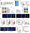 Figure 2
Intestinal epithelial cell-specific Nat10 deficiency attenuates colorectal tumorigenesis and enhances CD8+ T cell–mediated antitumor immunity. (A) Schematic illustration of AOM/DSS-induced CRC in Nat10fl/fl and Nat10cKO mice. (B and C) Representative colon tumor (B) and tumor number and tumor volume (C) in Nat10fl/fl and Nat10cKO mice (n = 8 mice/group). (D) scRNA-seq analysis of the cell types from colon tumors in Nat10fl/fl and Nat10cKO mice. (E and F) UMAP plot of tumor-infiltrating CD8+ T cells subset and proportions of subset. Tex, exhausted T; Teff, effector T; Trm, tissue-resident memory T. (G) Representative mIHC staining of CD4+ T cells, CD8+ T cells, and macrophages in tumors from Nat10fl/fl and Nat10cKO mice (n = 5 mice/group). Scale bar: 50 μm. (H and I) Flow cytometric analysis of total CD8+ T cells (H) and GzmB+ or IFN-γ+ cytotoxic subsets (I) (n = 5 mice/group). (J) ESTIMATE algorithm analysis of the correlation between Nat10 expression and the immune score in a TCGA CRC cohort (https://tcga-data.nci.nih.gov/tcga/) (n = 471). (K) Differential NAT10 expression in tumors from immunotherapy-responsive versus nonresponsive patients with CRC (GSE205506 dataset). (L) Representative mIHC staining of NAT10, GzmB, CD4+ T cells, CD8+ T cells, and macrophages in human CRC tissue microarrays (n = 536). Scale bar: 50 μm. (M) Proportions of infiltrated CD8+ T cells in human CRC tumors with high or low NAT10 expression (n = 536) (mean ± SEM). (N) Kaplan-Meier survival curves stratified by NAT10 and CD8 coexpression. Data are shown as the mean ± SD of indicated mice per group (C, H, and I). Statistical analysis was performed by 2-tailed Student’s t test (C, H, I, and M) and log-rank test (N). ns, P ≥ 0.05. P < 0.05 was considered to indicate statistical significance.
To further investigate the effect of Nat10 on the CRC TME, we isolated colorectal tumor tissues from Nat10fl/fl and Nat10cKO mice and performed single-cell RNA-seq (scRNA-seq). Compared with that in the Nat10fl/fl group, CD8+ T cell infiltration was markedly greater in tumors from Nat10cKO mice (Figure 2D and Supplemental Figure 2D). Using classical markers, we further classified CD8+ T cells into 5 distinct subsets: exhausted T cells, effector T cells, tissue-resident memory T cells, naive T cells, and memory T cells (Figure 2E). Notably, the proportion of effector T cells was substantially elevated and that of exhausted T cells was reduced in tumors from Nat10cKO mice (Figure 2F), accompanied by enhanced CD4+ and CD8+ T cell infiltration (Figure 2, G and H) and elevated GzmB and IFN-γ production by CD8+ T cells (Figure 2I).
To validate the correlation between NAT10 expression and immune responses in human CRC, an analysis of patient cohorts revealed an inverse correlation between NAT10 expression and immune scores, activated CD8+ T cell infiltration, and effector memory CD8+ T cell infiltration (Figure 2J and Supplemental Figure 3, A–D). Moreover, single-cell data from tumor tissues of patients with clinical CRC receiving anti–PD-1 therapy demonstrated that responders exhibited lower NAT10 expression levels (Figure 2K). Furthermore, multiplex immunohistochemical staining of primary human CRC tissue microarrays confirmed negative correlation between NAT10 protein expression and CD8+ T cell infiltration (Figure 2, L and M), with high NAT10 protein expression and low CD8+ T cell infiltration predicting poor patient survival (Figure 2N). Using CellChat, an algorithm for mapping cell-cell communications within scRNA-seq datasets, we demonstrated enhanced ligand-receptor interactions between Nat10-KO tumor cells and TME components, particularly CD8+ T cells (Supplemental Figure 3, E–G).
Furthermore, immunocompetent mice bearing MC38 Nat10-KO tumors were treated with neutralizing antibodies to eliminate CD8+ or CD4+ T cells (Supplemental Figure 4A), which markedly reduced the respective abundances of CD8+ and CD4+ T cells in the spleen (Supplemental Figure 4B). Depletion of CD8+ T cells markedly reversed Nat10-KO–mediated tumor suppression (Supplemental Figure 4, C–E), whereas CD4+ T cell or macrophage depletion showed no effect (Supplemental Figure 4, C–J). Collectively, these findings establish intestinal Nat10 as a regulator of CD8+ T cell–dependent antitumor immunity, with its ablation creating an immunogenic TME that restricts CRC progression.
NAT10 deficiency in tumor cells increases the infiltration of CD8+ T cells and enhances their cytotoxic functions. Given the observed enrichment of cytotoxic CD8+ T cells in Nat10-deficient tumors, we investigated direct tumor-T cell interactions using in vitro models. Using an in vitro T cell migration assay, we confirmed that knockdown of Nat10 in murine MC38 and CT-26 cells enhanced CD8+ T cell chemotaxis, whereas overexpression of Nat10 in MC38 cells suppressed the migratory capacity of these cells (Figure 3, A and B, and Supplemental Figure 5, A–C). Flow cytometric analysis of tumor–T cell cocultures revealed that, compared with their Nat10-KO counterparts, WT tumor cells markedly inhibited CD8+ T cell proliferation (Figure 3, C and D). Next, we established an in vitro coculture assay involving OVA-specific OT1 CD8+ T cells (isolated from OT1 transgenic mice) cocultured with OVA-modified tumor cells to evaluate the effect of WT or Nat10-KO tumor cells on the cytotoxic function of CD8+ T cells. OT1 CD8+ T cells produced substantially higher levels of GzmB and IFN-γ when cocultured with Nat10-KO MC38-OVA/CT-26-OVA cells compared with OVA-WT counterparts (Figure 3, E and F, and Supplemental Figure 5, D and E). This phenotype was also observed in melanoma cell models, where Nat10-KO B16F10-OVA cells exhibited a milder suppression on CD8+ T cell cytotoxicity (Supplemental Figure 5, F–H). Moreover, quantitative cytotoxic T lymphocyte assays demonstrated that the proportion of apoptotic Nat10-KO MC38-OVA cells increased across different effector-to-target ratios (Figure 3, G–I). Correspondingly, Nat10 depletion promoted the lactate dehydrogenase A (LDHA) release in OVA-WT tumor cells cocultured with OT1 CD8+ T cells (Figure 3J and Supplemental Figure 5I). Furthermore, 3D coculture systems revealed that Nat10-KO–OVA tumor spheroids exhibited a lower structural integrity and a higher caspase-3 activation compared with WT-OVA controls (Figure 3, K and L, and Supplemental Figure 5, J and K). Additionally, coculture of human CRC organoids with autologous CD8+ T cells showed that NAT10-KO organoids enhanced the killing capacity and cytotoxic function of CD8+ T cells (Figure 3, M and N, and Supplemental Figure 5, L and N). These data establish NAT10 as a tumor-intrinsic regulator that constrains recruitment of CD8+ T cell and impairs their cytotoxic function across multiple cancer models.
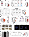 Figure 3
NAT10 deficiency in tumor cells enhances CD8+ T cell infiltration and cytotoxic functions. (A) Schematic of the in vitro CD8+ T cell migration assay. (B) Flow cytometric analysis of CD8+ T cell migration toward conditioned media from MC38 WT/Nat10-KO tumor cells. (C and D) Flow cytometric analysis of CFSE-labeled CD8+ T cell proliferation following 72-hour coculture with WT/Nat10-KO tumor cells at a 1:1 ratio. (E–J) OT-1 CD8+ T cells cocultured with OVA-modified WT/Nat10-KO tumor cells at different E:T ratios for 24 hours. (E and F) Flow cytometry of GzmB+/IFN-γ+ CD8+ T cells. (G and H) Tumor cell apoptosis and OT-1 CD8+ T cell cytotoxicity were assessed by (I) crystal violet staining and (J) LDHA release assay. (K and L) OT-1 CD8+ T cells cocultured with OVA-modified MC38 WT/Nat10-KO tumor cells (1:1, 24 hours) in a 3D coculture system. (K) Representative images of tumor spheroid disintegration and (L) mIHC staining (apoptotic tumor cells: cleaved caspase-3, red; CD8+ T cell infiltration, green). Scale bar: 50 μm. (M and N) Human CRC organoids cocultured with autologous peripheral blood–derived CD8+ T cells. Representative fluorescence images of apoptotic cells in WT/NAT10-KO organoids (M). Scale bar: 100 μm. Flow cytometry of GzmB+/IFN-γ+ CD8+ T cells (N) (n = 3). The data are presented as the mean ± SD of 3 independent experiments (B, D, F, H, J, and N). One of 3 representative experiments is shown (I and K–M). Statistical analysis was performed by 1-way ANOVA (B, D, F, H, and J) and 2-tailed Student’s t test (N). ns, P ≥ 0.05. P < 0.05 was considered to indicate statistical significance.
DKK2 mRNA is a direct target of NAT10-mediated ac4C modification. To further explore the molecular mechanism by which Nat10 regulates antitumor immunity, ac4C RNA immunoprecipitation sequencing (acRIP-seq) and RNA-seq were conducted in Nat10-deficient MC38 cells and control cells. Sequence motif analysis revealed enrichment of “CxxCxxCxx” patterns at ac4C-modified sites (Figure 4A), with ac4C peaks predominantly localized in coding sequences and 3′-UTRs (Figure 4B and Supplemental Figure 6A). Gene ontology analysis further revealed that ac4C-modified genes were substantially enriched in the Wnt signaling pathway (Figure 4C). Integration of RNA-seq and acRIP-seq data identified 8 candidate genes exhibiting both ac4C modification and reduced expression upon Nat10 KO, with the Wnt antagonist Dkk2 displaying the most pronounced ac4C peak (Figure 4D). Furthermore, Integrated Genomics Viewer (IGV) visualization showed that the ac4C peaks were distributed in the 3′-UTR of Dkk2 mRNA in WT cells but were diminished in Nat10-KO cells (Figure 4E). Since acRIP-seq may overestimate the prevalence of ac4C modification on mRNAs, we further employed NaCNBH3-based chemical ac4C sequencing (ac4C-seq) for a rigorous validation. IGV visualization confirmed that ac4C modifications identified via chemical sequencing were also enriched in the 3′-UTR of the Dkk2 transcript in WT cells but were absent in Nat10-KO cells (Figure 4F). Moreover, nucleotide-resolution mapping identified ac4C sites showing NaCNBH3-dependent misincorporation exclusively in WT samples (Supplemental Figure 6B).
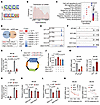 Figure 4
NAT10 directly targets DKK2 mRNA for ac4C modification to stabilize its expression. (A) Consensus motif analysis of ac4C-modified transcripts by HOMER in MC38 WT/Nat10-KO cells. (B) Metagene plot showing the distribution of ac4C-containing peaks across mRNAs. (C) Gene ontology (GO) analysis highlighting pathways significantly enriched in ac4C-modified genes. (D) Venn diagram of candidate genes with altered ac4C peaks (acRIP-seq) and transcript levels (mRNA-seq) upon Nat10 KO. Dkk2 exhibited the most pronounced reduction in ac4C modification. (E) Integrative Genomics Viewer (IGV) tracks of ac4C peak on Dkk2 mRNA based on acRIP-seq data in both MC38 WT/Nat10-KO cells. (F) IGV diagrams displaying read distributions and chemical ac4C sequencing–derived ac4C altered abundance across the 3′-UTR of DKK2 mRNA. (G and H) acRIP-qPCR quantification of Dkk2 mRNA using anti-ac4C in WT/Nat10-KO cells. (I) Schematic of the dual-luciferase reporter containing the WT and mutant (MUT) Dkk2 ac4C motif. (J) 293T cells were cotransfected with Myc-Nat10 plasmid and Dkk2 WT or MUT reporters for 48 hours. Luciferase activity in each group was detected. (K and L) Luciferase activity of WT/MUT reporters in Nat10-KO versus control cells. (M) qRT–PCR analysis of Dkk2 mRNA levels in Nat10-KO/Nat10-overexpressing (OE) CRC cells. (N) qRT–PCR analysis of Dkk2 mRNA levels in MC38 Nat10-KO/OE cells treated with actinomycin D (2.5 μg/mL) at the indicated time points. The data are presented as the mean ± SD of 3 independent experiments (G, H, and J–N). Statistical analysis was performed by 2-way ANOVA (G, H, J, and N), 1-way ANOVA (M, left), and 2-tailed Student’s t test (K, L, and M, right). ns, P ≥ 0.05. P < 0.05 was considered to indicate statistical significance.
Moreover, ac4C-RNA immunoprecipitation-quantitative PCR (acRIP-qPCR) confirmed reduced ac4C enrichment on Dkk2 mRNA in Nat10-KO MC38 and CT-26 cells compared with WT controls (Figure 4, G and H). Moreover, dual-luciferase reporter assays demonstrated that Nat10 overexpression increased WT Dkk2 3′-UTR-driven luciferase activity but had no effect on ac4C-motif mutants (MUTs) (Figure 4, I and J). Conversely, Nat10-KO markedly decreased the luciferase activity of the WT reporter gene, whereas it had no effect on the MUT reporter gene (Figure 4, K and L). Additionally, Dkk2 mRNA levels were decreased in the Nat10-KO cells and increased in Nat10-overexpression cells (Figure 4M and Supplemental Figure 6C). Given that the enrichment of the ac4C peak in the 3′-UTR of Dkk2 mRNA was markedly reduced in Nat10-KO cells, we hypothesized that Nat10 regulates Dkk2 mRNA stability. Thus, CRC cells were treated with actinomycin D (2.5 μg/mL) to examine RNA decay, and the stability of Dkk2 mRNA was markedly reduced in Nat10-KO cells, whereas the opposite effect was observed in cells overexpressing Nat10 (Figure 4N and Supplemental Figure 6D). Therefore, we identified Dkk2 as a direct target of Nat10, whose mRNA stability is regulated through ac4C modification in the 3′-UTR.
NAT10 modulates CD8+ T cell recruitment and cytotoxicity through DKK2 regulation. To further investigate the NAT10-DKK2 regulatory axis, we analyzed DKK2 protein expression following genetic manipulation of NAT10. Nat10-KO cells exhibited markedly reduced DKK2 levels, whereas Nat10 overexpression increased DKK2 levels (Figure 5A and Supplemental Figure 7A). Consistently, DKK2 expression decreased in NAT10-depleted human HCT116 cells but increased in NAT10-overexpressing SW620 cells (Supplemental Figure 7B). Since DKK2 is secreted, we quantified its levels in tumor cell conditioned media (CM). NAT10 deficiency markedly reduced DKK2 levels in the CM of mouse CRC cell lines (MC38 and CT-26) and the human HCT116 cell line (Figure 5B and Supplemental Figure 7D). Comparable results were obtained in B16F10 cells, while Nat10 overexpression increased secretion (Figure 5C and Supplemental Figure 7, C and D). Clinical correlation analyses revealed a positive association between NAT10 and DKK2 protein levels in human CRC tissues, and patients with high expression of both NAT10 and DKK2 had poorer prognosis (Figure 5, D–F). Consistently, the expression of Dkk2 was decreased in the intestinal tumors from Nat10cKO mice (Figure 5G and Supplemental Figure 7E).
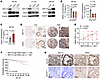 Figure 5
NAT10 positively regulates DKK2 expression with high expression of both associated with poor prognosis in patients with CRC. (A) Western blot analysis of Dkk2 protein levels in Nat10-KO/OE murine CRC cells. (B and C) ELISA quantification of secreted DKK2 in conditioned media from Nat10-KO/OE cells. (D) Representative IHC staining of NAT10 and DKK2 in human CRC tissues (n = 541). Scale bar: 25 μm; enlargement: original magnification, x40. (E) Positive correlation between NAT10 and DKK2 expression in CRC samples (n = 541). (F) Kaplan-Meier survival analysis of patients with CRC stratified by high versus low NAT10/DKK2 expression. (G) Representative IHC staining of Nat10 and Dkk2 expression in AOM/DSS-induced tumors from Nat10fl/fl and Nat10cKO mice (n = 5 mice/group). (B and C) The data are presented as the mean ± SD of 3 independent experiments. Statistical analysis was performed by 1-way ANOVA (B), 2-tailed Student’s t test (C), Pearson’s correlation coefficient (E), and log-rank test (F). ns, P ≥ 0.05. P < 0.05 was considered to indicate statistical significance.
Next, we investigated whether NAT10-driven DKK2 secretion suppresses antitumor immunity in vitro. Compared with WT cell control CM, Nat10-KO cell CM enhanced CD8+ T cell chemotaxis. Notably, this effect was abrogated by recombinant Dkk2 (rDkk2) supplementation in a dose-dependent manner. (Figure 6, A and B). We further explored whether Dkk2 directly impairs CD8+ T cell effector functions. OT1 CD8+ T cells were cocultured with OVA-expressing tumor cells (MC38, CT-26, and B16F10) in the presence or absence of rDkk2 (400 ng/mL). The results revealed that rDkk2 treatment markedly suppressed the production of GzmB and IFN-γ by CD8+ T cells cocultured with Nat10-KO cells (Figure 6, C–F, and Supplemental Figure 7, F–K). Additionally, the enhanced tumor-killing effect of CD8+ T cells under Nat10-KO conditions was abrogated by rDkk2 supplementation (Figure 6, G and H, and Supplemental Figure 7L). Collectively, these results suggest that NAT10 induces the secretion of DKK2 by tumor cells and that DKK2 serves as an immune suppressive factor that restricts the recruitment of CD8+ T cells and impairs their cytotoxic function in CRC.
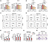 Figure 6
NAT10 orchestrates CD8+ T cell recruitment and cytotoxicity via aDKK2-dependent mechanism. (A and B) Flow cytometric analysis of CD8+ T cell migration toward conditioned media from WT/Nat10-KO tumor cells supplemented with or without recombinant Dkk2 (rDkk2) (μg/mL). (C–F) OT-1 CD8+ T cells were cocultured with OVA-modified MC38 WT/Nat10-KO tumor cells at 1:1 with or without rDkk2 for 24 hours. Flow cytometry analysis of GzmB+ and IFN-γ+ CD8+ T cell populations. (G and H) OT-1 CD8+ T cell cytotoxicity was assessed by LDHA release assay (G) and crystal violet staining assay (H). The data are presented as the mean ± SD of 3 independent experiments (A, B, and E–G). One of 3 representative experiments is shown (H). Statistical analysis was performed by 2-way ANOVA (A and B) and 2-tailed Student’s t test (E–G). ns, P ≥ 0.05. P < 0.05 was considered to indicate statistical significance.
DKK2 promotes cholesterol biosynthesis to suppress CD8+ T cell antitumor function. To elucidate the mechanism underlying the tumor-derived DKK2-mediated impairment of CD8+ T cell cytotoxicity, we investigated whether DKK2, a canonical Wnt signaling inhibitor (18), induced CD8+ T cell dysfunction by suppressing this pathway. Using SOST, a competitive Wnt antagonist (19), as a control, we found that SOST alone neither reduced GzmB and INF-γ secretion nor rescued rDkk2-induced CD8+ T cell dysfunction (Supplemental Figure 8, A and B). Transcriptomic profiling of CD8+ T cells exposed to rDkk2 revealed significant enrichment of pathways related to PPAR signaling, lipid digestion and absorption, and cholesterol metabolism (Supplemental Figure 8C). scRNA-seq data further revealed diminished cholesterol accumulation in tumor-infiltrating CD8+ T cells from Nat10cKO mice versus Nat10fl/fl controls (Supplemental Figure 8D), implicating Dkk2 in modulating CD8+ T cell cholesterol homeostasis. Intriguingly, we observed increased cholesterol content in CD8+ T cells treated with rDkk2 compared with control cells (Figure 7A). This finding is consistent with the established roles of cholesterol overload in T cell exhaustion (13, 20). Conversely, CD8+ T cells cultured in Nat10-KO tumor cell CM exhibited reduced cholesterol accumulation (Figure 7B and Supplemental Figure 8, E and F). Moreover, cholesterol supplementation in Nat10-KO CM suppressed GzmB and IFN-γ production by CD8+ T cells (Figure 7C and Supplemental Figure 8, G and H), whereas cholesterol depletion in CD8+ T cells via methyl-β-cyclodextrin (MβCD) (13, 21) increased GzmB and IFN-γ production and ameliorated rDkk2-induced cytotoxicity impairment (Figure 7D).
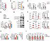 Figure 7
DKK2 promotes CD8+ T cell dysfunction via cholesterol accumulation. (A) Filipin III staining of cholesterol in CD8+ T cells with or without rDkk2 for 24 hours. (B) Cholesterol levels in activated CD8+ T cells cultured with MC38 WT/Nat10-KO cell conditioned media (CM) for 24 hours. (C) Flow cytometry of GzmB+/IFN-γ+ CD8+ T cell with cholesterol-supplemented tumor CM for 24 hours. (D) Flow cytometry of GzmB+/IFN-γ+ CD8+ T cell populations after 24 hours of culture with rDkk2 and MβCD. (E) Western blot of p-AKT/AKT, p-mTOR/mTOR, and p-S6K levels in CD8+ T cells. (F and G) Flow cytometry of p-AKT/p-mTOR levels in CD8+ T cells cultured with tumor CM for 24 hours. (H) Flow cytometry of GzmB+/IFN-γ+ CD8+ T cell populations treated with rDkk2 plus rapamycin or cholesterol. (I) qRT-PCR of cholesterol-related genes in activated CD8+ T cells after treatment with rDkk2 and rapamycin. The data are presented as the mean ± SD of 3 independent experiments (A–D and F–I). Statistical analysis was performed by 2-tailed Student’s t test (A and C), 1-way ANOVA (B, F, G, and I), and 2-way ANOVA (D and H). ns, P ≥ 0.05. P < 0.05 was considered to indicate statistical significance.
Mechanistically, given the established link between AKT-mTOR signaling and cholesterol biosynthesis (20, 22), we observed rDkk2-induced activation of AKT-mTOR-S6K signaling in activated CD8+ T cells (Figure 7E). This pathway was activated in WT tumor cell CM-cultured CD8+ T cells but inhibited in Nat10-KO tumor cell CM-cultured ones (Figure 7, F and G, and Supplemental Figure 8I). In addition, pharmacological mTOR inhibition (rapamycin, 10 nM) rescued rDkk2-impaired cytotoxicity in CD8+ T cells (Figure 7H) and downregulated cholesterol biosynthesis-related genes (Figure 7I and Supplemental Figure 9A). However, exogenous cholesterol supplementation abrogated rapamycin-mediated restoration of CD8+ T cell cytotoxicity (Figure 7H). Furthermore, in vivo data showed that anti-Dkk2 antibody (5F8) not only inhibited tumor proliferation and growth (Figure 8, A–D, and Supplemental Figure 9B), but also notably reversed the acceleration of Nat10-OE tumor growth (Figure 8, B–D, and Supplemental Figure 9B). Meanwhile, 5F8 treatment substantially increased the numbers of tumor-infiltrating CD8+ T cells and effector T cells in Nat10-OE tumors (Figure 8, E and F). Additionally, 5F8 markedly reversed the elevation in the cholesterol levels and enhanced AKT-mTOR signaling activation in these infiltrating CD8+ T cells (Figure 8, G and H, and Supplemental Figure 9C). These results indicate that NAT10 promotes cholesterol accumulation in CD8+ T cells via DKK2 to impair their cytotoxicity.
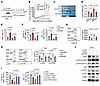 Figure 8
NAT10 drives CD8+ T cell dysfunction via DKK2/LRP6/AKT-mTOR axis–mediated cholesterol accumulation. (A) Schematic of anti-DKK2 antibody (5F8) treatment in MC38 control (Con) or Nat10-OE cell-bearing tumor mice. (B–D) Tumor growth curves (B) (mean ± SEM), representative tumor images (C), and tumor weights (D) (n = 8 mice/group). (E and F) Flow cytometry of tumor-infiltrating CD8+ T cells (E) and GzmB+/IFN-γ+ CD8+ T cell populations (F) in tumors (n = 8 mice/group). (G and H) Filipin III staining of cholesterol (G) and flow cytometry of p-AKT/p-mTOR levels (H) in tumor-infiltrating CD8+ T cells (n = 8 mice/group). (I) Flow cytometry of GzmB+/IFN-γ+ CD8+ T cells treated with rDkk2 after Lrp5 or Lrp6 knockdown. (J) Western blot of p-AKT/AKT, p-mTOR/mTOR, and p-S6K expression in Lrp6-knockdown CD8+ T cells. Data are shown as the mean ± SD of per group (B–H). The data are presented as the mean ± SD of 3 independent experiments (I). All statistical analysis was performed by 2-way ANOVA. ns, P ≥ 0.05. P < 0.05 was considered to indicate statistical significance.
As DKK2 binds to LRP5/6 to modulate glucose uptake and mTOR activation (22, 23), we next investigated receptor specificity. rDkk2 further exacerbated the reduction in GzmB and IFN-γ expression in Lrp5-knockdown CD8+ T cells, while knockdown of Lrp6 in CD8+ T cells reversed the rDkk2-induced suppression of cytotoxicity (Figure 8I and Supplemental Figure 9D). Interestingly, dual Lrp5/6 knockdown in CD8+ T cells partially restored cytotoxicity (Figure 8I), suggesting that LRP6 plays an important role in DKK2-mediated signaling. Moreover, DKK2-mediated activation of AKT-mTOR signaling in CD8+ T cells was alleviated by LRP6 knockdown (Figure 8J). Collectively, DKK2 engages LRP6 to hyperactivate AKT-mTOR-driven cholesterol biosynthesis, thereby suppressing the metabolism of CD8+ T cells in the TME.
Targeting NAT10 or DKK2 augments anti-PD1 therapy to suppress CRC growth. Given the role of NAT10 in driving DKK2 secretion to suppress CD8+ T cell function within the CRC microenvironment, we evaluated whether inhibiting NAT10 or neutralizing DKK2 could potentiate immune checkpoint blockade (ICB) efficacy. In C57BL/6 mice bearing MC38 syngeneic tumors, Remodelin (5 mg/kg, intraperitoneal injection every 2 days), a selective NAT10 inhibitor, was combined with anti–PD-1 or IgG isotype control for 14 days (Supplemental Figure 10A). Compared with the control treatment, Remodelin monotherapy markedly attenuated tumor growth (Figure 9A and Supplemental Figure 10, B and C). Strikingly, Remodelin synergized with anti–PD-1 therapy, resulting in superior tumor growth suppression compared with either agent alone (Figure 9A and Supplemental Figure 10, B and C). Furthermore, Remodelin combined with anti–PD-1 treatment substantially increased the infiltration of CD8+ T cells into the tumor (Figure 9B), accompanied by a high percentage of GzmB+ and IFN-γ+ CD8+ T cells (Figure 9C), demonstrating that NAT10 inhibition enhances the effects of PD-1 blockade.
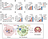 Figure 9
Dual targeting of NAT10 or DKK2 synergizes with PD1 blockade to suppress CRC. (A) Tumor growth curves. Data are shown as the mean ± SEM of 7 mice/group. (B and C) Flow cytometry analysis of tumor-infiltrating CD8+ T cells (B) and GzmB+ and IFN-γ+CD8+ T cell populations (C) in tumors form each group (n = 7 mice/group). (D) Tumor growth curves. Data are shown as the mean ± SEM of 7 mice/group. (E and F) Flow cytometry analysis of tumor-infiltrating CD8+ T cells (E) and GzmB+ and IFN-γ+CD8+ T cell populations (F) in tumors form each group (n = 5 mice/group). (G) Proposed model: tumor-intrinsic NAT10 stabilizes DKK2 mRNA via ac4C modification, enabling DKK2-LRP6/AKT-mTOR signaling to drive cholesterol accumulation and dysfunction in CD8+ T cells, thereby promoting CRC progression. Dual targeting of NAT10 or DKK2 synergizes with anti–PD-1 therapy to suppress CRC. Data are shown as the mean ± SD of indicated mice per group. All statistical analysis was performed by 2-way ANOVA. ns, P ≥ 0.05. P < 0.05 was considered to indicate statistical significance.
To further determine whether DKK2 neutralization similarly enhances ICB responsiveness, MC38 tumor-bearing mice were treated with anti-Dkk2 (5F8) alone or in combination with anti–PD-1 (Supplemental Figure 10D). Both 5F8 and anti–PD-1 monotherapies markedly reduced tumor burden (Figure 9D and Supplemental Figure 10, E and F), while their combination exhibited additive effects on tumor growth compared with the control or monotherapies (Figure 9D and Supplemental Figure 10, E and F). The dual-treatment group showed the greatest CD8+ T cell infiltration in MC38 tumors (Figure 9E) and the highest percentage of IFN-γ+GZMB+ CD8+ T cells (Figure 9F), mirroring NAT10-targeted outcomes. These findings indicate that disrupting the NAT10/DKK2 axis synergizes with PD-1 blockade to reinvigorate CD8+ T cell–mediated antitumor immunity, proposing a potential combinatorial immunotherapy strategy for CRC.


Copyright © 2026 American Society for Clinical Investigation
ISSN: 0021-9738 (print), 1558-8238 (online)