
Advertisement
Research ArticleCardiologyVascular biology
Open Access |  10.1172/JCI194175
Vascular smooth muscle cell–derived KIF13B inhibits proinflammatory responses to protect against atherosclerosis
Guolin Miao,1,2 Yufei Han,2 Jingxuan Chen,2 Yiran Liu,3 Ge Zhang,4 Shaotong Pei,4 Yinqi Zhao,2 Yitong Xu,2 Liwen Zheng,2 Zhaoling Li,2 Xiangru Liu,2 Sijing Shi,2 Xuya Kang,2 Yahan Liu,2 Ling Zhang,2 Wei Huang,2 Yuhui Wang,2 Junnan Tang,4 Erdan Dong,1,2,5,6 and Xunde Xian2,7,8
1Department of Cardiology and Institute of Vascular Medicine, Peking University Third Hospital, Beijing, China.
2Institute of Cardiovascular Sciences, State Key Laboratory of Vascular Homeostasis and Remodeling, School of Basic Medical Sciences, and
3Department of Biomedical Informatics, School of Basic Medical Sciences, Peking University, Beijing, China.
4Department of Cardiology, The First Affiliated Hospital of Zhengzhou University, Zhengzhou, China.
5Research Center for Cardiopulmonary Rehabilitation, University of Health and Rehabilitation Sciences Qingdao Hospital (Qingdao Municipal Hospital), School of Health and Life Sciences, University of Health and Rehabilitation Sciences, Qingdao, China.
6Research Unit of Medical Science Research Management/Basic and Clinical Research of Metabolic Cardiovascular Diseases, Chinese Academy of Medical Sciences, Beijing, China.
7Department of Human Anatomy and Histology and Embryology, School of Basic Medical Sciences, Peking University, Beijing, China.
8Beijing Key Laboratory of Cardiovascular Receptors Research, Peking University Third Hospital, Beijing, China.
Address correspondence to: Xunde Xian, Institute of Cardiovascular Sciences, Peking University, No. 38 Xueyuan Rd., Haidian District, Beijing, 100191, China. Phone: 86.10.82802769; Email: xianxunde@bjmu.edu.cn. Or to: Erdan Dong, Department of Cardiology and Institute of Vascular Medicine, Peking University Third Hospital, Beijing, 100191, China. Phone: 86.10.82265811; Email: donged@bjmu.edu.cn.
Authorship note: GM, YH, and JC contributed equally to this work.
Find articles by Miao, G. in: PubMed | Google Scholar
1Department of Cardiology and Institute of Vascular Medicine, Peking University Third Hospital, Beijing, China.
2Institute of Cardiovascular Sciences, State Key Laboratory of Vascular Homeostasis and Remodeling, School of Basic Medical Sciences, and
3Department of Biomedical Informatics, School of Basic Medical Sciences, Peking University, Beijing, China.
4Department of Cardiology, The First Affiliated Hospital of Zhengzhou University, Zhengzhou, China.
5Research Center for Cardiopulmonary Rehabilitation, University of Health and Rehabilitation Sciences Qingdao Hospital (Qingdao Municipal Hospital), School of Health and Life Sciences, University of Health and Rehabilitation Sciences, Qingdao, China.
6Research Unit of Medical Science Research Management/Basic and Clinical Research of Metabolic Cardiovascular Diseases, Chinese Academy of Medical Sciences, Beijing, China.
7Department of Human Anatomy and Histology and Embryology, School of Basic Medical Sciences, Peking University, Beijing, China.
8Beijing Key Laboratory of Cardiovascular Receptors Research, Peking University Third Hospital, Beijing, China.
Address correspondence to: Xunde Xian, Institute of Cardiovascular Sciences, Peking University, No. 38 Xueyuan Rd., Haidian District, Beijing, 100191, China. Phone: 86.10.82802769; Email: xianxunde@bjmu.edu.cn. Or to: Erdan Dong, Department of Cardiology and Institute of Vascular Medicine, Peking University Third Hospital, Beijing, 100191, China. Phone: 86.10.82265811; Email: donged@bjmu.edu.cn.
Authorship note: GM, YH, and JC contributed equally to this work.
Find articles by Han, Y. in: PubMed | Google Scholar
1Department of Cardiology and Institute of Vascular Medicine, Peking University Third Hospital, Beijing, China.
2Institute of Cardiovascular Sciences, State Key Laboratory of Vascular Homeostasis and Remodeling, School of Basic Medical Sciences, and
3Department of Biomedical Informatics, School of Basic Medical Sciences, Peking University, Beijing, China.
4Department of Cardiology, The First Affiliated Hospital of Zhengzhou University, Zhengzhou, China.
5Research Center for Cardiopulmonary Rehabilitation, University of Health and Rehabilitation Sciences Qingdao Hospital (Qingdao Municipal Hospital), School of Health and Life Sciences, University of Health and Rehabilitation Sciences, Qingdao, China.
6Research Unit of Medical Science Research Management/Basic and Clinical Research of Metabolic Cardiovascular Diseases, Chinese Academy of Medical Sciences, Beijing, China.
7Department of Human Anatomy and Histology and Embryology, School of Basic Medical Sciences, Peking University, Beijing, China.
8Beijing Key Laboratory of Cardiovascular Receptors Research, Peking University Third Hospital, Beijing, China.
Address correspondence to: Xunde Xian, Institute of Cardiovascular Sciences, Peking University, No. 38 Xueyuan Rd., Haidian District, Beijing, 100191, China. Phone: 86.10.82802769; Email: xianxunde@bjmu.edu.cn. Or to: Erdan Dong, Department of Cardiology and Institute of Vascular Medicine, Peking University Third Hospital, Beijing, 100191, China. Phone: 86.10.82265811; Email: donged@bjmu.edu.cn.
Authorship note: GM, YH, and JC contributed equally to this work.
Find articles by Chen, J. in: PubMed | Google Scholar
1Department of Cardiology and Institute of Vascular Medicine, Peking University Third Hospital, Beijing, China.
2Institute of Cardiovascular Sciences, State Key Laboratory of Vascular Homeostasis and Remodeling, School of Basic Medical Sciences, and
3Department of Biomedical Informatics, School of Basic Medical Sciences, Peking University, Beijing, China.
4Department of Cardiology, The First Affiliated Hospital of Zhengzhou University, Zhengzhou, China.
5Research Center for Cardiopulmonary Rehabilitation, University of Health and Rehabilitation Sciences Qingdao Hospital (Qingdao Municipal Hospital), School of Health and Life Sciences, University of Health and Rehabilitation Sciences, Qingdao, China.
6Research Unit of Medical Science Research Management/Basic and Clinical Research of Metabolic Cardiovascular Diseases, Chinese Academy of Medical Sciences, Beijing, China.
7Department of Human Anatomy and Histology and Embryology, School of Basic Medical Sciences, Peking University, Beijing, China.
8Beijing Key Laboratory of Cardiovascular Receptors Research, Peking University Third Hospital, Beijing, China.
Address correspondence to: Xunde Xian, Institute of Cardiovascular Sciences, Peking University, No. 38 Xueyuan Rd., Haidian District, Beijing, 100191, China. Phone: 86.10.82802769; Email: xianxunde@bjmu.edu.cn. Or to: Erdan Dong, Department of Cardiology and Institute of Vascular Medicine, Peking University Third Hospital, Beijing, 100191, China. Phone: 86.10.82265811; Email: donged@bjmu.edu.cn.
Authorship note: GM, YH, and JC contributed equally to this work.
Find articles by Liu, Y. in: PubMed | Google Scholar
1Department of Cardiology and Institute of Vascular Medicine, Peking University Third Hospital, Beijing, China.
2Institute of Cardiovascular Sciences, State Key Laboratory of Vascular Homeostasis and Remodeling, School of Basic Medical Sciences, and
3Department of Biomedical Informatics, School of Basic Medical Sciences, Peking University, Beijing, China.
4Department of Cardiology, The First Affiliated Hospital of Zhengzhou University, Zhengzhou, China.
5Research Center for Cardiopulmonary Rehabilitation, University of Health and Rehabilitation Sciences Qingdao Hospital (Qingdao Municipal Hospital), School of Health and Life Sciences, University of Health and Rehabilitation Sciences, Qingdao, China.
6Research Unit of Medical Science Research Management/Basic and Clinical Research of Metabolic Cardiovascular Diseases, Chinese Academy of Medical Sciences, Beijing, China.
7Department of Human Anatomy and Histology and Embryology, School of Basic Medical Sciences, Peking University, Beijing, China.
8Beijing Key Laboratory of Cardiovascular Receptors Research, Peking University Third Hospital, Beijing, China.
Address correspondence to: Xunde Xian, Institute of Cardiovascular Sciences, Peking University, No. 38 Xueyuan Rd., Haidian District, Beijing, 100191, China. Phone: 86.10.82802769; Email: xianxunde@bjmu.edu.cn. Or to: Erdan Dong, Department of Cardiology and Institute of Vascular Medicine, Peking University Third Hospital, Beijing, 100191, China. Phone: 86.10.82265811; Email: donged@bjmu.edu.cn.
Authorship note: GM, YH, and JC contributed equally to this work.
Find articles by Zhang, G. in: PubMed | Google Scholar
1Department of Cardiology and Institute of Vascular Medicine, Peking University Third Hospital, Beijing, China.
2Institute of Cardiovascular Sciences, State Key Laboratory of Vascular Homeostasis and Remodeling, School of Basic Medical Sciences, and
3Department of Biomedical Informatics, School of Basic Medical Sciences, Peking University, Beijing, China.
4Department of Cardiology, The First Affiliated Hospital of Zhengzhou University, Zhengzhou, China.
5Research Center for Cardiopulmonary Rehabilitation, University of Health and Rehabilitation Sciences Qingdao Hospital (Qingdao Municipal Hospital), School of Health and Life Sciences, University of Health and Rehabilitation Sciences, Qingdao, China.
6Research Unit of Medical Science Research Management/Basic and Clinical Research of Metabolic Cardiovascular Diseases, Chinese Academy of Medical Sciences, Beijing, China.
7Department of Human Anatomy and Histology and Embryology, School of Basic Medical Sciences, Peking University, Beijing, China.
8Beijing Key Laboratory of Cardiovascular Receptors Research, Peking University Third Hospital, Beijing, China.
Address correspondence to: Xunde Xian, Institute of Cardiovascular Sciences, Peking University, No. 38 Xueyuan Rd., Haidian District, Beijing, 100191, China. Phone: 86.10.82802769; Email: xianxunde@bjmu.edu.cn. Or to: Erdan Dong, Department of Cardiology and Institute of Vascular Medicine, Peking University Third Hospital, Beijing, 100191, China. Phone: 86.10.82265811; Email: donged@bjmu.edu.cn.
Authorship note: GM, YH, and JC contributed equally to this work.
Find articles by Pei, S. in: PubMed | Google Scholar
1Department of Cardiology and Institute of Vascular Medicine, Peking University Third Hospital, Beijing, China.
2Institute of Cardiovascular Sciences, State Key Laboratory of Vascular Homeostasis and Remodeling, School of Basic Medical Sciences, and
3Department of Biomedical Informatics, School of Basic Medical Sciences, Peking University, Beijing, China.
4Department of Cardiology, The First Affiliated Hospital of Zhengzhou University, Zhengzhou, China.
5Research Center for Cardiopulmonary Rehabilitation, University of Health and Rehabilitation Sciences Qingdao Hospital (Qingdao Municipal Hospital), School of Health and Life Sciences, University of Health and Rehabilitation Sciences, Qingdao, China.
6Research Unit of Medical Science Research Management/Basic and Clinical Research of Metabolic Cardiovascular Diseases, Chinese Academy of Medical Sciences, Beijing, China.
7Department of Human Anatomy and Histology and Embryology, School of Basic Medical Sciences, Peking University, Beijing, China.
8Beijing Key Laboratory of Cardiovascular Receptors Research, Peking University Third Hospital, Beijing, China.
Address correspondence to: Xunde Xian, Institute of Cardiovascular Sciences, Peking University, No. 38 Xueyuan Rd., Haidian District, Beijing, 100191, China. Phone: 86.10.82802769; Email: xianxunde@bjmu.edu.cn. Or to: Erdan Dong, Department of Cardiology and Institute of Vascular Medicine, Peking University Third Hospital, Beijing, 100191, China. Phone: 86.10.82265811; Email: donged@bjmu.edu.cn.
Authorship note: GM, YH, and JC contributed equally to this work.
Find articles by Zhao, Y. in: PubMed | Google Scholar
1Department of Cardiology and Institute of Vascular Medicine, Peking University Third Hospital, Beijing, China.
2Institute of Cardiovascular Sciences, State Key Laboratory of Vascular Homeostasis and Remodeling, School of Basic Medical Sciences, and
3Department of Biomedical Informatics, School of Basic Medical Sciences, Peking University, Beijing, China.
4Department of Cardiology, The First Affiliated Hospital of Zhengzhou University, Zhengzhou, China.
5Research Center for Cardiopulmonary Rehabilitation, University of Health and Rehabilitation Sciences Qingdao Hospital (Qingdao Municipal Hospital), School of Health and Life Sciences, University of Health and Rehabilitation Sciences, Qingdao, China.
6Research Unit of Medical Science Research Management/Basic and Clinical Research of Metabolic Cardiovascular Diseases, Chinese Academy of Medical Sciences, Beijing, China.
7Department of Human Anatomy and Histology and Embryology, School of Basic Medical Sciences, Peking University, Beijing, China.
8Beijing Key Laboratory of Cardiovascular Receptors Research, Peking University Third Hospital, Beijing, China.
Address correspondence to: Xunde Xian, Institute of Cardiovascular Sciences, Peking University, No. 38 Xueyuan Rd., Haidian District, Beijing, 100191, China. Phone: 86.10.82802769; Email: xianxunde@bjmu.edu.cn. Or to: Erdan Dong, Department of Cardiology and Institute of Vascular Medicine, Peking University Third Hospital, Beijing, 100191, China. Phone: 86.10.82265811; Email: donged@bjmu.edu.cn.
Authorship note: GM, YH, and JC contributed equally to this work.
Find articles by Xu, Y. in: PubMed | Google Scholar
1Department of Cardiology and Institute of Vascular Medicine, Peking University Third Hospital, Beijing, China.
2Institute of Cardiovascular Sciences, State Key Laboratory of Vascular Homeostasis and Remodeling, School of Basic Medical Sciences, and
3Department of Biomedical Informatics, School of Basic Medical Sciences, Peking University, Beijing, China.
4Department of Cardiology, The First Affiliated Hospital of Zhengzhou University, Zhengzhou, China.
5Research Center for Cardiopulmonary Rehabilitation, University of Health and Rehabilitation Sciences Qingdao Hospital (Qingdao Municipal Hospital), School of Health and Life Sciences, University of Health and Rehabilitation Sciences, Qingdao, China.
6Research Unit of Medical Science Research Management/Basic and Clinical Research of Metabolic Cardiovascular Diseases, Chinese Academy of Medical Sciences, Beijing, China.
7Department of Human Anatomy and Histology and Embryology, School of Basic Medical Sciences, Peking University, Beijing, China.
8Beijing Key Laboratory of Cardiovascular Receptors Research, Peking University Third Hospital, Beijing, China.
Address correspondence to: Xunde Xian, Institute of Cardiovascular Sciences, Peking University, No. 38 Xueyuan Rd., Haidian District, Beijing, 100191, China. Phone: 86.10.82802769; Email: xianxunde@bjmu.edu.cn. Or to: Erdan Dong, Department of Cardiology and Institute of Vascular Medicine, Peking University Third Hospital, Beijing, 100191, China. Phone: 86.10.82265811; Email: donged@bjmu.edu.cn.
Authorship note: GM, YH, and JC contributed equally to this work.
Find articles by Zheng, L. in: PubMed | Google Scholar
1Department of Cardiology and Institute of Vascular Medicine, Peking University Third Hospital, Beijing, China.
2Institute of Cardiovascular Sciences, State Key Laboratory of Vascular Homeostasis and Remodeling, School of Basic Medical Sciences, and
3Department of Biomedical Informatics, School of Basic Medical Sciences, Peking University, Beijing, China.
4Department of Cardiology, The First Affiliated Hospital of Zhengzhou University, Zhengzhou, China.
5Research Center for Cardiopulmonary Rehabilitation, University of Health and Rehabilitation Sciences Qingdao Hospital (Qingdao Municipal Hospital), School of Health and Life Sciences, University of Health and Rehabilitation Sciences, Qingdao, China.
6Research Unit of Medical Science Research Management/Basic and Clinical Research of Metabolic Cardiovascular Diseases, Chinese Academy of Medical Sciences, Beijing, China.
7Department of Human Anatomy and Histology and Embryology, School of Basic Medical Sciences, Peking University, Beijing, China.
8Beijing Key Laboratory of Cardiovascular Receptors Research, Peking University Third Hospital, Beijing, China.
Address correspondence to: Xunde Xian, Institute of Cardiovascular Sciences, Peking University, No. 38 Xueyuan Rd., Haidian District, Beijing, 100191, China. Phone: 86.10.82802769; Email: xianxunde@bjmu.edu.cn. Or to: Erdan Dong, Department of Cardiology and Institute of Vascular Medicine, Peking University Third Hospital, Beijing, 100191, China. Phone: 86.10.82265811; Email: donged@bjmu.edu.cn.
Authorship note: GM, YH, and JC contributed equally to this work.
Find articles by Li, Z. in: PubMed | Google Scholar
1Department of Cardiology and Institute of Vascular Medicine, Peking University Third Hospital, Beijing, China.
2Institute of Cardiovascular Sciences, State Key Laboratory of Vascular Homeostasis and Remodeling, School of Basic Medical Sciences, and
3Department of Biomedical Informatics, School of Basic Medical Sciences, Peking University, Beijing, China.
4Department of Cardiology, The First Affiliated Hospital of Zhengzhou University, Zhengzhou, China.
5Research Center for Cardiopulmonary Rehabilitation, University of Health and Rehabilitation Sciences Qingdao Hospital (Qingdao Municipal Hospital), School of Health and Life Sciences, University of Health and Rehabilitation Sciences, Qingdao, China.
6Research Unit of Medical Science Research Management/Basic and Clinical Research of Metabolic Cardiovascular Diseases, Chinese Academy of Medical Sciences, Beijing, China.
7Department of Human Anatomy and Histology and Embryology, School of Basic Medical Sciences, Peking University, Beijing, China.
8Beijing Key Laboratory of Cardiovascular Receptors Research, Peking University Third Hospital, Beijing, China.
Address correspondence to: Xunde Xian, Institute of Cardiovascular Sciences, Peking University, No. 38 Xueyuan Rd., Haidian District, Beijing, 100191, China. Phone: 86.10.82802769; Email: xianxunde@bjmu.edu.cn. Or to: Erdan Dong, Department of Cardiology and Institute of Vascular Medicine, Peking University Third Hospital, Beijing, 100191, China. Phone: 86.10.82265811; Email: donged@bjmu.edu.cn.
Authorship note: GM, YH, and JC contributed equally to this work.
Find articles by Liu, X. in: PubMed | Google Scholar
1Department of Cardiology and Institute of Vascular Medicine, Peking University Third Hospital, Beijing, China.
2Institute of Cardiovascular Sciences, State Key Laboratory of Vascular Homeostasis and Remodeling, School of Basic Medical Sciences, and
3Department of Biomedical Informatics, School of Basic Medical Sciences, Peking University, Beijing, China.
4Department of Cardiology, The First Affiliated Hospital of Zhengzhou University, Zhengzhou, China.
5Research Center for Cardiopulmonary Rehabilitation, University of Health and Rehabilitation Sciences Qingdao Hospital (Qingdao Municipal Hospital), School of Health and Life Sciences, University of Health and Rehabilitation Sciences, Qingdao, China.
6Research Unit of Medical Science Research Management/Basic and Clinical Research of Metabolic Cardiovascular Diseases, Chinese Academy of Medical Sciences, Beijing, China.
7Department of Human Anatomy and Histology and Embryology, School of Basic Medical Sciences, Peking University, Beijing, China.
8Beijing Key Laboratory of Cardiovascular Receptors Research, Peking University Third Hospital, Beijing, China.
Address correspondence to: Xunde Xian, Institute of Cardiovascular Sciences, Peking University, No. 38 Xueyuan Rd., Haidian District, Beijing, 100191, China. Phone: 86.10.82802769; Email: xianxunde@bjmu.edu.cn. Or to: Erdan Dong, Department of Cardiology and Institute of Vascular Medicine, Peking University Third Hospital, Beijing, 100191, China. Phone: 86.10.82265811; Email: donged@bjmu.edu.cn.
Authorship note: GM, YH, and JC contributed equally to this work.
Find articles by Shi, S. in: PubMed | Google Scholar
1Department of Cardiology and Institute of Vascular Medicine, Peking University Third Hospital, Beijing, China.
2Institute of Cardiovascular Sciences, State Key Laboratory of Vascular Homeostasis and Remodeling, School of Basic Medical Sciences, and
3Department of Biomedical Informatics, School of Basic Medical Sciences, Peking University, Beijing, China.
4Department of Cardiology, The First Affiliated Hospital of Zhengzhou University, Zhengzhou, China.
5Research Center for Cardiopulmonary Rehabilitation, University of Health and Rehabilitation Sciences Qingdao Hospital (Qingdao Municipal Hospital), School of Health and Life Sciences, University of Health and Rehabilitation Sciences, Qingdao, China.
6Research Unit of Medical Science Research Management/Basic and Clinical Research of Metabolic Cardiovascular Diseases, Chinese Academy of Medical Sciences, Beijing, China.
7Department of Human Anatomy and Histology and Embryology, School of Basic Medical Sciences, Peking University, Beijing, China.
8Beijing Key Laboratory of Cardiovascular Receptors Research, Peking University Third Hospital, Beijing, China.
Address correspondence to: Xunde Xian, Institute of Cardiovascular Sciences, Peking University, No. 38 Xueyuan Rd., Haidian District, Beijing, 100191, China. Phone: 86.10.82802769; Email: xianxunde@bjmu.edu.cn. Or to: Erdan Dong, Department of Cardiology and Institute of Vascular Medicine, Peking University Third Hospital, Beijing, 100191, China. Phone: 86.10.82265811; Email: donged@bjmu.edu.cn.
Authorship note: GM, YH, and JC contributed equally to this work.
Find articles by Kang, X. in: PubMed | Google Scholar
1Department of Cardiology and Institute of Vascular Medicine, Peking University Third Hospital, Beijing, China.
2Institute of Cardiovascular Sciences, State Key Laboratory of Vascular Homeostasis and Remodeling, School of Basic Medical Sciences, and
3Department of Biomedical Informatics, School of Basic Medical Sciences, Peking University, Beijing, China.
4Department of Cardiology, The First Affiliated Hospital of Zhengzhou University, Zhengzhou, China.
5Research Center for Cardiopulmonary Rehabilitation, University of Health and Rehabilitation Sciences Qingdao Hospital (Qingdao Municipal Hospital), School of Health and Life Sciences, University of Health and Rehabilitation Sciences, Qingdao, China.
6Research Unit of Medical Science Research Management/Basic and Clinical Research of Metabolic Cardiovascular Diseases, Chinese Academy of Medical Sciences, Beijing, China.
7Department of Human Anatomy and Histology and Embryology, School of Basic Medical Sciences, Peking University, Beijing, China.
8Beijing Key Laboratory of Cardiovascular Receptors Research, Peking University Third Hospital, Beijing, China.
Address correspondence to: Xunde Xian, Institute of Cardiovascular Sciences, Peking University, No. 38 Xueyuan Rd., Haidian District, Beijing, 100191, China. Phone: 86.10.82802769; Email: xianxunde@bjmu.edu.cn. Or to: Erdan Dong, Department of Cardiology and Institute of Vascular Medicine, Peking University Third Hospital, Beijing, 100191, China. Phone: 86.10.82265811; Email: donged@bjmu.edu.cn.
Authorship note: GM, YH, and JC contributed equally to this work.
Find articles by Liu, Y. in: PubMed | Google Scholar
1Department of Cardiology and Institute of Vascular Medicine, Peking University Third Hospital, Beijing, China.
2Institute of Cardiovascular Sciences, State Key Laboratory of Vascular Homeostasis and Remodeling, School of Basic Medical Sciences, and
3Department of Biomedical Informatics, School of Basic Medical Sciences, Peking University, Beijing, China.
4Department of Cardiology, The First Affiliated Hospital of Zhengzhou University, Zhengzhou, China.
5Research Center for Cardiopulmonary Rehabilitation, University of Health and Rehabilitation Sciences Qingdao Hospital (Qingdao Municipal Hospital), School of Health and Life Sciences, University of Health and Rehabilitation Sciences, Qingdao, China.
6Research Unit of Medical Science Research Management/Basic and Clinical Research of Metabolic Cardiovascular Diseases, Chinese Academy of Medical Sciences, Beijing, China.
7Department of Human Anatomy and Histology and Embryology, School of Basic Medical Sciences, Peking University, Beijing, China.
8Beijing Key Laboratory of Cardiovascular Receptors Research, Peking University Third Hospital, Beijing, China.
Address correspondence to: Xunde Xian, Institute of Cardiovascular Sciences, Peking University, No. 38 Xueyuan Rd., Haidian District, Beijing, 100191, China. Phone: 86.10.82802769; Email: xianxunde@bjmu.edu.cn. Or to: Erdan Dong, Department of Cardiology and Institute of Vascular Medicine, Peking University Third Hospital, Beijing, 100191, China. Phone: 86.10.82265811; Email: donged@bjmu.edu.cn.
Authorship note: GM, YH, and JC contributed equally to this work.
Find articles by Zhang, L. in: PubMed | Google Scholar
1Department of Cardiology and Institute of Vascular Medicine, Peking University Third Hospital, Beijing, China.
2Institute of Cardiovascular Sciences, State Key Laboratory of Vascular Homeostasis and Remodeling, School of Basic Medical Sciences, and
3Department of Biomedical Informatics, School of Basic Medical Sciences, Peking University, Beijing, China.
4Department of Cardiology, The First Affiliated Hospital of Zhengzhou University, Zhengzhou, China.
5Research Center for Cardiopulmonary Rehabilitation, University of Health and Rehabilitation Sciences Qingdao Hospital (Qingdao Municipal Hospital), School of Health and Life Sciences, University of Health and Rehabilitation Sciences, Qingdao, China.
6Research Unit of Medical Science Research Management/Basic and Clinical Research of Metabolic Cardiovascular Diseases, Chinese Academy of Medical Sciences, Beijing, China.
7Department of Human Anatomy and Histology and Embryology, School of Basic Medical Sciences, Peking University, Beijing, China.
8Beijing Key Laboratory of Cardiovascular Receptors Research, Peking University Third Hospital, Beijing, China.
Address correspondence to: Xunde Xian, Institute of Cardiovascular Sciences, Peking University, No. 38 Xueyuan Rd., Haidian District, Beijing, 100191, China. Phone: 86.10.82802769; Email: xianxunde@bjmu.edu.cn. Or to: Erdan Dong, Department of Cardiology and Institute of Vascular Medicine, Peking University Third Hospital, Beijing, 100191, China. Phone: 86.10.82265811; Email: donged@bjmu.edu.cn.
Authorship note: GM, YH, and JC contributed equally to this work.
Find articles by Huang, W. in: PubMed | Google Scholar
1Department of Cardiology and Institute of Vascular Medicine, Peking University Third Hospital, Beijing, China.
2Institute of Cardiovascular Sciences, State Key Laboratory of Vascular Homeostasis and Remodeling, School of Basic Medical Sciences, and
3Department of Biomedical Informatics, School of Basic Medical Sciences, Peking University, Beijing, China.
4Department of Cardiology, The First Affiliated Hospital of Zhengzhou University, Zhengzhou, China.
5Research Center for Cardiopulmonary Rehabilitation, University of Health and Rehabilitation Sciences Qingdao Hospital (Qingdao Municipal Hospital), School of Health and Life Sciences, University of Health and Rehabilitation Sciences, Qingdao, China.
6Research Unit of Medical Science Research Management/Basic and Clinical Research of Metabolic Cardiovascular Diseases, Chinese Academy of Medical Sciences, Beijing, China.
7Department of Human Anatomy and Histology and Embryology, School of Basic Medical Sciences, Peking University, Beijing, China.
8Beijing Key Laboratory of Cardiovascular Receptors Research, Peking University Third Hospital, Beijing, China.
Address correspondence to: Xunde Xian, Institute of Cardiovascular Sciences, Peking University, No. 38 Xueyuan Rd., Haidian District, Beijing, 100191, China. Phone: 86.10.82802769; Email: xianxunde@bjmu.edu.cn. Or to: Erdan Dong, Department of Cardiology and Institute of Vascular Medicine, Peking University Third Hospital, Beijing, 100191, China. Phone: 86.10.82265811; Email: donged@bjmu.edu.cn.
Authorship note: GM, YH, and JC contributed equally to this work.
Find articles by Wang, Y. in: PubMed | Google Scholar
1Department of Cardiology and Institute of Vascular Medicine, Peking University Third Hospital, Beijing, China.
2Institute of Cardiovascular Sciences, State Key Laboratory of Vascular Homeostasis and Remodeling, School of Basic Medical Sciences, and
3Department of Biomedical Informatics, School of Basic Medical Sciences, Peking University, Beijing, China.
4Department of Cardiology, The First Affiliated Hospital of Zhengzhou University, Zhengzhou, China.
5Research Center for Cardiopulmonary Rehabilitation, University of Health and Rehabilitation Sciences Qingdao Hospital (Qingdao Municipal Hospital), School of Health and Life Sciences, University of Health and Rehabilitation Sciences, Qingdao, China.
6Research Unit of Medical Science Research Management/Basic and Clinical Research of Metabolic Cardiovascular Diseases, Chinese Academy of Medical Sciences, Beijing, China.
7Department of Human Anatomy and Histology and Embryology, School of Basic Medical Sciences, Peking University, Beijing, China.
8Beijing Key Laboratory of Cardiovascular Receptors Research, Peking University Third Hospital, Beijing, China.
Address correspondence to: Xunde Xian, Institute of Cardiovascular Sciences, Peking University, No. 38 Xueyuan Rd., Haidian District, Beijing, 100191, China. Phone: 86.10.82802769; Email: xianxunde@bjmu.edu.cn. Or to: Erdan Dong, Department of Cardiology and Institute of Vascular Medicine, Peking University Third Hospital, Beijing, 100191, China. Phone: 86.10.82265811; Email: donged@bjmu.edu.cn.
Authorship note: GM, YH, and JC contributed equally to this work.
Find articles by Tang, J. in: PubMed | Google Scholar
1Department of Cardiology and Institute of Vascular Medicine, Peking University Third Hospital, Beijing, China.
2Institute of Cardiovascular Sciences, State Key Laboratory of Vascular Homeostasis and Remodeling, School of Basic Medical Sciences, and
3Department of Biomedical Informatics, School of Basic Medical Sciences, Peking University, Beijing, China.
4Department of Cardiology, The First Affiliated Hospital of Zhengzhou University, Zhengzhou, China.
5Research Center for Cardiopulmonary Rehabilitation, University of Health and Rehabilitation Sciences Qingdao Hospital (Qingdao Municipal Hospital), School of Health and Life Sciences, University of Health and Rehabilitation Sciences, Qingdao, China.
6Research Unit of Medical Science Research Management/Basic and Clinical Research of Metabolic Cardiovascular Diseases, Chinese Academy of Medical Sciences, Beijing, China.
7Department of Human Anatomy and Histology and Embryology, School of Basic Medical Sciences, Peking University, Beijing, China.
8Beijing Key Laboratory of Cardiovascular Receptors Research, Peking University Third Hospital, Beijing, China.
Address correspondence to: Xunde Xian, Institute of Cardiovascular Sciences, Peking University, No. 38 Xueyuan Rd., Haidian District, Beijing, 100191, China. Phone: 86.10.82802769; Email: xianxunde@bjmu.edu.cn. Or to: Erdan Dong, Department of Cardiology and Institute of Vascular Medicine, Peking University Third Hospital, Beijing, 100191, China. Phone: 86.10.82265811; Email: donged@bjmu.edu.cn.
Authorship note: GM, YH, and JC contributed equally to this work.
Find articles by Dong, E. in: PubMed | Google Scholar
1Department of Cardiology and Institute of Vascular Medicine, Peking University Third Hospital, Beijing, China.
2Institute of Cardiovascular Sciences, State Key Laboratory of Vascular Homeostasis and Remodeling, School of Basic Medical Sciences, and
3Department of Biomedical Informatics, School of Basic Medical Sciences, Peking University, Beijing, China.
4Department of Cardiology, The First Affiliated Hospital of Zhengzhou University, Zhengzhou, China.
5Research Center for Cardiopulmonary Rehabilitation, University of Health and Rehabilitation Sciences Qingdao Hospital (Qingdao Municipal Hospital), School of Health and Life Sciences, University of Health and Rehabilitation Sciences, Qingdao, China.
6Research Unit of Medical Science Research Management/Basic and Clinical Research of Metabolic Cardiovascular Diseases, Chinese Academy of Medical Sciences, Beijing, China.
7Department of Human Anatomy and Histology and Embryology, School of Basic Medical Sciences, Peking University, Beijing, China.
8Beijing Key Laboratory of Cardiovascular Receptors Research, Peking University Third Hospital, Beijing, China.
Address correspondence to: Xunde Xian, Institute of Cardiovascular Sciences, Peking University, No. 38 Xueyuan Rd., Haidian District, Beijing, 100191, China. Phone: 86.10.82802769; Email: xianxunde@bjmu.edu.cn. Or to: Erdan Dong, Department of Cardiology and Institute of Vascular Medicine, Peking University Third Hospital, Beijing, 100191, China. Phone: 86.10.82265811; Email: donged@bjmu.edu.cn.
Authorship note: GM, YH, and JC contributed equally to this work.
Find articles by
Xian, X.
in:
PubMed
|
Google Scholar
|

Published January 29, 2026 - More info
J Clin Invest. 2026;136(6):e194175. https://doi.org/10.1172/JCI194175.
© 2026 Miao et al. This work is licensed under the Creative Commons Attribution 4.0 International License. To view a copy of this license, visit http://creativecommons.org/licenses/by/4.0/.
Received: April 3, 2025; Accepted: January 22, 2026


-
Results
Decreased expression of KIF13B in VSMCs is linked to the progression of atherosclerosis in humans. To investigate the potential role of KIF13B in atherogenesis, we first measured KIF13B mRNA and protein levels in advanced human aortic plaques and adjacent control tissues. We observed a significant reduction in KIF13B expression in advanced plaques (Figure 1, A and B). We further examined KIF13B expression in stable versus unstable plaques using datasets from the Gene Expression Omnibus (GEO) database. Consistent with our initial findings, both mRNA and protein levels of KIF13B were significantly lower in unstable plaques (Figure 1, C and D). Recent independent research has investigated the involvement of DNA methylation in the etiopathogenesis of atherosclerosis, documenting the role of this mechanism in various aspects of the disease (23, 24). However, it is unclear whether regulation of KIF13B expression by DNA methylation occurs in aortic plaque tissues. Next, we integrated 3 independent public databases regarding gene methylation. Our findings revealed that the methylation level of KIF13B was higher in atherosclerotic plaques than in healthy arterial tissues (Supplemental Figure 1, A and B; supplemental material available online with this article; https://doi.org/10.1172/JCI194175DS1). Even more remarkably, when compared with the asymptomatic atherosclerotic plaques, the methylation level of KIF13B was also significantly increased in the symptomatic atherosclerotic plaques (Supplemental Figure 1, C and D). Together, these results establish a negative correlation between KIF13B downregulation in plaques and the progression of human ASCVD.
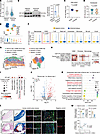 Figure 1
Decreased expression of KIF13B in VSMCs is associated with more severe human atherosclerosis. (A) scRNA-seq analysis of KIF13B mRNA levels in advanced plaques versus adjacent control tissues (n = 4 per group). (B) Western blotting for KIF13B protein expression in advanced plaques versus adjacent control tissues (n = 6 per group). (C and D) Violin plot of KIF13B mRNA (C) and KIF13B protein (D) expression in stable versus unstable human plaques from GEO GSE120521 (n = 4 per group) and ProteomeXchange PXD062283 (stable n = 19, unstable n = 38), respectively. (E) Uniform manifold approximation and projection (UMAP) visualization of scRNA-seq profiles in human atherosclerotic plaque tissues. Scatter plot shows cell-type scale of KIF13B mRNA expression among different cells in human atherosclerotic plaque tissues. (F) Violin plots of KIF13B mRNA expression among different cells in human atherosclerotic plaque tissues. (G) Left: Human plaque overlaid with capture spot cluster annotations. Right: UMAP of capture spots colored by human plaque-cluster affiliation. (H) Dot plot showing expression of cell-type markers in clusters. Dots are colored by minimum-maximum scaled gene expression level. (I) Dot plot showing KIF13B mRNA expression among the different cell clusters annotated in G. (J) Volcano plot of DEGs between fibrous cap 1 and cap 2 VSMCs, with upregulated genes in red, downregulated genes in blue, and nonsignificant genes in gray. (K) GO enrichment analysis of DEGs between fibrous cap 1 and cap 2 VSMCs. (L) Representative images of H&E staining, Masson’s staining, and immunofluorescence staining for α-SMA and KIF13B in human carotid artery plaque tissues (n = 6 per group). Scale bars: 1 mm and 50 μm; original magnification, ×2.09. (M) Quantitative analysis of KIF13B and α-SMA colocalization and collagen content and their correlation. The lesion areas are outlined by white dashed lines, and the white arrows indicate the positive staining of KIF13B in smooth muscle cells. Data are presented as the mean ± SEM and were analyzed by 2-tailed, unpaired Student’s t test (B–D, and M) or the Wilcoxon rank-sum test (A and F).
Given that the stability of atherosclerotic plaques is governed by the combined actions of multiple cell types, to identify the cell types in which KIF13B undergoes the most substantial changes, we conducted single-cell RNA-seq (scRNA-seq) on human atherosclerotic plaque tissues. The data indicated widespread expression of KIF13B in various types of atherosclerotic plaque cells (Figure 1E). Nevertheless, it was only in VSMCs that the expression of KIF13B mRNA significantly diminished in unstable plaques (Figure 1F). Hence, we hypothesized that KIF13B in VSMCs may play a crucial role in the stability of atherosclerotic plaques. Subsequently, we analyzed aortic samples containing fibrous caps of early plaques through spatial transcriptomics (25) to explore the expression of KIF13B in VSMCs and their spatial distribution, with the aim of clarifying the effect of KIF13B on the functions of VSMCs. Through in-depth analysis, we discovered that VSMCs were distributed within the fibrous cap, lipid core, and media of atherosclerotic plaques (Figure 1G). These cells not only expressed traditional contractile markers but also the marker genes related to the characteristics of fibroblasts and macrophages (Figure 1H). We found that the fibrous cap contained 2 types of VSMC-derived cells, which we named fibrous cap 1 VSMCs and fibrous cap 2 VSMCs (Figure 1G). KIF13B was highly expressed in fibrous cap 1 VSMCs expressing contraction markers (ACTA2, MYH11, TAGLN), fibroblast markers (FN1, LUM, COL1A2), and M2 macrophage markers (CD163, CD206), while it was expressed at very low level sin fibrous cap 2 VSMCs (Figure 1, H and I). Gene ontology (GO) enrichment analysis of differentially expressed genes (DEGs) between fibrous cap1 and fibrous cap 2 VSMCs showed that the upregulated genes in fibrous cap 1 VSMCs with elevated expression of KIF13B were primarily enriched in biological processes, including positive regulation of the immune system process, regulation of anatomical structure morphogenesis, and collagen fibril organization (Figure 1, J and K). Furthermore, histopathological analysis indicated that the content of KIF13B protein in VSMCs and collagen content were decreased in advanced atherosclerotic plaques compared with adjacent control (Figure 1, L and M). Notably, KIF13B expression in VSMCs showed a positive correlation with collagen content within human atherosclerotic plaques (Figure 1M). The above results collectively demonstrate that the expression of KIF13B in VSMCs was closely related to the stability of human atherosclerotic plaques.
Depletion of Kif13b in VSMCs exacerbates atherosclerosis in mice. To validate the effect of VSMC-derived KIF13B on atherosclerotic development and plaque stability, we generated VSMC-specific Kif13b-KO (Kif13bVSMCKO) mice (Figure 2A) and then confirmed the successful KO of the Kif13b gene in primary VSMCs isolated from mouse aortic tissues (Figure 2B). Next, Kif13bVSMCKO mice and littermate control Kif13bfl/fl mice were injected via the tail vein with adeno-associated virus type 8 (AAV8) expressing PCSK9-D377Y and then fed a Western diet (WD) or a chow diet (CD) for 20 weeks (Figure 2C). Intriguingly, we found that, compared with Kif13bfl/fl mice, Kif13bVSMCKO mice had identical body weights and plasma total cholesterol (TC) and triglyceride (TG) levels, but showed increased plasma levels of the proinflammatory cytokines IL-1β, IL-6, and TNF-α (Figure 2, D–F), indicating that depletion of VSMC-derived KIF13B promoted systemic inflammation. We then assessed the effect of VSMC Kif13b deficiency on adrenergic receptor alpha 1–mediated vasoconstriction using phenylephrine in aortic rings isolated from Kif13bVSMCKO and Kif13bfl/fl mice. We found that loss of KIF13B in VSMCs significantly reduced the maximal contraction response of the aortic ring to phenylephrine, independent of diet interventions (Figure 2, G–I). These findings suggest that KIF13B derived from VSMCs had a remarkably protective effect on vascular contractile function in a plasma lipid–independent manner.
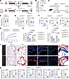 Figure 2
VSMC-specific Kif13b KO exacerbates atherosclerosis in mice. (A) Schematic of Kif13bVSMCKO mouse generation. (B) Kif13b mRNA levels in primary mouse VSMCs. (C) Schematic of the experimental timeline: 8-week-old male Kif13bfl/fl and Kif13bVSMCKO mice were injected with AAV8-PCSK9-D377Y and then fed a WD or CD for 20 weeks. (D–F) Body weight (D), plasma lipid levels (TC and TG) (E), and proinflammatory cytokines (IL-1β, IL-6, and TNF-α) (F) in mice fed a WD for 20 weeks (n = 8 per group). (G–I) Phenylephrine-induced vasoconstriction in the aortic ring from Kif13bfl/fl and Kif13bVSMCKO mice injected with AAV8-PCSK9-D377Y and then fed a WD or CD for 20 weeks (n = 4 per group). PE, phenylephrine. (J) Representative images of en face lesions in the whole aorta and representative images of H&E, Oil Red O (ORO), immunofluorescence staining (CD68, α-SMA, and TUNEL), and Masson’s trichrome staining of the aortic root. The necrotic core and total lesion area are outlined in black and white dashed lines, respectively; green arrowheads point to the fibrous cap (n = 8 per group). (K) Quantitative analyses of the data presented in J, including the percentage of lesion area in the entire aorta, the relative areas of the necrotic core, CD68+ area, α-SMA+ area, and TUNEL+ area within the aortic root sections, the lipid deposition area of the aortic root, the percentage of collagen content in the plaque area, and the plaque stability score. The lesion areas are outlined by white dashed lines, and the green arrowheads show the thickness of the fibrous cap in the atherosclerotic plaque. Data are presented as the mean ± SEM and were analyzed by 2-tailed, unpaired Student’s t test (B, D–F, and K) or 2-way ANOVA with Tukey post hoc test (H and I).
It is widely recognized that the attenuation of vascular contraction function can elicit atherogenesis (26). Therefore, we investigated the role of VSMC-derived KIF13B deficiency in atherosclerosis development. The results of our pathological analysis of atherosclerosis indicated an increase in atherosclerotic lesion areas in both the entire aorta and the aortic root, with larger necrotic core areas, enhanced CD68+ macrophage infiltration, and more TUNEL+ apoptotic cells in Kif13bVSMCKO mice; however, no difference in the α–smooth muscle actin (α-SMA) content was observed between the 2 groups (Figure 2, J and K). Moreover, Masson’s trichrome staining showed a significant reduction in collagen content in the fibrous cap of Kif13bVSMCKO mice (Figure 2, J and K), leading to plaque instability.
Loss of KIF13B in VSMC does not affect intestinal morphology or lipid absorption. Since VSMCs, as the principal contractile elements in blood vessels and hollow organs, such as the intestine and urinary bladder, have essential physiological functions in these systems, and TAGLN-Cre has been reported to delete floxed genes in the intestine and bladder (27, 28), we performed a histological examination of multiple intestinal segments. Our analysis revealed no significant morphological differences in the duodenum, jejunum, ileum, or large intestine between Kif13bfl/fl and Kif13bVSMCKO mice under either hypercholesterolemic or normocholesterolemic conditions (Figure 3A). To further evaluate whether VSMC-specific deletion of Kif13b affects intestinal lipid absorption, we measured TG levels in plasma from Kif13bfl/fl and Kif13bVSMCKO mice after an overnight fast followed by oral olive oil gavage. Results from this lipid tolerance test demonstrated that Kif13b deficiency in VSMCs did not alter circulating TG concentrations under either dietary condition (Figure 3, B and C), indicating that Kif13bVSMCKO mice had normal intestinal morphology and intact intestinal function.
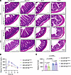 Figure 3
VSMC-specific Kif13b deletion does not affect intestinal morphology or lipid absorption. Eight-week-old male Kif13bfl/fl and Kif13bVSMCKO mice were injected with AAV8-PCSK9-D377Y for 2 weeks and then fed a WD (n = 7 per group) or CD (n = 6 per group) for 20 weeks. (A) Representative images of H&E staining of intestinal segments (duodenum, jejunum, ileum, colon) show no morphological differences between the 2 genotypes. Scale bars: 200 μm. (B) Oral lipid tolerance test. Mice were fasted for 4 hours, followed by oral gavage of olive oil (10 μL/g), and then plasma TG levels were measured at each subsequent time point. (C) Quantitative analyses of the AUC in B. Data are presented as the mean ± SEM and were analyzed by 2-way ANOVA with Tukey post hoc test (C).
Depletion of KIF13B in VSMC does not induce maladaptive bladder remodeling resembling megabladder. Previous studies have also reported that the decline in VSMC contractile function caused by VSMC phenotypic remodeling can lead to maladaptive bladder remodeling resembling megabladder in humans and mice (27–29). To determine whether VSMC-restricted KIF13B deficiency has any effects on bladder function, we examined bladder morphology using MRI of Kif13bfl/fl and Kif13bVSMCKO mice under CD and WD conditions. Strikingly, we found that VSMC-specific Kif13b deficiency had urinary bladder volume identical to that of Kif13bfl/fl mice fed a CD and WD (Figure 4, A and B). Further characterization of the grossly dilated bladder revealed similarities in bladder wall thickness and collagen deposition in both Kif13bfl/fl and Kif13bVSMCKO mice (Figure 4, C–F). Collectively, these data show that KIF13B in VSMCs had no influence on maintaining the structural integrity of the bladder under hypercholesterolemic and normocholesterolemic conditions.
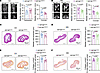 Figure 4
Kif13b deficiency in VSMCs does not cause bladder remodeling. Eight-week-old male Kif13bfl/fl and Kif13bVSMCKO mice were injected with AAV8-PCSK9-D377Y for 2 weeks and then fed a WD or CD for 20 weeks (n = 7 per group). (A and B) Representative images and quantitative analysis of bladder volume shown by MRI of Kif13bfl/fl and Kif13bVSMCKO mice. (C and D) Representative images of H&E staining and quantitative analysis of bladder wall thickness in Kif13bfl/fl and Kif13bVSMCKO mice. Scale bars: 800 μm. (E and F) Representative images of Picrosirius red staining and quantitative analysis of collagen deposition in Kif13bfl/fl and Kif13bVSMCKO mice. Scale bars: 800 μm. Data are presented as the mean ± SEM and were analyzed by 2-tailed, unpaired Student’s t test (A–F).
Inhibiting KIF13B promotes proinflammatory fibroblast–like VSMC transition. To elucidate the characteristics of atherosclerotic plaques in the setting of KIF13B deficiency, we conducted in vivo scRNA-seq on Ldlr–/– and Ldlr–/– Kif13b–/– mice after 20 weeks on a WD, which revealed that KIF13B inactivation markedly decreased collagen content in plaques with thinner fibrous caps (Supplemental Figure 2). Furthermore, our scRNA-seq data indicated that VSMC-derived fibroblast-like cells (VSMC-Fibs) were the predominant cell type in advanced-stage plaques (Figure 5A). Leiden clustering analysis further delineated that VSMCs could switch into 5 subtypes of VSMC-Fibs in advanced stages of mouse atherosclerosis (Figure 5B and Supplemental Figure 3A). Subsequently, we reconstructed a pseudotime trajectory aligned with VSMCs and VSMC-Fibs to model their phenotypic transition. These cells were categorized into 9 distinct states (Figure 5C). Among them, VSMC-Fib1 mapped to state 9, which was strongly associated with extracellular matrix stability, collagen fibril secretion, and vascular structural homeostasis, collectively conferring plaque-stabilizing characteristics. In contrast, VSMC-Fib4 localized to state 7 and exhibited a heightened proinflammatory response, indicative of a vulnerability-prone plaque phenotype (Figure 5, C–F). Gene set enrichment analysis (GSEA) further demonstrated that pathways related to apoptosis and inflammatory responses were significantly activated in Kif13b-deficient VSMC-Fibs, whereas extracellular matrix assembly and collagen fibril organization were markedly suppressed (Figure 5G). These findings demonstrate the observed vulnerable plaque phenotype in Kif13bVSMCKO mice. Consistent with this model, Kif13b deletion led to a marked reduction in VSMC-Fib1 cell numbers, alongside a notable increase in VSMC-Fib4 cells (Figures 5, H and I). To further validate these findings, we examined the VSMC-Fib1 cluster in Ldlr–/– Kif13b–/– mice and observed significant downregulation of key stability-related genes (e.g., Col2a1, Col3a1, Vnt), along with a marked upregulation of the extracellular matrix-degrading protease Mmp2 when compared with Ldlr–/– controls (Figure 5J). Conversely, the VSMC-Fib4 cluster from Ldlr–/– Kif13b–/– mice showed significantly elevated expression of proinflammatory mediators, including Ccl2, Il1b, Il6, Il18, and Trap1 (Figure 5K).
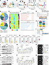 Figure 5
KIF13B directs VSMC transition to a matrix-stabilizing phenotype. (A) UMAP visualization of aorta scRNA-seq samples in Ldlr–/– and Ldlr–/– Kif13b–/– mice fed a WD for 20 weeks (mix n = 5 into 1 sample). (B) UMAP visualization of VSMC-Fib subclustering analysis derived from A. (C) Monocle analysis derived from A, showing the ordering of VSMC-Fibs along pseudotime marked by the VSMC-Fibs state. (D) Distribution of VSMC-Fibs from different cell states based on the data in C. (E) Heatmap of different blocks of DEGs along the pseudotime trajectory. (F) Selected top GO terms related to the corresponding DEGs in E. (G) GSEA of VSMC-Fib aorta scRNA-seq data for Ldlr–/– and Ldlr–/– Kif13b–/– mice, derived from A. (H) Stacked bar plot showing the proportion of aortic cells in Ldlr–/– and Ldlr–/– Kif13b–/– mice from scRNA-seq data derived from A. (I) Bar plot showing the difference in cell proportions (log2 fold change [FC]) between Ldlr–/– and Ldlr–/– Kif13b–/– mice for aorta scRNA-seq data derived from A. (J) Expression of Col2a1, Col3a1, Vnt, and Mmp2 mRNA in VSMC-Fib1 cells from the scRNA-seq data on aortic cells from Ldlr–/– and Ldlr–/– Kif13b–/– mice from A. (K) Expression of Ccl2, Il1b, Il6, Il18, and Trap1 mRN in VSMC-Fib1 cells from the scRNA-seq data of aortic cells from Ldlr–/– and Ldlr–/– Kif13b–/– mice from A. (L) Representative Western blot images and quantitative analysis of KIF13B, TAGLN, and fibroblast-specific protein 1 (FSP1) protein expression in HASMCs transfected with scramble or siKIF13B and subsequently treated with oxidized low-density lipoprotein (oxLDL) (50 μg/mL) for 48 hours (n = 3 independent biological replicates). (M) ELISA of supernatant COL1α1, COL2α1, COL3α1, CCL2, IL-1β, and IL-6 in HASMCs transfected with scramble or siKIF13B and subsequently treated with oxLDL (50 μg/mL) for 48 hours (n = 3 independent biological replicates). (N) HASMCs were transfected with scramble or siKIF13B and subsequently treated with oxLDL (50 μg/mL) for 48 hours, mixed with ice-cold collagen at a 1:4 ratio, and allowed to polymerize for 24 hours. Collagen gel size was subsequently measured (n = 5 independent biological replicates). (O) Representative Western blots and quantitative analysis of KIF13B, TAGLN, and FSP1 protein expression in HASMCs infected with lentivirus (LV) expressing GFP-HA or KIF13B-HA and subsequently treated with oxLDL (50 μg/mL) for 48 hours (n = 3 independent biological replicates). (P) ELISA of supernatant COL1α1, COL2α1, COL3α1, CCL2, IL-1β, and IL-6 content in HASMCs infected with LV expressing GFP-HA or KIF13B-HA and subsequently treated with oxLDL (50 μg/mL) for 48 hours (n = 3 independent biological replicates). (Q) HASMCs were infected with LV expressing GFP-HA or KIF13B-HA and subsequently treated with oxLDL (50 μg/mL) for 48 hours, mixed with ice-cold collagen at a 1:4 ratio, and allowed to polymerize for 24 hours. Collagen gel size was subsequently measured (n = 6 independent biological replicates). Data are presented as the mean ± SEM and were analyzed by Wilcoxon rank-sum test (J and K), 2-tailed, unpaired Student’s t test (L, N, O, and Q), or multiple unpaired t tests (M and P).
To directly assess the functional effect of KIF13B on VSMC phenotypic switching, we conducted complementary gain- and loss-of-function studies in primary human aortic smooth muscle cells (HASMCs). siRNA-mediated KIF13B knockdown promoted the phenotypic switch of HASMCs toward proinflammatory fibroblast-like cells (VSMC-Fibs), whereas KIF13B overexpression drove the transition toward a collagen-secreting, matrix-stabilizing phenotype (Figure 5, L and O). Consistent with these morphological changes, KIF13B knockdown significantly reduced collagen secretion and cell contractility, while enhancing the expression and secretion of inflammatory factors (Figure 5, M and N, and Supplemental Figure 3B). Conversely, KIF13B overexpression elevated collagen production and cell contractility (Figure 5, P and Q, and Supplemental Figure 3C). Taken together, our findings delineate an essential role of KIF13B in atherosclerotic plaque stabilization by directing VSMCs toward a matrix-preserving VSMC-Fib phenotype.
KLF4 mediates KIF13B-dependent proinflammatory VSMC reprogramming. To explore the detailed molecular mechanism by which KIF13B governs the phenotypic transition of VSMCs, we performed a single-cell regulatory network inference and clustering (SCENIC) analysis of the DEGs in VSMC-Fibs from Ldlr–/– and Ldlr–/– Kif13b–/– mice. The findings revealed that depletion of Kif13b in VSMCs significantly enhanced the transcriptional activity of KLF4, a key transcription factor for phenotypic switching in atherosclerosis and cancer (Figure 6, A and B). Western blot analysis demonstrated that knockdown of KIF13B in HASMCs upregulated the expression of KLF4, whereas overexpression of KIF13B-suppressed KLF4 expression (Figure 6C). Immunofluorescence staining further validated our observations, showing that KIF13B exerted an inverse regulatory effect on the levels of KLF4 in HASMCs (Figure 6, D and E).
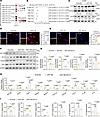 Figure 6
KLF4 acts downstream of KIF13B to drive proinflammatory reprogramming in VSMCs. (A) SCENIC analysis identified KLF4 as a key transcription factor activated in KIF13B-deficient VSMC-Fibs. (B) Visualization of forest plots of KLF4 transcriptional activity proportions in the VSMC-Fib subcluster of aorta scRNA-seq samples from Ldlr–/– and Ldlr–/– Kif13b–/– mice fed a WD for 12 weeks. (C) Top: Representative Western blots and quantitative analysis of KLF4 protein expression in HASMCs transfected with scramble or siKIF13B and subsequently treated with oxLDL (50 μg/mL) for 48 hours (n = 3 independent biological replicate). Bottom: Representative Western blots and quantitative analysis of KLF4 protein expression in HASMCs infected with LV expressing GFP-HA or KIF13B-HA and subsequently treated with oxLDL (50 μg/mL) for 48 hours (n = 3 independent biological replicates). (D and E) Representative images and quantitative analyses of immunofluorescence staining for KLF4 and distribution in HASMCs transfected with scramble or siKIF13B (D) or infected with LV expressing GFP-HA or KIF13B-HA (E) for 48 hours (n = 10 independent biological replicates). Scale bars: 10 µm, original magnification, ×2.06. (F and G) Representative Western blots and quantitative analysis of KIF13B, KLF4, TAGLN, and FSP1 protein expression in HASMCs transfected with scramble, siKIF13B, or siKIF13B + siKLF4 and subsequently treated with oxLDL (50 μg/mL) for 48 hours (n = 3 independent biological replicates). (H) Quantitative PCR analysis of VSMC-Fib phenotypic switching (FSP1, TAGLN, MYH11), collagen production (COL1A1, COL2A1, COL3A1), and inflammatory factor expression (CCL2, IL1B, IL6) in HASMCs transfected with scramble, siKIF13B, or siKIF13B +s iKLF4 and subsequently treated with oxLDL (50 μg/mL) for 24 hours (n = 3 independent biological replicates). (I) ELISA detected supernatant COL1α1, COL2α1, COL3α1, CCL2, IL-1β, and IL-6 levels in HASMCs transfected with scramble or siKIF13B or siKIF13B + siKLF4 and subsequently treated with oxLDL (50 μg/mL) for 48 hours (n = 3 independent biological replicates). Data are presented as the mean ± SEM and were analyzed by 2-tailed, unpaired Student’s t test (C–E) or 2-way ANOVA with Tukey post hoc test (G–I).
Next, to determine whether KIF13B regulates the VSMC phenotypic transition through KLF4, we used a dual-knockdown strategy in HASMCs. Silencing KIF13B alone significantly reduced collagen production and increased the secretion of inflammatory factors, which were rescued by concomitant knockdown of KLF4 (Figure 6, F–I), demonstrating that KLF4 is a crucial downstream mediator of KIF13B signaling in VSMC phenotypic switching.
KIF13B facilitates KCTD10-mediated ubiquitination and degradation of KLF4. Given that KIF13B does not directly regulate KLF4 transcription (Supplemental Figure 4A), we hypothesized that it may modulate KLF4 at the posttranslational level. To test this, we performed a cycloheximide (CHX) chase assay with or without the proteasome inhibitor MG132. Our results demonstrated that KIF13B knockdown stabilized the KLF4 protein by attenuating its proteasomal degradation (Figure 7A), which was further supported by co-IP experiments showing a reduction in KLF4 ubiquitination upon KIF13B knockdown (Figure 7B).
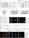 Figure 7
KIF13B drives KLF4 degradation in atherosclerosis through KCTD10-mediated ubiquitination. (A) Representative Western blots and quantitative analysis of KLF4 in HASMCs transfected with scramble or siKIF13B and treated with PBS or 10 μM MG132 in the presence of 50 μg/mL CHX for the indicated durations (n = 3 independent biological replicates). (B) Co-IP analysis and quantification of the ubiquitination of KLF4 (n = 3 independent biological replicates). (C) Venn diagram illustrates the intersection of commonly downregulated genes across DEGs in VSMCs from Ldlr–/– Kif13b–/– versus Ldlr–/– mice fed a WD for 12 weeks, DEGs from unstable versus stable atherosclerotic plaques (GSE120521), and E3 ligase. DEGs with an FDR of less than 0.05 and a log2 FC of less than 0 were included. (D) Left: Representative Western blots of KCTD10 and SKP1 protein expression in HASMCs transfected with scramble or siKIF13B and subsequently treated with oxLDL (50 μg/mL) for 48 hours (n = 3 independent biological replicates). Right: Representative Western blots of KCTD10 and SKP1 protein expression in HASMCs infected with LV expressing GFP-HA or KIF13B-HA and subsequently treated with oxLDL (50 μg/mL) for 48 hours (n = 3 independent biological replicates). (E) Quantitative analysis of KCTD10 and SKP1 protein expression shown in D. (F) Immunofluorescence staining and quantification of α-SMA and KCTD10 in the aortic root of Kif13bfl/fl and Kif13bVSMCKO mice injected with AAV8-PCSK9-D377Y and then fed a WD for 20 weeks (n = 8 per group). (G) Violin plots of KCTD10 mRNA expression in different cells in human atherosclerotic plaque tissues. (H) Immunofluorescence staining and quantification of α-SMA and KCTD10 expression in human carotid artery plaque tissues (n = 6 per group). White arrows indicate the content of KCTD10 in atherosclerotic plaque. Scale bars: 50 μm and 100 μm. Data are presented as the mean ± SEM and were analyzed by 2-way ANOVA with Tukey’s post hoc test (A), 2-tailed, unpaired Student’s t test (B, F, and H), or Wilcoxon rank-sum test (G).
We next sought to identify the mechanism by which KIF13B regulates KLF4 ubiquitination. Integrated transcriptomics analysis of VSMCs from Ldlr–/– Kif13b–/– versus Ldlr–/– mouse atherosclerotic tissues and of human unstable versus stable plaques revealed 2 downregulated ubiquitin ligase components: potassium channel tetramerization domain containing 10 (KCTD10) and S-phase kinase–associated protein 1 (SKP1) (Figure 7C). The Western blots showed that knockdown or overexpression of KIF13B specifically altered the expression of KCTD10, but not SKP1, in HASMCs (Figure 7, D and E). KCTD10 acts as a substrate receptor for really interesting new gene-type (RING-type) ubiquitin ligase complexes, and it has been implicated in cardiovascular disease (CVD) (30); however, its role in VSMCs and atherosclerosis remains unknown. We found that Kif13b deficiency also markedly reduced KCTD10 expression in VSMCs within mouse atherosclerotic plaques (Figure 7F). Analysis of human plaques further confirmed significantly reduced KCTD10 expression levels in VSMCs of advanced atherosclerotic plaques (Figure 7, G and H).
To determine whether KIF13B regulates KLF4 stability through KCTD10, we first predicted a direct KCTD10-KLF4 interaction via molecular docking (Figure 8A). Co-IP assays then confirmed that KCTD10 binds to KLF4 and promotes its ubiquitination, thereby reducing KLF4 protein levels (Figure 8, B and C). Furthermore, the stabilization of KLF4 resulting from KIF13B knockdown was effectively reversed by KCTD10 overexpression (Figure 8D). Both Western blot and immunofluorescence analyses confirmed an inverse correlation between KCTD10 and KLF4 protein levels in HASMCs (Figure 8, E and F). Finally, the proinflammatory and collagen-suppressive phenotype induced by KIF13B knockdown was significantly rescued by KCTD10 overexpression (Figure 8G and Supplemental Figure 4B).
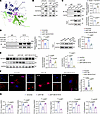 Figure 8
KIF13B promotes KLF4 ubiquitination and degradation via KCTD10. (A) Molecular docking prediction of KCTD10 and KLF4 interaction. (B) Quantitative analysis of co-IP assays confirmed the interaction between KCTD10 and KLF4 in HASMCs (n = 3 independent biological replicates). (C) Quantitative analysis of co-IP assays of the ubiquitination of KLF4 in HASMCs transfected with KCTD10 plasmid for 48 hours (n = 3 independent biological replicates). (D) Quantitative analysis of co-IP assays of the ubiquitination of KLF4 in HASMCs transfected with scramble, siKIF13B, or siKIF13B + KCTD10 for 48 hours (n = 3 independent biological replicates). (E) Representative Western blots and quantitative analysis of KLF4 in HASMCs transfected with scramble, siKIF13B, or siKIF13B + KCTD10 and subsequently treated with oxLDL (50 μg/mL) for 48 hours (n = 3 independent biological replicates). (F) Representative images and quantitative analyses of immunofluorescence staining of KLF4 expression and distribution in HASMCs transfected with scramble, siKIF13B, or siKIF13B + KCTD10 for 48 hours (n = 10 independent biological replicates). Scale bars: 10 µm; original magnification, ×2.06. (G) ELISA of supernatant COL1α1, COL2α1, COL3α1, CCL2, IL-1β, and IL-6 levels in HASMCs transfected with scramble, siKIF13B, or siKIF13B + KCTD10 for 48 hours (n = 3 independent biological replicates). Data are presented as the mean ± SEM and were analyzed by 2-tailed, unpaired Student’s t test (C) or 1-way ANOVA with Tukey post hoc test (D–G).
Pharmacological inhibition of KLF4 with Kenpaullone alleviates atherosclerosis in mice. Finally, to explore a potential therapeutic strategy for atherosclerosis stemming from VSMC-specific Kif13b deficiency, we screened a small-molecule compound library and identified Kenpaullone as a KLF4 inhibitor (Figure 9A). Our reporter assays showed that Kenpaullone treatment significantly suppressed KLF4 promoter activity to reduce the mRNA expression level of KLF4 (Supplemental Figure 5), suggesting that its downregulatory effect on KLF4 occurred at the transcriptional level. In HASMCs, Kenpaullone treatment effectively counteracted the effects of KIF13B knockdown by significantly reducing KLF4 protein levels and then enhancing collagen production and suppressing the secretion of inflammatory factors (Figure 9, B–D, and Supplemental Figure 6).
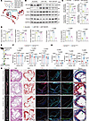 Figure 9
Pharmacological inhibition of KLF4 effectively ameliorates atherosclerosis in mice. (A) Schematic representation of the structure of Kenpaullone and its working model. (B) Representative Western blots of KIF13B, KLF4, FSP1, and TAGLN expression in HASMCs transfected with scramble, siKIF13B, or siKIF13B + Kenpaullone (10 μM) and subsequently treated with oxLDL (50 μg/mL) for 48 hours. (C) Quantitative analyses of the Western blotting in B (n = 3 independent biological replicates). (D) ELISA of supernatant COL1α1, COL2α1, COL3α1, CCL2, IL-1β, and IL-6 levels in HASMCs transfected with scramble, siKIF13B, or siKIF13B + Kenpaullone (10 μM) for 48 hours (n = 3 independent biological replicates). (E) Schematic of the experimental timeline: 8-week-old male Kif13bfl/fl and Kif13bVSMCKO mice received a single tail vein injection of AAV8-PCSK9-D377Y, followed 2 weeks later by feeding with a Western diet (WD) for another 20 weeks to induce advanced atherosclerosis. The mice received oral administration of Kenpaullone (1 mg/kg/d) for the last 12 weeks (n = 6 per group). (F) Body weights and plasma lipid levels (TC and TG) of the mice in E. (G) Proinflammatory cytokines (IL-1β, IL-6, TNF-α) of the mice in E. (H) Representative images of the en face lesions in the whole aorta and representative images of H&E staining, ORO staining, immunofluorescence staining (CD68, α-SMA, and TUNEL), and Masson’s trichrome staining of the aortic root. The necrotic core and total lesion area are outlined with black and white dashed lines, respectively; green arrowheads indicate the fibrous cap (n = 6 per group). Scale bars: 5 mm, 100 μm, and 300 μm. Data are presented as the mean ± SEM and were analyzed by 1-way ANOVA with Tukey post hoc test (C and D) or 2-way ANOVA with Tukey’s post hoc tests (F and G). KPL, Kenpaullone.
We next evaluated the in vivo therapeutic efficacy of Kenpaullone. Advanced atherosclerosis was induced in 8-week-old male Kif13bVSMCKO and Kif13bfl/fl mice injected with AAV8-PCSK9-D377Y via the tail vein and then fed a WD for 20 weeks, followed by oral administration of Kenpaullone (1 mg/kg/d) for the last 12 weeks (Figure 9E). Kenpaullone treatment had no obvious influence on body weight, plasma lipid profiles, or markers of liver toxicity, such as ALT and AST (Figure 9F and Supplemental Figure 7, A and B). However, it significantly reduced systemic inflammation, as evidenced by decreased plasma levels of IL-1β, IL-6, and TNF-α (Figure 9G). Histological analyses demonstrated that Kenpaullone attenuated the atherosclerotic lesion burden in both the aorta and aortic root, concomitant with a reduction in the necrotic core area, CD68+ macrophage infiltration, and TUNEL+ apoptotic cells, alongside an increase in collagen content (Figure 9H and Supplemental Figure 7C).
Collectively, these findings indicate that Kenpaullone impeded atherosclerosis progression by inhibiting KLF4 and the associated proinflammatory VSMC reprogramming without any detectable adverse effects, highlighting its potential as a promising therapeutic strategy.


Copyright © 2026 American Society for Clinical Investigation
ISSN: 0021-9738 (print), 1558-8238 (online)