
Advertisement
Research ArticleAgingNeuroscience
Open Access |  10.1172/JCI190646
Neuromuscular junction failure in sarcopenia is linked to NaV1.4 loss and reversed by ClC-1 inhibition
W. David Arnold,1,2 Jeanette Jeppesen Morgen,3 Pernille Bogetofte Thomasen,3 Martin Broch-Lips,3 Leatha A. Clark,4,5 Thomas Groennebaek,3 Martin Skov,3 Jeppe Blichfeldt Winther,3 Abdullah Ramadan,6,7 Philippa A. Rust,6 Jessica H. Myers,8 Fereshteh B. Darvishi,1,2 Anna R. Dashtmian,1,2 Lauren A. Fish,9 Deepti Chugh,10 Jane Bold,3 Jorge Quiroz,3 John Hutchison,3 Hiroshi Nishimune,1,2 Ross A. Jones,6 Xueyong Wang,10 Justin R. Fallon,9 Thomas H. Gillingwater,6 Mark M. Rich,8 Thomas Holm Pedersen,3,11 and Brian C. Clark4,5
1NextGen Precision Health and
2Department of Physical Medicine and Rehabilitation, University of Missouri, Columbia, Missouri, USA.
3NMD Pharma A/S, Aarhus, Denmark.
4Ohio Musculoskeletal and Neurological Institute and
5Department of Biomedical Sciences, Ohio University Heritage College of Osteopathic Medicine, Athens, Ohio, USA.
6Edinburgh Medical School, Biomedical Sciences, College of Medicine and Veterinary Medicine, University of Edinburgh, Edinburgh, United Kingdom.
7College of Sciences and Health Professions, King Saud bin Abdulaziz for Health Sciences, Jeddah, Saudi Arabia. .
8Department of Neuroscience, Cell Biology and Physiology, Wright State University, Dayton, Ohio, USA.
9Department of Neuroscience and Carney Institute for Brain Science, Brown University, Providence, Rhode Island, USA.
10Indian Institute of Technology, Kanpur, India
11Department of Biomedicine, Aarhus University, Aarhus, Denmark.
Address correspondence to: W. David Arnold, NextGen Precision Health, Department of Physical Medicine and Rehabilitation, University of Missouri, 1030 Hitt Street, Columbia, Missouri 652011, USA. Email: wdavidarnold@health.missouri.edu. Or to: Brian C. Clark, Ohio Musculoskeletal and Neurological Institute, Department of Biomedical Sciences, Ohio University Heritage College of Osteopathic Medicine, 250 Irvine Hall, Athens, Ohio 45701, USA. Email: clarkb2@ohio.edu.
Find articles by Arnold, W. in: PubMed | Google Scholar
1NextGen Precision Health and
2Department of Physical Medicine and Rehabilitation, University of Missouri, Columbia, Missouri, USA.
3NMD Pharma A/S, Aarhus, Denmark.
4Ohio Musculoskeletal and Neurological Institute and
5Department of Biomedical Sciences, Ohio University Heritage College of Osteopathic Medicine, Athens, Ohio, USA.
6Edinburgh Medical School, Biomedical Sciences, College of Medicine and Veterinary Medicine, University of Edinburgh, Edinburgh, United Kingdom.
7College of Sciences and Health Professions, King Saud bin Abdulaziz for Health Sciences, Jeddah, Saudi Arabia. .
8Department of Neuroscience, Cell Biology and Physiology, Wright State University, Dayton, Ohio, USA.
9Department of Neuroscience and Carney Institute for Brain Science, Brown University, Providence, Rhode Island, USA.
10Indian Institute of Technology, Kanpur, India
11Department of Biomedicine, Aarhus University, Aarhus, Denmark.
Address correspondence to: W. David Arnold, NextGen Precision Health, Department of Physical Medicine and Rehabilitation, University of Missouri, 1030 Hitt Street, Columbia, Missouri 652011, USA. Email: wdavidarnold@health.missouri.edu. Or to: Brian C. Clark, Ohio Musculoskeletal and Neurological Institute, Department of Biomedical Sciences, Ohio University Heritage College of Osteopathic Medicine, 250 Irvine Hall, Athens, Ohio 45701, USA. Email: clarkb2@ohio.edu.
Find articles by Jeppesen Morgen, J. in: PubMed | Google Scholar
1NextGen Precision Health and
2Department of Physical Medicine and Rehabilitation, University of Missouri, Columbia, Missouri, USA.
3NMD Pharma A/S, Aarhus, Denmark.
4Ohio Musculoskeletal and Neurological Institute and
5Department of Biomedical Sciences, Ohio University Heritage College of Osteopathic Medicine, Athens, Ohio, USA.
6Edinburgh Medical School, Biomedical Sciences, College of Medicine and Veterinary Medicine, University of Edinburgh, Edinburgh, United Kingdom.
7College of Sciences and Health Professions, King Saud bin Abdulaziz for Health Sciences, Jeddah, Saudi Arabia. .
8Department of Neuroscience, Cell Biology and Physiology, Wright State University, Dayton, Ohio, USA.
9Department of Neuroscience and Carney Institute for Brain Science, Brown University, Providence, Rhode Island, USA.
10Indian Institute of Technology, Kanpur, India
11Department of Biomedicine, Aarhus University, Aarhus, Denmark.
Address correspondence to: W. David Arnold, NextGen Precision Health, Department of Physical Medicine and Rehabilitation, University of Missouri, 1030 Hitt Street, Columbia, Missouri 652011, USA. Email: wdavidarnold@health.missouri.edu. Or to: Brian C. Clark, Ohio Musculoskeletal and Neurological Institute, Department of Biomedical Sciences, Ohio University Heritage College of Osteopathic Medicine, 250 Irvine Hall, Athens, Ohio 45701, USA. Email: clarkb2@ohio.edu.
Find articles by Thomasen, P. in: PubMed | Google Scholar
1NextGen Precision Health and
2Department of Physical Medicine and Rehabilitation, University of Missouri, Columbia, Missouri, USA.
3NMD Pharma A/S, Aarhus, Denmark.
4Ohio Musculoskeletal and Neurological Institute and
5Department of Biomedical Sciences, Ohio University Heritage College of Osteopathic Medicine, Athens, Ohio, USA.
6Edinburgh Medical School, Biomedical Sciences, College of Medicine and Veterinary Medicine, University of Edinburgh, Edinburgh, United Kingdom.
7College of Sciences and Health Professions, King Saud bin Abdulaziz for Health Sciences, Jeddah, Saudi Arabia. .
8Department of Neuroscience, Cell Biology and Physiology, Wright State University, Dayton, Ohio, USA.
9Department of Neuroscience and Carney Institute for Brain Science, Brown University, Providence, Rhode Island, USA.
10Indian Institute of Technology, Kanpur, India
11Department of Biomedicine, Aarhus University, Aarhus, Denmark.
Address correspondence to: W. David Arnold, NextGen Precision Health, Department of Physical Medicine and Rehabilitation, University of Missouri, 1030 Hitt Street, Columbia, Missouri 652011, USA. Email: wdavidarnold@health.missouri.edu. Or to: Brian C. Clark, Ohio Musculoskeletal and Neurological Institute, Department of Biomedical Sciences, Ohio University Heritage College of Osteopathic Medicine, 250 Irvine Hall, Athens, Ohio 45701, USA. Email: clarkb2@ohio.edu.
Find articles by Broch-Lips, M. in: PubMed | Google Scholar
1NextGen Precision Health and
2Department of Physical Medicine and Rehabilitation, University of Missouri, Columbia, Missouri, USA.
3NMD Pharma A/S, Aarhus, Denmark.
4Ohio Musculoskeletal and Neurological Institute and
5Department of Biomedical Sciences, Ohio University Heritage College of Osteopathic Medicine, Athens, Ohio, USA.
6Edinburgh Medical School, Biomedical Sciences, College of Medicine and Veterinary Medicine, University of Edinburgh, Edinburgh, United Kingdom.
7College of Sciences and Health Professions, King Saud bin Abdulaziz for Health Sciences, Jeddah, Saudi Arabia. .
8Department of Neuroscience, Cell Biology and Physiology, Wright State University, Dayton, Ohio, USA.
9Department of Neuroscience and Carney Institute for Brain Science, Brown University, Providence, Rhode Island, USA.
10Indian Institute of Technology, Kanpur, India
11Department of Biomedicine, Aarhus University, Aarhus, Denmark.
Address correspondence to: W. David Arnold, NextGen Precision Health, Department of Physical Medicine and Rehabilitation, University of Missouri, 1030 Hitt Street, Columbia, Missouri 652011, USA. Email: wdavidarnold@health.missouri.edu. Or to: Brian C. Clark, Ohio Musculoskeletal and Neurological Institute, Department of Biomedical Sciences, Ohio University Heritage College of Osteopathic Medicine, 250 Irvine Hall, Athens, Ohio 45701, USA. Email: clarkb2@ohio.edu.
Find articles by Clark, L. in: PubMed | Google Scholar
1NextGen Precision Health and
2Department of Physical Medicine and Rehabilitation, University of Missouri, Columbia, Missouri, USA.
3NMD Pharma A/S, Aarhus, Denmark.
4Ohio Musculoskeletal and Neurological Institute and
5Department of Biomedical Sciences, Ohio University Heritage College of Osteopathic Medicine, Athens, Ohio, USA.
6Edinburgh Medical School, Biomedical Sciences, College of Medicine and Veterinary Medicine, University of Edinburgh, Edinburgh, United Kingdom.
7College of Sciences and Health Professions, King Saud bin Abdulaziz for Health Sciences, Jeddah, Saudi Arabia. .
8Department of Neuroscience, Cell Biology and Physiology, Wright State University, Dayton, Ohio, USA.
9Department of Neuroscience and Carney Institute for Brain Science, Brown University, Providence, Rhode Island, USA.
10Indian Institute of Technology, Kanpur, India
11Department of Biomedicine, Aarhus University, Aarhus, Denmark.
Address correspondence to: W. David Arnold, NextGen Precision Health, Department of Physical Medicine and Rehabilitation, University of Missouri, 1030 Hitt Street, Columbia, Missouri 652011, USA. Email: wdavidarnold@health.missouri.edu. Or to: Brian C. Clark, Ohio Musculoskeletal and Neurological Institute, Department of Biomedical Sciences, Ohio University Heritage College of Osteopathic Medicine, 250 Irvine Hall, Athens, Ohio 45701, USA. Email: clarkb2@ohio.edu.
Find articles by Groennebaek, T. in: PubMed | Google Scholar
1NextGen Precision Health and
2Department of Physical Medicine and Rehabilitation, University of Missouri, Columbia, Missouri, USA.
3NMD Pharma A/S, Aarhus, Denmark.
4Ohio Musculoskeletal and Neurological Institute and
5Department of Biomedical Sciences, Ohio University Heritage College of Osteopathic Medicine, Athens, Ohio, USA.
6Edinburgh Medical School, Biomedical Sciences, College of Medicine and Veterinary Medicine, University of Edinburgh, Edinburgh, United Kingdom.
7College of Sciences and Health Professions, King Saud bin Abdulaziz for Health Sciences, Jeddah, Saudi Arabia. .
8Department of Neuroscience, Cell Biology and Physiology, Wright State University, Dayton, Ohio, USA.
9Department of Neuroscience and Carney Institute for Brain Science, Brown University, Providence, Rhode Island, USA.
10Indian Institute of Technology, Kanpur, India
11Department of Biomedicine, Aarhus University, Aarhus, Denmark.
Address correspondence to: W. David Arnold, NextGen Precision Health, Department of Physical Medicine and Rehabilitation, University of Missouri, 1030 Hitt Street, Columbia, Missouri 652011, USA. Email: wdavidarnold@health.missouri.edu. Or to: Brian C. Clark, Ohio Musculoskeletal and Neurological Institute, Department of Biomedical Sciences, Ohio University Heritage College of Osteopathic Medicine, 250 Irvine Hall, Athens, Ohio 45701, USA. Email: clarkb2@ohio.edu.
Find articles by Skov, M. in: PubMed | Google Scholar
1NextGen Precision Health and
2Department of Physical Medicine and Rehabilitation, University of Missouri, Columbia, Missouri, USA.
3NMD Pharma A/S, Aarhus, Denmark.
4Ohio Musculoskeletal and Neurological Institute and
5Department of Biomedical Sciences, Ohio University Heritage College of Osteopathic Medicine, Athens, Ohio, USA.
6Edinburgh Medical School, Biomedical Sciences, College of Medicine and Veterinary Medicine, University of Edinburgh, Edinburgh, United Kingdom.
7College of Sciences and Health Professions, King Saud bin Abdulaziz for Health Sciences, Jeddah, Saudi Arabia. .
8Department of Neuroscience, Cell Biology and Physiology, Wright State University, Dayton, Ohio, USA.
9Department of Neuroscience and Carney Institute for Brain Science, Brown University, Providence, Rhode Island, USA.
10Indian Institute of Technology, Kanpur, India
11Department of Biomedicine, Aarhus University, Aarhus, Denmark.
Address correspondence to: W. David Arnold, NextGen Precision Health, Department of Physical Medicine and Rehabilitation, University of Missouri, 1030 Hitt Street, Columbia, Missouri 652011, USA. Email: wdavidarnold@health.missouri.edu. Or to: Brian C. Clark, Ohio Musculoskeletal and Neurological Institute, Department of Biomedical Sciences, Ohio University Heritage College of Osteopathic Medicine, 250 Irvine Hall, Athens, Ohio 45701, USA. Email: clarkb2@ohio.edu.
Find articles by Winther, J. in: PubMed | Google Scholar
1NextGen Precision Health and
2Department of Physical Medicine and Rehabilitation, University of Missouri, Columbia, Missouri, USA.
3NMD Pharma A/S, Aarhus, Denmark.
4Ohio Musculoskeletal and Neurological Institute and
5Department of Biomedical Sciences, Ohio University Heritage College of Osteopathic Medicine, Athens, Ohio, USA.
6Edinburgh Medical School, Biomedical Sciences, College of Medicine and Veterinary Medicine, University of Edinburgh, Edinburgh, United Kingdom.
7College of Sciences and Health Professions, King Saud bin Abdulaziz for Health Sciences, Jeddah, Saudi Arabia. .
8Department of Neuroscience, Cell Biology and Physiology, Wright State University, Dayton, Ohio, USA.
9Department of Neuroscience and Carney Institute for Brain Science, Brown University, Providence, Rhode Island, USA.
10Indian Institute of Technology, Kanpur, India
11Department of Biomedicine, Aarhus University, Aarhus, Denmark.
Address correspondence to: W. David Arnold, NextGen Precision Health, Department of Physical Medicine and Rehabilitation, University of Missouri, 1030 Hitt Street, Columbia, Missouri 652011, USA. Email: wdavidarnold@health.missouri.edu. Or to: Brian C. Clark, Ohio Musculoskeletal and Neurological Institute, Department of Biomedical Sciences, Ohio University Heritage College of Osteopathic Medicine, 250 Irvine Hall, Athens, Ohio 45701, USA. Email: clarkb2@ohio.edu.
Find articles by Ramadan, A. in: PubMed | Google Scholar
1NextGen Precision Health and
2Department of Physical Medicine and Rehabilitation, University of Missouri, Columbia, Missouri, USA.
3NMD Pharma A/S, Aarhus, Denmark.
4Ohio Musculoskeletal and Neurological Institute and
5Department of Biomedical Sciences, Ohio University Heritage College of Osteopathic Medicine, Athens, Ohio, USA.
6Edinburgh Medical School, Biomedical Sciences, College of Medicine and Veterinary Medicine, University of Edinburgh, Edinburgh, United Kingdom.
7College of Sciences and Health Professions, King Saud bin Abdulaziz for Health Sciences, Jeddah, Saudi Arabia. .
8Department of Neuroscience, Cell Biology and Physiology, Wright State University, Dayton, Ohio, USA.
9Department of Neuroscience and Carney Institute for Brain Science, Brown University, Providence, Rhode Island, USA.
10Indian Institute of Technology, Kanpur, India
11Department of Biomedicine, Aarhus University, Aarhus, Denmark.
Address correspondence to: W. David Arnold, NextGen Precision Health, Department of Physical Medicine and Rehabilitation, University of Missouri, 1030 Hitt Street, Columbia, Missouri 652011, USA. Email: wdavidarnold@health.missouri.edu. Or to: Brian C. Clark, Ohio Musculoskeletal and Neurological Institute, Department of Biomedical Sciences, Ohio University Heritage College of Osteopathic Medicine, 250 Irvine Hall, Athens, Ohio 45701, USA. Email: clarkb2@ohio.edu.
Find articles by Rust, P. in: PubMed | Google Scholar
1NextGen Precision Health and
2Department of Physical Medicine and Rehabilitation, University of Missouri, Columbia, Missouri, USA.
3NMD Pharma A/S, Aarhus, Denmark.
4Ohio Musculoskeletal and Neurological Institute and
5Department of Biomedical Sciences, Ohio University Heritage College of Osteopathic Medicine, Athens, Ohio, USA.
6Edinburgh Medical School, Biomedical Sciences, College of Medicine and Veterinary Medicine, University of Edinburgh, Edinburgh, United Kingdom.
7College of Sciences and Health Professions, King Saud bin Abdulaziz for Health Sciences, Jeddah, Saudi Arabia. .
8Department of Neuroscience, Cell Biology and Physiology, Wright State University, Dayton, Ohio, USA.
9Department of Neuroscience and Carney Institute for Brain Science, Brown University, Providence, Rhode Island, USA.
10Indian Institute of Technology, Kanpur, India
11Department of Biomedicine, Aarhus University, Aarhus, Denmark.
Address correspondence to: W. David Arnold, NextGen Precision Health, Department of Physical Medicine and Rehabilitation, University of Missouri, 1030 Hitt Street, Columbia, Missouri 652011, USA. Email: wdavidarnold@health.missouri.edu. Or to: Brian C. Clark, Ohio Musculoskeletal and Neurological Institute, Department of Biomedical Sciences, Ohio University Heritage College of Osteopathic Medicine, 250 Irvine Hall, Athens, Ohio 45701, USA. Email: clarkb2@ohio.edu.
Find articles by Myers, J. in: PubMed | Google Scholar
1NextGen Precision Health and
2Department of Physical Medicine and Rehabilitation, University of Missouri, Columbia, Missouri, USA.
3NMD Pharma A/S, Aarhus, Denmark.
4Ohio Musculoskeletal and Neurological Institute and
5Department of Biomedical Sciences, Ohio University Heritage College of Osteopathic Medicine, Athens, Ohio, USA.
6Edinburgh Medical School, Biomedical Sciences, College of Medicine and Veterinary Medicine, University of Edinburgh, Edinburgh, United Kingdom.
7College of Sciences and Health Professions, King Saud bin Abdulaziz for Health Sciences, Jeddah, Saudi Arabia. .
8Department of Neuroscience, Cell Biology and Physiology, Wright State University, Dayton, Ohio, USA.
9Department of Neuroscience and Carney Institute for Brain Science, Brown University, Providence, Rhode Island, USA.
10Indian Institute of Technology, Kanpur, India
11Department of Biomedicine, Aarhus University, Aarhus, Denmark.
Address correspondence to: W. David Arnold, NextGen Precision Health, Department of Physical Medicine and Rehabilitation, University of Missouri, 1030 Hitt Street, Columbia, Missouri 652011, USA. Email: wdavidarnold@health.missouri.edu. Or to: Brian C. Clark, Ohio Musculoskeletal and Neurological Institute, Department of Biomedical Sciences, Ohio University Heritage College of Osteopathic Medicine, 250 Irvine Hall, Athens, Ohio 45701, USA. Email: clarkb2@ohio.edu.
Find articles by Darvishi, F. in: PubMed | Google Scholar
1NextGen Precision Health and
2Department of Physical Medicine and Rehabilitation, University of Missouri, Columbia, Missouri, USA.
3NMD Pharma A/S, Aarhus, Denmark.
4Ohio Musculoskeletal and Neurological Institute and
5Department of Biomedical Sciences, Ohio University Heritage College of Osteopathic Medicine, Athens, Ohio, USA.
6Edinburgh Medical School, Biomedical Sciences, College of Medicine and Veterinary Medicine, University of Edinburgh, Edinburgh, United Kingdom.
7College of Sciences and Health Professions, King Saud bin Abdulaziz for Health Sciences, Jeddah, Saudi Arabia. .
8Department of Neuroscience, Cell Biology and Physiology, Wright State University, Dayton, Ohio, USA.
9Department of Neuroscience and Carney Institute for Brain Science, Brown University, Providence, Rhode Island, USA.
10Indian Institute of Technology, Kanpur, India
11Department of Biomedicine, Aarhus University, Aarhus, Denmark.
Address correspondence to: W. David Arnold, NextGen Precision Health, Department of Physical Medicine and Rehabilitation, University of Missouri, 1030 Hitt Street, Columbia, Missouri 652011, USA. Email: wdavidarnold@health.missouri.edu. Or to: Brian C. Clark, Ohio Musculoskeletal and Neurological Institute, Department of Biomedical Sciences, Ohio University Heritage College of Osteopathic Medicine, 250 Irvine Hall, Athens, Ohio 45701, USA. Email: clarkb2@ohio.edu.
Find articles by Dashtmian, A. in: PubMed | Google Scholar
1NextGen Precision Health and
2Department of Physical Medicine and Rehabilitation, University of Missouri, Columbia, Missouri, USA.
3NMD Pharma A/S, Aarhus, Denmark.
4Ohio Musculoskeletal and Neurological Institute and
5Department of Biomedical Sciences, Ohio University Heritage College of Osteopathic Medicine, Athens, Ohio, USA.
6Edinburgh Medical School, Biomedical Sciences, College of Medicine and Veterinary Medicine, University of Edinburgh, Edinburgh, United Kingdom.
7College of Sciences and Health Professions, King Saud bin Abdulaziz for Health Sciences, Jeddah, Saudi Arabia. .
8Department of Neuroscience, Cell Biology and Physiology, Wright State University, Dayton, Ohio, USA.
9Department of Neuroscience and Carney Institute for Brain Science, Brown University, Providence, Rhode Island, USA.
10Indian Institute of Technology, Kanpur, India
11Department of Biomedicine, Aarhus University, Aarhus, Denmark.
Address correspondence to: W. David Arnold, NextGen Precision Health, Department of Physical Medicine and Rehabilitation, University of Missouri, 1030 Hitt Street, Columbia, Missouri 652011, USA. Email: wdavidarnold@health.missouri.edu. Or to: Brian C. Clark, Ohio Musculoskeletal and Neurological Institute, Department of Biomedical Sciences, Ohio University Heritage College of Osteopathic Medicine, 250 Irvine Hall, Athens, Ohio 45701, USA. Email: clarkb2@ohio.edu.
Find articles by Fish, L. in: PubMed | Google Scholar
1NextGen Precision Health and
2Department of Physical Medicine and Rehabilitation, University of Missouri, Columbia, Missouri, USA.
3NMD Pharma A/S, Aarhus, Denmark.
4Ohio Musculoskeletal and Neurological Institute and
5Department of Biomedical Sciences, Ohio University Heritage College of Osteopathic Medicine, Athens, Ohio, USA.
6Edinburgh Medical School, Biomedical Sciences, College of Medicine and Veterinary Medicine, University of Edinburgh, Edinburgh, United Kingdom.
7College of Sciences and Health Professions, King Saud bin Abdulaziz for Health Sciences, Jeddah, Saudi Arabia. .
8Department of Neuroscience, Cell Biology and Physiology, Wright State University, Dayton, Ohio, USA.
9Department of Neuroscience and Carney Institute for Brain Science, Brown University, Providence, Rhode Island, USA.
10Indian Institute of Technology, Kanpur, India
11Department of Biomedicine, Aarhus University, Aarhus, Denmark.
Address correspondence to: W. David Arnold, NextGen Precision Health, Department of Physical Medicine and Rehabilitation, University of Missouri, 1030 Hitt Street, Columbia, Missouri 652011, USA. Email: wdavidarnold@health.missouri.edu. Or to: Brian C. Clark, Ohio Musculoskeletal and Neurological Institute, Department of Biomedical Sciences, Ohio University Heritage College of Osteopathic Medicine, 250 Irvine Hall, Athens, Ohio 45701, USA. Email: clarkb2@ohio.edu.
Find articles by Chugh, D. in: PubMed | Google Scholar
1NextGen Precision Health and
2Department of Physical Medicine and Rehabilitation, University of Missouri, Columbia, Missouri, USA.
3NMD Pharma A/S, Aarhus, Denmark.
4Ohio Musculoskeletal and Neurological Institute and
5Department of Biomedical Sciences, Ohio University Heritage College of Osteopathic Medicine, Athens, Ohio, USA.
6Edinburgh Medical School, Biomedical Sciences, College of Medicine and Veterinary Medicine, University of Edinburgh, Edinburgh, United Kingdom.
7College of Sciences and Health Professions, King Saud bin Abdulaziz for Health Sciences, Jeddah, Saudi Arabia. .
8Department of Neuroscience, Cell Biology and Physiology, Wright State University, Dayton, Ohio, USA.
9Department of Neuroscience and Carney Institute for Brain Science, Brown University, Providence, Rhode Island, USA.
10Indian Institute of Technology, Kanpur, India
11Department of Biomedicine, Aarhus University, Aarhus, Denmark.
Address correspondence to: W. David Arnold, NextGen Precision Health, Department of Physical Medicine and Rehabilitation, University of Missouri, 1030 Hitt Street, Columbia, Missouri 652011, USA. Email: wdavidarnold@health.missouri.edu. Or to: Brian C. Clark, Ohio Musculoskeletal and Neurological Institute, Department of Biomedical Sciences, Ohio University Heritage College of Osteopathic Medicine, 250 Irvine Hall, Athens, Ohio 45701, USA. Email: clarkb2@ohio.edu.
Find articles by Bold, J. in: PubMed | Google Scholar
1NextGen Precision Health and
2Department of Physical Medicine and Rehabilitation, University of Missouri, Columbia, Missouri, USA.
3NMD Pharma A/S, Aarhus, Denmark.
4Ohio Musculoskeletal and Neurological Institute and
5Department of Biomedical Sciences, Ohio University Heritage College of Osteopathic Medicine, Athens, Ohio, USA.
6Edinburgh Medical School, Biomedical Sciences, College of Medicine and Veterinary Medicine, University of Edinburgh, Edinburgh, United Kingdom.
7College of Sciences and Health Professions, King Saud bin Abdulaziz for Health Sciences, Jeddah, Saudi Arabia. .
8Department of Neuroscience, Cell Biology and Physiology, Wright State University, Dayton, Ohio, USA.
9Department of Neuroscience and Carney Institute for Brain Science, Brown University, Providence, Rhode Island, USA.
10Indian Institute of Technology, Kanpur, India
11Department of Biomedicine, Aarhus University, Aarhus, Denmark.
Address correspondence to: W. David Arnold, NextGen Precision Health, Department of Physical Medicine and Rehabilitation, University of Missouri, 1030 Hitt Street, Columbia, Missouri 652011, USA. Email: wdavidarnold@health.missouri.edu. Or to: Brian C. Clark, Ohio Musculoskeletal and Neurological Institute, Department of Biomedical Sciences, Ohio University Heritage College of Osteopathic Medicine, 250 Irvine Hall, Athens, Ohio 45701, USA. Email: clarkb2@ohio.edu.
Find articles by Quiroz, J. in: PubMed | Google Scholar
1NextGen Precision Health and
2Department of Physical Medicine and Rehabilitation, University of Missouri, Columbia, Missouri, USA.
3NMD Pharma A/S, Aarhus, Denmark.
4Ohio Musculoskeletal and Neurological Institute and
5Department of Biomedical Sciences, Ohio University Heritage College of Osteopathic Medicine, Athens, Ohio, USA.
6Edinburgh Medical School, Biomedical Sciences, College of Medicine and Veterinary Medicine, University of Edinburgh, Edinburgh, United Kingdom.
7College of Sciences and Health Professions, King Saud bin Abdulaziz for Health Sciences, Jeddah, Saudi Arabia. .
8Department of Neuroscience, Cell Biology and Physiology, Wright State University, Dayton, Ohio, USA.
9Department of Neuroscience and Carney Institute for Brain Science, Brown University, Providence, Rhode Island, USA.
10Indian Institute of Technology, Kanpur, India
11Department of Biomedicine, Aarhus University, Aarhus, Denmark.
Address correspondence to: W. David Arnold, NextGen Precision Health, Department of Physical Medicine and Rehabilitation, University of Missouri, 1030 Hitt Street, Columbia, Missouri 652011, USA. Email: wdavidarnold@health.missouri.edu. Or to: Brian C. Clark, Ohio Musculoskeletal and Neurological Institute, Department of Biomedical Sciences, Ohio University Heritage College of Osteopathic Medicine, 250 Irvine Hall, Athens, Ohio 45701, USA. Email: clarkb2@ohio.edu.
Find articles by Hutchison, J. in: PubMed | Google Scholar
1NextGen Precision Health and
2Department of Physical Medicine and Rehabilitation, University of Missouri, Columbia, Missouri, USA.
3NMD Pharma A/S, Aarhus, Denmark.
4Ohio Musculoskeletal and Neurological Institute and
5Department of Biomedical Sciences, Ohio University Heritage College of Osteopathic Medicine, Athens, Ohio, USA.
6Edinburgh Medical School, Biomedical Sciences, College of Medicine and Veterinary Medicine, University of Edinburgh, Edinburgh, United Kingdom.
7College of Sciences and Health Professions, King Saud bin Abdulaziz for Health Sciences, Jeddah, Saudi Arabia. .
8Department of Neuroscience, Cell Biology and Physiology, Wright State University, Dayton, Ohio, USA.
9Department of Neuroscience and Carney Institute for Brain Science, Brown University, Providence, Rhode Island, USA.
10Indian Institute of Technology, Kanpur, India
11Department of Biomedicine, Aarhus University, Aarhus, Denmark.
Address correspondence to: W. David Arnold, NextGen Precision Health, Department of Physical Medicine and Rehabilitation, University of Missouri, 1030 Hitt Street, Columbia, Missouri 652011, USA. Email: wdavidarnold@health.missouri.edu. Or to: Brian C. Clark, Ohio Musculoskeletal and Neurological Institute, Department of Biomedical Sciences, Ohio University Heritage College of Osteopathic Medicine, 250 Irvine Hall, Athens, Ohio 45701, USA. Email: clarkb2@ohio.edu.
Find articles by Nishimune, H. in: PubMed | Google Scholar
1NextGen Precision Health and
2Department of Physical Medicine and Rehabilitation, University of Missouri, Columbia, Missouri, USA.
3NMD Pharma A/S, Aarhus, Denmark.
4Ohio Musculoskeletal and Neurological Institute and
5Department of Biomedical Sciences, Ohio University Heritage College of Osteopathic Medicine, Athens, Ohio, USA.
6Edinburgh Medical School, Biomedical Sciences, College of Medicine and Veterinary Medicine, University of Edinburgh, Edinburgh, United Kingdom.
7College of Sciences and Health Professions, King Saud bin Abdulaziz for Health Sciences, Jeddah, Saudi Arabia. .
8Department of Neuroscience, Cell Biology and Physiology, Wright State University, Dayton, Ohio, USA.
9Department of Neuroscience and Carney Institute for Brain Science, Brown University, Providence, Rhode Island, USA.
10Indian Institute of Technology, Kanpur, India
11Department of Biomedicine, Aarhus University, Aarhus, Denmark.
Address correspondence to: W. David Arnold, NextGen Precision Health, Department of Physical Medicine and Rehabilitation, University of Missouri, 1030 Hitt Street, Columbia, Missouri 652011, USA. Email: wdavidarnold@health.missouri.edu. Or to: Brian C. Clark, Ohio Musculoskeletal and Neurological Institute, Department of Biomedical Sciences, Ohio University Heritage College of Osteopathic Medicine, 250 Irvine Hall, Athens, Ohio 45701, USA. Email: clarkb2@ohio.edu.
Find articles by Jones, R. in: PubMed | Google Scholar
1NextGen Precision Health and
2Department of Physical Medicine and Rehabilitation, University of Missouri, Columbia, Missouri, USA.
3NMD Pharma A/S, Aarhus, Denmark.
4Ohio Musculoskeletal and Neurological Institute and
5Department of Biomedical Sciences, Ohio University Heritage College of Osteopathic Medicine, Athens, Ohio, USA.
6Edinburgh Medical School, Biomedical Sciences, College of Medicine and Veterinary Medicine, University of Edinburgh, Edinburgh, United Kingdom.
7College of Sciences and Health Professions, King Saud bin Abdulaziz for Health Sciences, Jeddah, Saudi Arabia. .
8Department of Neuroscience, Cell Biology and Physiology, Wright State University, Dayton, Ohio, USA.
9Department of Neuroscience and Carney Institute for Brain Science, Brown University, Providence, Rhode Island, USA.
10Indian Institute of Technology, Kanpur, India
11Department of Biomedicine, Aarhus University, Aarhus, Denmark.
Address correspondence to: W. David Arnold, NextGen Precision Health, Department of Physical Medicine and Rehabilitation, University of Missouri, 1030 Hitt Street, Columbia, Missouri 652011, USA. Email: wdavidarnold@health.missouri.edu. Or to: Brian C. Clark, Ohio Musculoskeletal and Neurological Institute, Department of Biomedical Sciences, Ohio University Heritage College of Osteopathic Medicine, 250 Irvine Hall, Athens, Ohio 45701, USA. Email: clarkb2@ohio.edu.
Find articles by Wang, X. in: PubMed | Google Scholar
1NextGen Precision Health and
2Department of Physical Medicine and Rehabilitation, University of Missouri, Columbia, Missouri, USA.
3NMD Pharma A/S, Aarhus, Denmark.
4Ohio Musculoskeletal and Neurological Institute and
5Department of Biomedical Sciences, Ohio University Heritage College of Osteopathic Medicine, Athens, Ohio, USA.
6Edinburgh Medical School, Biomedical Sciences, College of Medicine and Veterinary Medicine, University of Edinburgh, Edinburgh, United Kingdom.
7College of Sciences and Health Professions, King Saud bin Abdulaziz for Health Sciences, Jeddah, Saudi Arabia. .
8Department of Neuroscience, Cell Biology and Physiology, Wright State University, Dayton, Ohio, USA.
9Department of Neuroscience and Carney Institute for Brain Science, Brown University, Providence, Rhode Island, USA.
10Indian Institute of Technology, Kanpur, India
11Department of Biomedicine, Aarhus University, Aarhus, Denmark.
Address correspondence to: W. David Arnold, NextGen Precision Health, Department of Physical Medicine and Rehabilitation, University of Missouri, 1030 Hitt Street, Columbia, Missouri 652011, USA. Email: wdavidarnold@health.missouri.edu. Or to: Brian C. Clark, Ohio Musculoskeletal and Neurological Institute, Department of Biomedical Sciences, Ohio University Heritage College of Osteopathic Medicine, 250 Irvine Hall, Athens, Ohio 45701, USA. Email: clarkb2@ohio.edu.
Find articles by Fallon, J. in: PubMed | Google Scholar
1NextGen Precision Health and
2Department of Physical Medicine and Rehabilitation, University of Missouri, Columbia, Missouri, USA.
3NMD Pharma A/S, Aarhus, Denmark.
4Ohio Musculoskeletal and Neurological Institute and
5Department of Biomedical Sciences, Ohio University Heritage College of Osteopathic Medicine, Athens, Ohio, USA.
6Edinburgh Medical School, Biomedical Sciences, College of Medicine and Veterinary Medicine, University of Edinburgh, Edinburgh, United Kingdom.
7College of Sciences and Health Professions, King Saud bin Abdulaziz for Health Sciences, Jeddah, Saudi Arabia. .
8Department of Neuroscience, Cell Biology and Physiology, Wright State University, Dayton, Ohio, USA.
9Department of Neuroscience and Carney Institute for Brain Science, Brown University, Providence, Rhode Island, USA.
10Indian Institute of Technology, Kanpur, India
11Department of Biomedicine, Aarhus University, Aarhus, Denmark.
Address correspondence to: W. David Arnold, NextGen Precision Health, Department of Physical Medicine and Rehabilitation, University of Missouri, 1030 Hitt Street, Columbia, Missouri 652011, USA. Email: wdavidarnold@health.missouri.edu. Or to: Brian C. Clark, Ohio Musculoskeletal and Neurological Institute, Department of Biomedical Sciences, Ohio University Heritage College of Osteopathic Medicine, 250 Irvine Hall, Athens, Ohio 45701, USA. Email: clarkb2@ohio.edu.
Find articles by
Gillingwater, T.
in:
PubMed
|
Google Scholar
|

1NextGen Precision Health and
2Department of Physical Medicine and Rehabilitation, University of Missouri, Columbia, Missouri, USA.
3NMD Pharma A/S, Aarhus, Denmark.
4Ohio Musculoskeletal and Neurological Institute and
5Department of Biomedical Sciences, Ohio University Heritage College of Osteopathic Medicine, Athens, Ohio, USA.
6Edinburgh Medical School, Biomedical Sciences, College of Medicine and Veterinary Medicine, University of Edinburgh, Edinburgh, United Kingdom.
7College of Sciences and Health Professions, King Saud bin Abdulaziz for Health Sciences, Jeddah, Saudi Arabia. .
8Department of Neuroscience, Cell Biology and Physiology, Wright State University, Dayton, Ohio, USA.
9Department of Neuroscience and Carney Institute for Brain Science, Brown University, Providence, Rhode Island, USA.
10Indian Institute of Technology, Kanpur, India
11Department of Biomedicine, Aarhus University, Aarhus, Denmark.
Address correspondence to: W. David Arnold, NextGen Precision Health, Department of Physical Medicine and Rehabilitation, University of Missouri, 1030 Hitt Street, Columbia, Missouri 652011, USA. Email: wdavidarnold@health.missouri.edu. Or to: Brian C. Clark, Ohio Musculoskeletal and Neurological Institute, Department of Biomedical Sciences, Ohio University Heritage College of Osteopathic Medicine, 250 Irvine Hall, Athens, Ohio 45701, USA. Email: clarkb2@ohio.edu.
Find articles by Rich, M. in: PubMed | Google Scholar
1NextGen Precision Health and
2Department of Physical Medicine and Rehabilitation, University of Missouri, Columbia, Missouri, USA.
3NMD Pharma A/S, Aarhus, Denmark.
4Ohio Musculoskeletal and Neurological Institute and
5Department of Biomedical Sciences, Ohio University Heritage College of Osteopathic Medicine, Athens, Ohio, USA.
6Edinburgh Medical School, Biomedical Sciences, College of Medicine and Veterinary Medicine, University of Edinburgh, Edinburgh, United Kingdom.
7College of Sciences and Health Professions, King Saud bin Abdulaziz for Health Sciences, Jeddah, Saudi Arabia. .
8Department of Neuroscience, Cell Biology and Physiology, Wright State University, Dayton, Ohio, USA.
9Department of Neuroscience and Carney Institute for Brain Science, Brown University, Providence, Rhode Island, USA.
10Indian Institute of Technology, Kanpur, India
11Department of Biomedicine, Aarhus University, Aarhus, Denmark.
Address correspondence to: W. David Arnold, NextGen Precision Health, Department of Physical Medicine and Rehabilitation, University of Missouri, 1030 Hitt Street, Columbia, Missouri 652011, USA. Email: wdavidarnold@health.missouri.edu. Or to: Brian C. Clark, Ohio Musculoskeletal and Neurological Institute, Department of Biomedical Sciences, Ohio University Heritage College of Osteopathic Medicine, 250 Irvine Hall, Athens, Ohio 45701, USA. Email: clarkb2@ohio.edu.
Find articles by Pedersen, T. in: PubMed | Google Scholar
1NextGen Precision Health and
2Department of Physical Medicine and Rehabilitation, University of Missouri, Columbia, Missouri, USA.
3NMD Pharma A/S, Aarhus, Denmark.
4Ohio Musculoskeletal and Neurological Institute and
5Department of Biomedical Sciences, Ohio University Heritage College of Osteopathic Medicine, Athens, Ohio, USA.
6Edinburgh Medical School, Biomedical Sciences, College of Medicine and Veterinary Medicine, University of Edinburgh, Edinburgh, United Kingdom.
7College of Sciences and Health Professions, King Saud bin Abdulaziz for Health Sciences, Jeddah, Saudi Arabia. .
8Department of Neuroscience, Cell Biology and Physiology, Wright State University, Dayton, Ohio, USA.
9Department of Neuroscience and Carney Institute for Brain Science, Brown University, Providence, Rhode Island, USA.
10Indian Institute of Technology, Kanpur, India
11Department of Biomedicine, Aarhus University, Aarhus, Denmark.
Address correspondence to: W. David Arnold, NextGen Precision Health, Department of Physical Medicine and Rehabilitation, University of Missouri, 1030 Hitt Street, Columbia, Missouri 652011, USA. Email: wdavidarnold@health.missouri.edu. Or to: Brian C. Clark, Ohio Musculoskeletal and Neurological Institute, Department of Biomedical Sciences, Ohio University Heritage College of Osteopathic Medicine, 250 Irvine Hall, Athens, Ohio 45701, USA. Email: clarkb2@ohio.edu.
Find articles by Clark, B. in: PubMed | Google Scholar
Published July 9, 2026 - More info
J Clin Invest. 2026;136(17):e190646. https://doi.org/10.1172/JCI190646.
© 2026 Arnold et al. This work is licensed under the Creative Commons Attribution 4.0 International License. To view a copy of this license, visit http://creativecommons.org/licenses/by/4.0/.
Received: January 27, 2025; Accepted: July 2, 2026

-
Results
Clinical assessment of NMJ transmission failure
Older adults with clinically meaningful weakness exhibit notable impairments in NMJ transmission. We first investigated whether older humans with clinically meaningful weakness indicative of sarcopenia exhibited impairments in NMJ transmission. Ten older adults who self-reported physical limitations were recruited (mean age: 85.9 ± 4.4 years; range: 83–96 years; 40% female). A control group of 8 young and middle-aged adults was tested for comparison (mean age: 29.5 ± 8.7 years; range: 20–45 years; 38% female). Descriptive statistics of study participants are detailed in Table 1 (inclusion and exclusion criteria are in Supplemental Table 1; supplemental material available online with this article; https://doi.org/10.1172/JCI190646DS1). To assess muscle strength, we measured participants’ isokinetic leg extensor strength at 60°/sec. We also normalized this strength measure to their quadriceps fat-subtracted muscle volume on MRI (35). To further characterize neuromuscular health in study participants, we also quantified handgrip strength, overall physical function, and body composition using dual-energy x-ray absorptiometry scans to quantify lean mass (Table 1).
Stimulated SFEMG was used to assess NMJ transmission. During SFEMG, a pair of monopolar needle electrodes were inserted in the motor point region of the vastus lateralis to allow stimulation of terminal nerve twigs of the femoral nerve. A highly selective SFEMG recording needle electrode was inserted into the vastus lateralis approximately 2 cm distal to the stimulation electrodes. Nerve stimulation was delivered at 10 Hz, and single muscle fiber AP responses (all-or-none responses ≥200 μV amplitude and < 300 μs rise times) were recorded following 50–100 consecutive stimulations of each synapse (Figure 1A). SFEMG parameters of jitter, which is the variability in the arrival time of APs to the recording electrode between consecutive electrical discharges, and intermittent blocking (i.e., complete NMJ transmission failure) were quantified (Figure 1A). One of the older adults did not exhibit clinically meaningful leg extensor weakness, whereas 9 were classified as having clinically meaningful leg extensor weakness based on previously published cut points. Of these, 7 were classified as a high-risk and 2 as moderate-risk for subsequently developing severe mobility limitations defined as two consecutive reports of a lot of difficulty or inability to walk one-quarter of a mile or climb 10 steps) (36). In addition, 7 older participants were also classified as having handgrip weakness based on the most recent European consensus definition for sarcopenia (7). The older participants also exhibited low levels of physical performance, as assessed by the short physical performance battery (e.g., 50% scored ≤ 8) (Table 1). Descriptive characteristics of the participants’ leg extensor muscle strength (normalized to quadriceps femoris muscle volume) are provided in Figure 1B. Consistent with prior rodent findings (20, 21), we observed that, on average, older adults with leg extensor weakness (n = 9) exhibited approximately 250% higher mean jitter values than the individuals acting as controls (Figure 1C). Moreover, most of the older adults exhibited prominent levels of blocking (Figure 1D). The degree of jitter and blocking were both inversely associated with volitional muscle strength (normalized to muscle volume) (Figure 1, E and F).
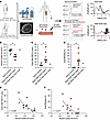 Figure 1
Older adults with clinically meaningful muscle weakness exhibit notable impairments in NMJ transmission. (A) Overview of key muscle and electrophysiological outcomes. Images on the far right represent example SFEMG traces from an adult (top) and a weak older adult (bottom). The arrows denote the negative peaks of the superimposed single fiber APs, and the red traces represent blocked APs. Note: The absolute latency between stimulation and single fiber AP generation is determined by the relative distance between the stimulating and recording electrodes. (B) Levels of isokinetic leg extensor muscle strength normalized to MRI-derived values of quadriceps muscle volume (fat subtracted) — referred to as Muscle Quality — were 44% lower in weak older adult cases compared with healthy adults acting as controls. *P < 0.001. (C) Jitter — the variability in the arrival time of APs to the recording electrode between consecutive electrical discharges — was greater in weak older adults when compared with that in adults acting as controls. *P = 0.003. (D) Neuromuscular junction blocking (indicated as the percentage of synapses with transmission failure) was prominently observed in weak older adults. *P = 0.005. (E and F) The degree of jitter (*P < 0.002) and blocking (#P = 0.007) were both associated with impaired muscle strength (normalized to muscle volume). P values reported in B–D are derived from independent sample t tests with Welch’s correction for equal variances not assumed, and R2 values reported in E and F are derived from quadratic regression. Solid lines in B–D represent the mean values. Triangles indicate female participants; circles indicate male participants.
Mechanistic investigation of age-related NMJ transmission failure
Age-related loss of postsynaptic excitability at the NMJ despite maintained whole muscle fiber excitability. To address the discrepancy between the unchanged or enhanced EP amplitude reported in older animals (20, 32), which imply an increased safety factor, and indications of NMJ transmission failure on SFEMG recordings, we conducted a series of experiments to assess the excitability of isolated single muscle fibers using intracellular microelectrodes in both adult and old C57BL/6 mice and found no discernible differences. Here, extensor digitorum longus (EDL) muscle fiber excitability was studied in 10 adult mice (6–7 months, 5 males, 5 females) and 9 old mice (26–27 months, 5 males, 4 females). No sex differences were observed, so data were pooled. No differences in resting potential, passive membrane properties, or AP parameters were observed between adult and old mice (Table 2). In a subset of old mice, we measured the ability of muscle fibers to sustain AP firing throughout a 2-second train of direct muscle stimulation delivered at 40 Hz. In all of the 7 old mice studied, all fibers were able to fire APs in response to all 80 stimuli delivered, demonstrating that there is no defect in the ability of whole fibers from old mice to fire APs repetitively following direct muscle stimulation.
After confirming no loss of excitability, at the whole-fiber level, in isolated muscle fibers, we next investigated alterations of excitability localized at the NMJ (Figure 2). Using intracellular electrodes, we triggered and recorded APs both near the endplate (junctional) and away from the endplate (extrajunctional) in both adult and old male C57BL/6 mice and Wistar rats (Figure 2). Previous studies have shown that the excitability of healthy muscle fibers is highest around the NMJ due to a larger current through NaV1.4 at the junction compared with extrajunctional regions (30). Junctional enhancement (gain) was indicated by the observation that a smaller stimulus was required to initiate an AP at the NMJ compared with regions away from the NMJ (localized excitability enhancement or “gain”). Such gain was more frequently observed in fibers from adult mice as compared with muscle fibers in older mice (Figure 2, A and B). We also extended these investigations to adult and old rats using a similar recording approach. Consistent with the findings in mice, older rats also demonstrated significantly reduced gain at NMJ (Figure 2, C and D). To address whether these age-related differences in endplate excitability could be related to loss of resting membrane potential during repeated electrode impalements, we compared the correlation between synaptic gain in individual fibers and the degree of depolarization across repeated impalements of muscle fibers (Supplemental Figure 1). These analyses showed no meaningful correlation between resting membrane potential and synaptic gain validating that the decreased gain in old muscle fibers could not be ascribed to an increased NaV1.4 inactivation caused by more pronounced depolarization induced by repeated electrode impalement. Collectively, low-gain fibers occurred predominantly in old mice, supporting that reduction of NaV1.4 current at NMJ could introduce age-related synaptic deficit (37).
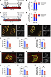 Figure 2
Age-related decline in localized postsynaptic excitability and reduced NaV1.4 channel distribution at the NMJ. (A and B) Excitability was assessed in adult and old mice using a 2-electrode approach in which electrodes E1 and E2 were positioned together near the NMJ (junctional, within 1,000 μm) and then repositioned farther from the NMJ (extrajunctional, >1,000 μm). Older mice exhibited significantly fewer fibers with localized enhanced excitability at the NMJ (gain) compared with adult mice. Gain was defined by a higher stimulation requirement in extrajunctional regions than at the NMJ to trigger an action potential (AP). Total fibers measured: adult, 81; old, 69. (C and D) Similar reductions in excitability gain were observed in old rats (23–24 months, n = 7) compared with adult rats (6–7 months, n = 5) using a 3-electrode approach. The centralstimulating electrode (Es) was positioned near the NMJ (junctional within 50 μm) and then farther from the NMJ (extrajunctional >800 μm), and APs were elicited by current injection through Es. Total fibers measured: adult, 36; old, 45. (E and F) Representative confocal images of sternomastoid NMJs from adult (3–4 months, n = 4) and old (26–28 months, n = 4) mice. AChRs were labeled with α-bungarotoxin (green) and NaV1.4 with specific antibodies (red). (G–I) Quantitative analysis in mouse NMJs: (G) AChR area, (H) NaV1.4 area, and (I) NaV1.4 area normalized to AChR area. Compared with adults, aged mice showed significant reductions across all measures. (J and K) Representative confocal images of lumbrical NMJs from adult (5.5–7 months, n = 5) and old (21–23 months, n = 5) rats. (L–N) Quantitative analysis in rat NMJs: (L) AChR area, (M) NaV1.4 area, and (N) NaV1.4 area normalized to AChR area. Old rats showed significant reductions in NaV1.4 area from adult rats. (G–I and L–N) Each data point represents mean values per animal from 25–27 NMJs per mouse and 23–25 NMJs per rat. Data are shown as mean ± SEM. *P < 0.05, **P < 0.01 using unpaired t tests. Scale bars: 10 μm.
Age-dependent reduction of NaV1.4 enrichment at the NMJ. Voltage-gated sodium channel NaV1.4 is highly enriched at the postsynaptic membrane of the NMJ, where its clustering at the endplate increases local membrane excitability and secures reliable AP initiation in response to EPs (30). Prior studies have demonstrated that NaV1.4 concentration within the perijunctional folds contributes critically to the safety factor of neuromuscular transmission (30). We therefore examined whether age-related alterations in NaV1.4 channel density at the NMJ could contribute to functional transmission defects observed with aging.
To quantify NaV1.4 distribution at the NMJ, immunohistochemical staining and confocal microscopy morphometric analysis were performed in muscles from adult and aged rodents. We measured acetylcholine receptor (AChR) area, NaV1.4+ area, and NaV1.4 area normalized to AChR area in sternomastoid muscles from mice and lumbrical muscles from rats. In aged mice, all 3 parameters were significantly reduced compared with those in adult controls (Figure 2, E–I), indicating both reduced NMJ size and diminished NaV1.4 enrichment. In aged rats, AChR area was preserved, whereas NaV1.4 area was significantly reduced. NaV1.4 area normalized to AChR area showed a similar downward trend but did not reach statistical significance (Figure 2, J–N). These findings suggest that aging preferentially affects NaV1.4 localization at the NMJ, with species- and muscle-dependent differences.
To determine whether these changes reflect fiber-type–specific regulation or redistribution along the myofiber, we analyzed longitudinal cryosections of fast-twitch EDL and slow-twitch soleus muscles from adult and aged mice. NaV1.4 expression was quantified at junctional and extrajunctional regions and normalized to AChR-defined endplate area. In the predominantly fast-twitch EDL, NaV1.4 enrichment at the NMJ was significantly reduced in aged mice, while extrajunctional NaV1.4 levels were unchanged (Supplemental Figure 2, A–D), indicating a selective junctional defect rather than global channel loss. In contrast, NaV1.4 distribution in the slow-twitch soleus was preserved with aging, with no evidence of redistribution or junctional depletion (Supplemental Figure 2, C–M). Thus, aging appears to alter NaV1.4 localization in a muscle-dependent manner, preferentially affecting fast-twitch muscle. To determine whether reduced junctional NaV1.4 reflects transcriptional downregulation, we quantified Scn4a transcript levels in EDL and soleus muscles using digital PCR (Supplemental Figure 3). Whole-muscle Scn4a mRNA levels were unchanged with age in EDL but showed a modest yet significant reduction in soleus. The absence of mRNA decline in EDL despite reduced junctional NaV1.4 protein supports a post-transcriptional or trafficking-based mechanism in fast-twitch muscle, whereas soleus demonstrates modest transcriptional regulation without detectable junctional protein loss. Together, these data indicate muscle-specific regulation of NaV1.4 with aging rather than uniform channel depletion.
NaV1.4 loss at the human NMJ occurs despite preserved morphology. To assess translational relevance, we examined NaV1.4 distribution in surgical biopsy samples from dorsal interosseous muscles obtained during reconstructive procedures (Supplemental Table 2). Consistent with prior analyses of lower-limb muscles in aging, detailed morphometric assessment revealed no overt structural denervation or gross disruption of NMJ architecture in older adults (Figure 3, A–G, and Supplemental Figure 4) (38).
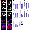 Figure 3
Absence of anatomical denervation but presence of junctional and parajunctional NaV1.4 disruption in human NMJs with aging. (A) Representative confocal images of NMJs from adult and old human dorsal interosseous muscles. AChRs were labeled with α-bungarotoxin (red), while neurofilament and synaptic vesicles were labeled with 2H3 and SV2 (cyan). Individual channels are shown in gray scale below. These representative NMJ images are also shown in Supplemental Figure 4A. Scale bars: 10 μm. (B–G) Morphology quantification in adult (blue) versus old (violet) human samples. (B) Nerve terminal area. (C) AChR area. (D) Endplate diameter. (E) Nerve-endplate overlap. (F) Postsynaptic fragmentation. (G) Muscle fiber diameter. In B–G, each data point represents the mean of ≈40 NMJs per individual (n = 3 per group); no age-related differences were detected. (H) Representative confocal images of NMJs from adult (left) and old (right) human dorsal interosseous muscles (same biopsies as in A). AChRs were labeled with α-bungarotoxin (red), and NaV1.4 is shown in blue. At adult NMJs, the NaV1.4 signal extends beyond the AChR-defined postsynaptic region, forming a parajunctional rim (denoted by yellow dashed lines). By contrast, in aged NMJs this NaV1.4 rim was markedly reduced, with NaV1.4 confined to the AChR territory (indicated by yellow bars). Scale bars: 10 μm (top); 2.5 μm (bottom). (I) Parajunctional NaV1.4 rim width (≈18 NMJs per individual, n = 3 per group) revealed a significant reduction in older individuals. (J) NaV1.4 signal area normalized to AChR area after matching adult and old NMJs. Each old NMJ was matched to the adult NMJ with the most comparable AChR area, using a nearest-neighbor algorithm; pairs differing by >10% were excluded. Lines connect matched NMJs from different individuals. Data are shown as mean ± SEM. Unpaired t tests or Mann-Whitney U tests were used for B–G and I, and paired t test were used for J. *P < 0.05, ***P < 0.001.
Despite preserved structural integrity, NaV1.4 protein localization at the NMJ was significantly reduced in older individuals. Quantification of the parajunctional NaV1.4 rim — reflecting channel enrichment within secondary postsynaptic folds extending beyond AChR-defined endplate boundaries — demonstrated a significant reduction in rim width in older samples (Figure 3, H and I). In a subset analysis (~18 NMJs per individual), total NaV1.4 area was similarly reduced. To control for potential confounding by NMJ size differences, we implemented a one-to-one nearest-neighbor matching strategy, pairing each older NMJ with a size-matched younger NMJ (AChR-defined area difference, <10%). Even after rigorous normalization, NaV1.4 area relative to AChR area remained significantly decreased in older adults (Figure 3J). These findings demonstrate selective loss of junctional NaV1.4 with aging in humans, independent of gross synaptic remodeling. Collectively, the rodent and human data indicate that aging is associated with reduced postsynaptic NaV1.4 enrichment at the NMJ despite preserved overall morphology. This supports a model in which age-related neuromuscular transmission failure reflects impaired postsynaptic excitability, consistent with increased jitter and impulse block observed clinically in older adults.
Acute NaV1.4 inhibition recapitulates age-related NMJ transmission failure. To test whether reduced NaV1.4 function is sufficient to reproduce age-related transmission defects, we acutely inhibited NaV1.4 in adult rats using μ-conotoxin GIIIB, a selective pore blocker of skeletal muscle NaV1.4. Adult rats (3–4 months) received intramuscular injection of μ-conotoxin GIIIB or vehicle in the gastrocnemius. Neuromuscular transmission was assessed using repetitive nerve stimulation (RNS) and stimulated SFEMG.
μ-Conotoxin–treated rats demonstrated significantly increased CMAP decrement during RNS, whereas vehicle-treated controls maintained stable responses (Supplemental Figure 5). SFEMG revealed a frequency-dependent increase in jitter and blocking following NaV1.4 inhibition, particularly at 10 and 20 Hz stimulation (P < 0.01), recapitulating electrophysiological features observed in aged animals (Figure 4). Thus, acute functional impairment of NaV1.4 is sufficient to induce NMJ transmission failure resembling the aging phenotype, supporting a causal link between reduced junctional NaV1.4 enrichment and age-associated decline in neuromuscular reliability.
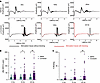 Figure 4
Selective pharmacologic inhibition of NaV1.4 with μ-conotoxin in muscles from adult rats replicates aging-like NMJ transmission defects. (A) Representative SFEMG traces recorded at 5 Hz, 10 Hz, and 20 Hz stimulation in saline- (top) and μ-conotoxin–injected (bottom) muscles. μ-Conotoxin markedly increased variability in latency and blocking compared with vehicle. Red traces indicate blocked responses. (B) Quantification of jitter demonstrated significant increases in μ-conotoxin–treated rats at 10 and 20 Hz stimulation. (C) Percentage blocking was also significantly elevated in the μ-conotoxin group, with effects most pronounced at 20 Hz. Each data point represents an individual fiber measurement. Data are shown as mean ± SEM. Statistical analyses were performed using 2-way ANOVA with Šidák post hoc correction for multiple comparisons. n = 3 rats per group (Wistar, 3–4 months old). *P < 0.05; **P < 0.01.
Investigation of ClC-1 inhibition to enhance muscle excitability, NMJ transmission, and muscle function
The findings of NMJ failure in older adults and rodents raised the possibility that treatment to enhance NMJ transmission could offer a useful approach to restore muscle function (e.g., strength) and, in turn, potentially improve mobility and quality of life. Skeletal muscle–specific Cl– (ClC-1) channels are well known to serve as the primary negative regulator of muscle excitability (39), and recent work has reported enhancement of muscle force and NMJ transmission via small-molecule inhibition of ClC-1 channels in primary and secondary NMJ deficits (e.g., myasthenia gravis, pharmacological NMJ disruption, hereditary peripheral nerve disease and denervation [Charcot Marie Tooth Disease]) (40–43). Based on this, we wanted to establish whether small-molecule inhibition of ClC-1 could improve muscle strength in old rats with SFEMG findings of jitter and blocking like those observed in older humans in the clinical study.
Characterization of small-molecule inhibitors of ClC-1. We conducted a series of experiments to characterize 2 small-molecule inhibitors: NMD1226 and NMD653. In this line of experiments we (a) confirmed that ClC-1 function remains intact in old age by showing that resting membrane conductance (Gm) was the same in muscle fibers from old and adult rats; (b) confirmed that the small-molecule NMD1226 inhibits ClC-1, first, through concentration-dependent reduction in the Gm of individual muscle fibers in intact muscles from adult rats and, second, by demonstrating that NMD1226 was able to decrease maximum ClC-1 current in Chinese hamster ovary (CHO) cells expressing ClC-1 channel on an automated patch clamping system; (c) confirmed that the ClC-1 inhibitor NMD1226 increases muscle fiber excitability; and (d) confirmed that NMD1226 restores muscle function in an experimental condition of compromised NMJ transmission (i.e., AChR antagonism via tubocurarine). More details on these results from both NMD1226 and NMD653 are described in the supplemental Methods, Supplemental Figure 6 and Supplemental Table 3.
ClC-1 chloride inhibition enhances muscle function in aged rodents
Old rats exhibit NMJ transmission failure similar to observations from older humans. Having confirmed the ability of the compounds to inhibit ClC-1, the next series of experiments returned to the core objective of evaluating whether enhancing NMJ transmission can restore muscle function in old animals with confirmed NMJ transmission failure. To this end, we first assessed NMJ function using stimulated SFEMG in old rats (Figure 5A, illustration of SFEMG setup) and confirmed increased jitter and blocking indicative of NMJ transmission failure, similar to prior observations in older adult humans. The old rats demonstrated close to a doubling of jitter (Figure 5B). Most old rats (9 of 12, 75%) showed blocking. In contrast, blocking was not seen in the adult group. On average old rats showed blocking at approximately 30.0% of synapses tested per animal (Figure 5C). These findings closely paralleled the clinical NMJ failure phenotype in older adults and provided a clear rationale to test ClC-1 inhibition in aged rats.
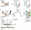 Figure 5
Older rats exhibit impaired NMJ transmission, and ClC-1 inhibition enhances muscle function. (A) Schematic of SFEMG measurements in anesthetized rat gastrocnemius muscles. (B and C) Jitter and NMJ blocking (percentages of synapses with transmission failure) were significantly higher in old rats compared with adult rats (jitter, *P = 0.0176; blocking, **P = 0.003; adult, n = 5; old, n = 12). Graphs show individual values, mean, and P values from t tests with Welch’s correction. (D) Schematic of nerve-stimulated triceps surae force measurements of anesthetized old rats, with representative 80 Hz force traces before dosing (black) and after 60 mg/kg NMD1226 (green). (E) Absolute stimulated muscle force (AUC) at 80 Hz measured before dosing (Baseline) and after NMD1226 or vehicle (After dose). NMD1226 acutely increased force in old rats by 14.4%, whereas force decreased by 3.6% in vehicle-treated old rats and was largely unchanged in NMD1226-treated adult rats (+2.96%), (adult with or without NMD1226, n = 3; old with or without vehicle, n = 3; old with or without NMD1226, n = 9). The graph shows individual values with mean; asterisks show 2-way repeated measures ANOVA (group × treatment interaction P = 0.0140) followed by Fisher’s LSD test (***P = 0.0002). (F) Grip strength (GS) normalized to body weight was assessed as percentage change from baseline in a blinded 7-day dosing study in 20-month-old rats treated with vehicle (n = 4) or NMD1226 (n = 5), with day 8 as a predefined washout period of approximately 26 hours. “Dose P.O.” in the timeline indicates the number of oral doses/day. Mean GS decreased by 9.7% in vehicle-treated rats and increased by 11.2% in NMD1226-treated rats from day 0 to 4. Two-way repeated measures analysis of covariance with baseline GS as a covariate showed significant interaction (group × time interaction P < 0.01) and effect size (η² = 0.536). LSD post-hoc testing indicated significant increases in GS with NMD1226 on days 4 and 7 (*P = 0.017 and 0.005) and a group difference on day 4 (#P = 0.03). Data are shown as mean ± SEM.
ClC-1 inhibition improves NMJ transmission and muscle contractile function in old rodents. After confirming SFEMG evidence for NMJ transmission failure in old rats, the effect of NMD1226 on muscle force was determined in anesthetized old rats. Rats were anesthetized and placed in an experimental setup that enabled recordings of contractile force from hind leg muscles in response to stimulation of the sciatic nerve (Figure 5D). At baseline, compared with adult rats (adult rats, 7.1 ± 0.9 months, 618 ± 98 grams, n = 3; vs. old rats, 22 ± 2 months, 668 ± 86 grams, n = 12), the force production during sustained contraction (1 s, 80 Hz stimulation) was 26% lower in the old rats (1,028 g × s vs. 764 g × s) (Figure 5E). As exemplified by the black trace in Figure 5D, it was furthermore observed that force production tended to decline during the 1-second train of 80 Hz stimuli in old rats. However, oral administration of NMD1226 increased force production and reduced the decline during the contraction (Figure 5D, green trace). Force enhancement was observed in all old rats, and the average force increase was 14% (average baseline force of 762 g × s increased to 870 × s after dosing, Figure 5E). Accordingly, ClC-1 inhibition resulted in rescue of >50% of the force deficit seen in old versus adult rats. In contrast, old rats treated with vehicle showed a slight reduction of force production (Figure 5E). Furthermore, ClC-1 inhibition showed no change in force production in the adult rats that did not present with NMJ transmission deficits on SFEMG (Figure 5E). Collectively, these observations suggested that ClC-1 inhibition can improve muscle function in old rat model with detectable NMJ transmission deficit.
ClC-1 inhibition improves grip strength in old rats. After observing restored force production in old rats in response to nerve stimulation, we were interested in understanding the effect that ClC-1 inhibition could have on voluntary motor function. To explore this, we conducted a blinded study in which old rats were treated with either NMD1226 or vehicle twice daily over 7 days. On day 0, rats underwent grip strength testing to evaluate voluntary muscle strength prior to treatment, and then grip strength was retested while on treatment (or vehicle) on days 4 and 7. At study end on day 8, grip strength was reevaluated when animals were taken off treatment (see insert in Figure 5F). At baseline (prior to treatment), the NMD1226 group exhibited numerically lower grip values, but this was not statistically significant (NMD1226 group, 1.84 ± 0.11 g/g BW; vehicle group: 2.13 ± 0.06 g/g BW, P = 0.1833). During the treatment period, the aged rats treated with the NMD1226 showed an increased grip strength (day 4 and 7) compared with a gradual decline in grip strength in the vehicle treated rats (Figure 5F). The positive effect of NMD1226 on grip strength was completely abolished following removal of treatment as grip strength reverted and became almost identical to that of the control group 1 day after terminating administration of NMD1226 (day 8). In a complementary experiment, aged C57BL/6 mice underwent baseline RNS assessment and were then randomized to receive acute dosing of a ClC-1 inhibitor (NMD653) (n = 11 [6 females, 5 males], mean age, 27.9 months; range, 27–29 months) or vehicle (n = 11 [4 females, 7 males], mean age, 28.3 months; range age, 27–29 months). Following treatment, mice underwent RNS for CMAP decrement as well as terminal SFEMG. Treatment with ClC-1 inhibition resulted in significantly improved function compared with vehicle treatment on RNS and mean SFEMG jitter (NMD653, 13.66 ± 5.26 μs vs. vehicle, 20.42 ± 8.93 μs), indicating improved NMJ transmission and aligning with findings in aged rats (Supplemental Figure 7).


Copyright © 2026 American Society for Clinical Investigation
ISSN: 0021-9738 (print), 1558-8238 (online)