Inflammation
Abstract
The interaction between cells and extracellular matrix (ECM) has been recognized in mechanism of fibrotic diseases. Collagen type VII (collagen VII) is an ECM component which plays an important role in cell-ECM interaction, particularly in cell anchoring and maintaining ECM integrity. Pleural mesothelial cells (PMCs) drive inflammatory reactions and ECM production in pleura. However, the role of collagen VII and PMCs in pleural fibrosis was poorly understood. In this study, collagen VII protein was found increase in pleura of patients with tuberculous pleural fibrosis. Investigation of cellular and animal models revealed that collagen VII began to increase at early stage in pleural fibrotic process. Increase of collagen VII occurred ahead of collagen I and α-SMA in PMCs and pleura of animal models. Inhibition of collagen VII by mesothelial cell-specific deletion of collagen VII gene (WT1-Cre+-COL7A1flox/flox) attenuated mouse experimental pleural fibrosis. At last, it was found that excessive collagen VII changed collagen conformation which resulted in elevation of ECM stiffness. Elevation of ECM stiffness activated integrin/PI3K-AKT/JUN signaling and promoted more ECM deposition, as well as mediated pleural fibrosis. In conclusion, excessive collagen VII mediated pleural fibrosis via increasing extracellular matrix stiffness.
Authors
Qian Li, Xin-Liang He, Shuai-Jun Chen, Qian Niu, Tan-Ze Cao, Xiao-Ling Cui, Zi-Heng Jia, He-De Zhang, Xiao Feng, Ye-Han Jiang, Li-Mei Liang, Pei-Pei Cheng, Shi-He Hu, Liang Xiong, Meng Wang, Hong Ye, Wan-Li Ma
Abstract
Deficits in the mitochondrial energy-generating machinery cause mitochondrial disease (MD), a group of untreatable and usually fatal disorders. Among many severe symptoms, refractory epileptic events are a common neurological presentation of MD. However, the neuronal substrates and circuits for MD-induced epilepsy remain unclear. Here, using mouse models of Leigh Syndrome, a severe form of MD associated to epilepsy, that lack mitochondrial complex I subunit NDUFS4 in a constitutive or conditional manner, we demonstrate that mitochondrial dysfunction leads to a reduction in the number of GABAergic neurons in the rostral external globus pallidus (GPe), and identify a specific affectation of pallidal Lhx6-expressing inhibitory neurons, contributing to altered GPe excitability. Our findings further reveal that viral vector-mediated Ndufs4 re-expression in the GPe effectively prevents seizures and improves the survival in the models. Additionally, we highlight the subthalamic nucleus (STN) as a critical structure in the neural circuit involved in mitochondrial epilepsy, as its inhibition effectively reduces epileptic events. Thus, we have identified a role for pallido-subthalamic projections in the development of epilepsy in the context of mitochondrial dysfunction. Our results suggest STN inhibition as a potential therapeutic intervention for refractory epilepsy in patients with MD providing promising leads in the quest to identify effective treatments.
Authors
Laura Sánchez-Benito, Melania González-Torres, Irene Fernández-González, Laura Cutando, María Royo, Joan Compte, Miquel Vila, Sandra Jurado, Elisenda Sanz, Albert Quintana
Abstract
Immune cells are constantly exposed to microbiota-derived compounds that can engage innate recognition receptors. How this constitutive stimulation is down-modulated to avoid systemic inflammation and auto-immunity is poorly understood. Here we show that Aryl hydrocarbon Receptor (AhR) deficiency in monocytes unleashes spontaneous cytokine responses in vivo, driven by STING-mediated tonic sensing of microbiota. This effect was specific to monocytes, as mice deficient for AhR specifically in macrophages did not show any dysregulation of tonic cytokine responses. AhR inhibition also increased tonic cytokine production in human monocytes. Finally, in patients with systemic juvenile idiopathic arthritis, low AhR activity in monocytes correlated with elevated cytokine responses. Our findings evidence an essential role for AhR in monocytes in restraining tonic microbiota sensing and in maintaining immune homeostasis.
Authors
Adeline Cros, Alessandra Rigamonti, Alba de Juan, Alice Coillard, Mathilde Rieux-Laucat, Darawan Tabtim-On, Emeline Papillon, Christel Goudot, Alma-Martina Cepika, Romain Banchereau, Virginia Pascual, Marianne Burbage, Burkhard Becher, Elodie Segura
Abstract
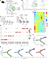
Bacterial infections, particularly uropathogenic E. coli (UPEC), contribute substantially to male infertility through tissue damage and subsequent fibrosis in the testis and epididymis. The role of testicular macrophages (TMs), a diverse cell population integral to tissue maintenance and immune balance, in fibrosis is not fully understood. Here, we used single-cell RNA sequencing in a murine model of epididymo-orchitis to analyze TM dynamics during UPEC infection. Our study identified a marked increase in S100a4+ macrophages, originating from monocytes, strongly associated with fibrotic changes. This association was validated in human testicular and epididymal samples. We further demonstrated that S100a4+ macrophages transition to a myofibroblast-like phenotype, producing extracellular matrix proteins such as collagen I and fibronectin. S100a4, both extracellular and intracellular, activated collagen synthesis through the TGF-β/STAT3 signaling pathway, highlighting this pathway as a therapeutic target. Inhibition of S100a4 with niclosamide or macrophage-specific S100a4 KO markedly reduced immune infiltration, tissue damage, and fibrosis in infected murine models. Our findings establish the critical role of S100a4+ macrophages in fibrosis during UPEC-induced epididymo-orchitis and propose them as potential targets for antifibrotic therapy development.
Authors
Ming Wang, Xu Chu, Zhongyu Fan, Lin Chen, Huafei Wang, Peng Wang, Zihao Wang, Yiming Zhang, Yihao Du, Sudhanshu Bhushan, Zhengguo Zhang
Abstract
BACKGROUND Inter- and intraindividual fluctuations in pain intensity pose a major challenge to treatment efficacy, with a majority of people perceiving their pain relief as inadequate. Recent preclinical studies have identified circadian rhythmicity as a potential contributor to these fluctuations and a therapeutic target.METHODS We therefore sought to determine the impact of circadian rhythms in people with chronic low back pain (CLBP) through a detailed characterization, including questionnaires to evaluate biopsychosocial characteristics, ecological momentary assessment (7 day e-diaries at 8:00/14:00/20:00) to observe pain fluctuations, and intraday blood transcriptomics (at 8:00/20:00) to identify genes/pathways of interest.RESULTS While most individuals displayed constant or variable/mixed pain phenotypes, a distinct subset had daily fluctuations of increasing pain scores (>30% change in intensity over 12 hours in ≥4/7 days). This population had no opioid users, better biopsychosocial profiles, and differentially expressed transcripts relative to other pain phenotypes. The circadian-governed neutrophil degranulation pathway was particularly enriched among arrhythmic individuals; the link between neutrophil degranulation and opioid use was further confirmed in a separate CLBP cohort.CONCLUSION Our findings identified pain rhythmicity and the circadian expression of neutrophil degranulation pathways as indicators of CLBP outcomes, which may help provide a personalized approach to phenotyping biopsychosocial characteristics and medication use. This highlights the need to better understand the impact of circadian rhythmicity across chronic pain conditions.FUNDING This work was funded by grants from the Canadian Institutes of Health Research (CIHR; grant PJT-190170, to NG and MGP) and the CIHR-Strategy for Patient-Oriented Research Chronic Pain Network (grant SCA-145102, to NG, QD, LD, MGP, and MC). DT was funded by a MS Canada endMS Doctoral Research Award, AMZ by an Ontario Graduate Scholarship, HGMG by a CIHR Doctoral Research Award, MGP by a Junior 2 Research Scholarship from the Fonds de recherche du Québec – Santé, and LD by a Canadian Excellence Research Chairs and Pfizer Canada Professorship in Pain Research.
Authors
Doriana Taccardi, Amanda M. Zacharias, Hailey G.M. Gowdy, Mitra Knezic, Marc Parisien, Etienne J. Bisson, Zhi Yi Fang, Sara A. Stickley, Elizabeth Brown, Daenis Camiré, Rosemary Wilson, Lesley N. Singer, Jennifer Daly-Cyr, Manon Choinière, Zihang Lu, M. Gabrielle Pagé, Luda Diatchenko, Qingling Duan, Nader Ghasemlou
Abstract
Metabolic dysfunction-associated steatohepatitis (MASH) is a progressive liver disease characterized by complex interactions between lipotoxicity, ER stress responses, and immune-mediated inflammation. We identified enrichment of the proinflammatory alarmin S100 calcium-binding protein A11 (S100A11) on extracellular vesicles stimulated by palmitate-induced lipotoxic ER stress with concomitant upregulation of hepatocellular S100A11 abundance in an IRE1A-XBP1s dependent manner. We next investigated the epigenetic mechanisms that regulate this stress response. Publicly available human liver ChIP-Seq GEO datasets demonstrated a region of histone H3 lysine 27 (H3K27) acetylation upstream to the S100A11 promoter. H3K27acetylation ChIP-qPCR demonstrated a positive correlation between lipotoxic ER stress and H3K27acetylation of the region, which we termed Lipotoxicity Influenced Enhancer (LIE) domain. CRISPR-mediated repression of the LIE domain reduced palmitate-induced H3K27acetylation and corresponding S100A11 upregulation in Huh7 cells and immortalized mouse hepatocytes. Silencing of the murine LIE in two independent steatohepatitis models demonstrated reduced S100a11 upregulation and attenuated liver injury. We confirmed H3K27acetylation and XBP1s occupancy at the LIE domain in human MASH liver samples and an increase in hepatocyte-derived S100A11-enriched extracellular vesicles in MASH patient plasma. Our studies demonstrate a LIE domain which mediates hepatic S100A11 upregulation. This pathway may be a potential therapeutic target in MASH.
Authors
P. Vineeth Daniel, Hanna L. Erickson, Daheui Choi, Feda H. Hamdan, Yasuhiko Nakao, Gyanendra Puri, Takahito Nishihara, Yeriel Yoon, Amy S. Mauer, Debanjali Dasgupta, Jill Thompson, Alexander Revzin, Harmeet Malhi
Abstract
Authors
Bowen Yan, Qingchen Yuan, Marco M. Buttigieg, Prabhjot Kaur, Annalisse R. McKee, Daniil E. Shabashvili, Caitlyn Vlasschaert, Alexander G. Bick, Michael J. Rauh, Olga A. Guryanova
Abstract
Endoplasmic reticulum (ER) stress through IRE1/XBP-1 is implicated in the onset and progression of graft-versus-host disease (GVHD), but the role of ER stress sensor PERK in T-cell allogeneic responses and GVHD remains unexplored. Here, we report that PERK is a key regulator in T-cell allogeneic response and GVHD induction. PERK augments GVHD through increasing Th1 and Th17 population, while reducing Treg differentiation by activating Nrf2 pathway. Genetical deletion or selective inhibition of PERK pharmacologically reduces GVHD while preserving graft-versus-leukemia (GVL) activity. At cellular level, PERK positively regulates CD4+ T-cell pathogenicity, while negatively regulating CD8+ T-cell pathogenicity in the induction of GVHD. At molecular level, PERK interacts with SEL1L and regulates SEL1L expression, leading to augmented T-cell allogeneic responses and GVHD development. In vivo, PERK deficiency in donor T cells alleviate GVHD through ER-associated degradation (ERAD). Furthermore, pharmacological inhibition of PERK with AMG44 significantly suppresses the severity of GVHD induced by murine or human T cells. In summary, our findings validate PERK as a potential therapeutic target for the prevention of GVHD while preserving GVL responses, and uncover the mechanism by which PERK differentially regulates CD4+ versus CD8+ T-cell allogeneic and anti-tumor responses.
Authors
Qiao Cheng, Hee-Jin Choi, Yongxia Wu, Xiaohong Yuan, Allison Pugel, Linlu Tian, Michael Hendrix, Denggang Fu, Reza Alimohammadi, Chen Liu, Xue-Zhong Yu
Abstract
Allergic diseases have reached epidemic proportions globally, calling attention to the need for better treatment and preventive approaches. Herein, we developed allergen-encoding messenger RNA (mRNA) lipid nanoparticle (LNP) strategies for both therapy and prevention of allergic responses. Immunization with allergen-encoded mRNA-LNPs modulated T cell differentiation, inhibiting the generation of T helper type 2 (Th2) and type 17 (Th17) cells upon allergen exposure in experimental asthma models induced by ovalbumin (OVA), and naturally occurring house dust mite (HDM) and the major HDM allergen Der p1. Allergen-specific mRNA-LNP treatment attenuated clinicopathology in both preventive and established allergy models, including reduction in eosinophilia, mucus production, and airway hypersensitivity, while enhancing production of allergen-specific IgG antibodies and maintaining low IgE levels. Additionally, allergen-specific mRNA-LNP vaccines in mice elicited a CD8+CD38+KLRG- T cell response as seen following SARS-CoV-2 mRNA vaccination in human, underscoring a conserved immune mechanism across species, regardless of the mRNA-encoded protein. Notably, mRNA-LNP vaccination in combination with an mTOR inhibitor reduced the CD8+ T cell response without affecting the vaccine-induced anti-allergic effect in the preventive model of asthma. This technology renders allergen-specific mRNA-LNP therapy as a promising approach for prevention and treatment of allergic diseases.
Authors
Yrina Rochman, Michael Kotliar, Andrea M. Klingler, Mark Rochman, Mohamad-Gabriel Alameh, Jilian R. Melamed, Garrett A. Osswald, Julie M. Caldwell, Jennifer M. Felton, Lydia E. Mack, Julie Hargis, Ian P. Lewkowich, Artem Barski, Drew Weissman, Marc E. Rothenberg
Abstract
Severe systemic inflammatory reactions, including sepsis, often lead to shock, organ failure and death, in part through an acute release of cytokines that promote vascular dysfunction. However, little is known about the vascular endothelial signaling pathways regulating the transcriptional profile in failing organs. This work focuses on signaling downstream of IL-6, due to its clinical importance as a biomarker for disease severity and predictor of mortality. Here, we show that loss of endothelial expression of the IL-6 pathway inhibitor, SOCS3, promoted a type I interferon (IFNI)-like gene signature in response to endotoxemia in mouse kidneys and brains. In cultured primary human endothelial cells, IL-6 induced a transient IFNI-like gene expression in a non-canonical, interferon-independent fashion. We further show that STAT3, which we had previously shown to control IL-6-driven endothelial barrier function, was dispensable for this activity. Instead, IL-6 promoted a transient increase in cytosolic mitochondrial DNA and required STAT1, cGAS, STING, and the IRFs 1, 3, and 4. Inhibition of this pathway in endothelial-specific STING knockout mice or global STAT1 knockout mice led to reduced severity of an acute endotoxemic challenge and prevented the endotoxin-induced IFNI-like gene signature. These results suggest that permeability and DNA sensing responses are driven by parallel pathways downstream of this cytokine, provide new insights into the complex response to acute inflammatory responses, and offer the possibility of potential novel therapeutic strategies for independently controlling the intracellular responses to IL-6 in order to tailor the inflammatory response.
Authors
Nina Martino, Erin K. Sanders, Ramon Bossardi Ramos, Iria Di John Portela, Fatma Awadalla, Shuhan Lu, Dareen Chuy, Neil Poddar, Mei Xing G Zuo, Uma Balasubramanian, Peter A. Vincent, Pilar Alcaide, Alejandro P. Adam
No posts were found with this tag.


Copyright © 2026 American Society for Clinical Investigation
ISSN: 0021-9738 (print), 1558-8238 (online)


