Advertisement
The genetic and neurobiologic compass points toward common signaling dysfunctions in autism spectrum disorders
Pat Levitt and Daniel B. Campbell
Vanderbilt Kennedy Center for Research on Human Development and Department of Pharmacology, Vanderbilt University Medical Center, Nashville, Tennessee, USA.
Address correspondence to: Pat Levitt, Zilkha Neurogenetic Institute, Keck School of Medicine of University of Southern California, 1501 San Pablo Street, Los Angeles, California 90087, USA. Phone: (323) 442-1509; Fax: (323) 442-2145; E-mail: plevitt@usc.edu.
Find articles by Levitt, P. in: PubMed | Google Scholar
Vanderbilt Kennedy Center for Research on Human Development and Department of Pharmacology, Vanderbilt University Medical Center, Nashville, Tennessee, USA.
Address correspondence to: Pat Levitt, Zilkha Neurogenetic Institute, Keck School of Medicine of University of Southern California, 1501 San Pablo Street, Los Angeles, California 90087, USA. Phone: (323) 442-1509; Fax: (323) 442-2145; E-mail: plevitt@usc.edu.
Find articles by Campbell, D. in: PubMed | Google Scholar
Published April 1, 2009 - More info
J Clin Invest. 2009;119(4):747–754. https://doi.org/10.1172/JCI37934.
© 2009 The American Society for Clinical Investigation
Autism spectrum disorder (ASD) is a syndrome characterized by a triad of core deficits: disturbances in social behavior, atypical verbal and nonverbal communication, and restricted interests that can be accompanied by repetitive behavior. The clinical diagnosis, which includes individuals with any one of a spectrum of neurodevelopmental conditions (including autism, Rett syndrome, pervasive developmental disorder–not otherwise specified, and Asperger syndrome), is made in 1 of every 150 individuals and is four times more prevalent in boys than girls (1). While ASD is among the most heritable psychiatric disorders defined in the Diagnostic and statistical manual of mental disorders (4th edition) (2), it is not a static or simple disorder with fixed effects on a circumscribed age. Instead, equally fundamental facets of pathology emerge at different points of maturation of the child. Moreover, the disorder does not result in immutable social and cognitive deficits, but rather the core symptoms typically change over time and to different degrees. Co-occurring medical conditions (sleep problems, epilepsy, and gastrointestinal symptoms) and psychiatric disturbances (anxiety, obsessive-compulsive disorder, and aggression) are common and can appear at different ages in children on the spectrum.
Contemporary hypotheses of the causes of ASD often include experience-dependent processes through which atypical gene-by-environment (G X E) interactions yield pathophysiology in later emerging systems that underlie social and communication competencies. The later emergence of symptoms is consistent with the concept that developmental differentiation, whether at the cellular, circuit, or systems level, occurs from the bottom up; behavior develops from basic sensory and perceptual systems that feed into higher integration centers (3–5). Impairments in initial basic processes become expressed in ever more complex systems, with the population heterogeneity of the clinical features of ASD expected to increase from infancy to childhood and through adolescence. However, it is not clear whether the factors that contribute to the developmental diversification and phenotypic heterogeneity of ASD are related to a complex genetic etiology of ASD itself or whether they also involve the interaction between ASD-specific and -nonspecific functional features.
Clinical researchers have noted the importance of addressing disorder heterogeneity in the study of ASD (6–8). In this regard, the conundrum facing investigators is connecting the well-defined, highly heritable nature of ASD with the striking differences in the initial expression of core symptoms, progressive changes over time, and differential response to interventions. Thus, a major goal of the current interdisciplinary research agenda is not only to explain the etiologies of ASD but also to understand the syndrome-specific and -nonspecific factors that influence variability in the relative risk of developing ASD, in the developmental course of symptom presentation, in the responsiveness to treatment, and in the co-occurrence of other medical dysfunctions (6, 8–11). This Review highlights the current understanding of ASD genetics, key pathophysiological findings from behavior and imaging studies, and potential G X E interactions that may be at the core of ASD expression. The Review ends with what we believe to be a novel hypothesis that combines recent genetic findings to propose one potential mechanism of heterogeneity in ASD.
-
Distinct genetic mechanisms can result in ASD
Based on studies in mono- and dizygotic twins (12, 13), the estimated heritability of ASD is approximately 0.90. This far exceeds the estimated heritability of other common polygenic diseases, including cancer, heart disease, schizophrenia, and depression. The focus, therefore, on defining the underlying genetic etiology of ASD has escalated dramatically during the current decade, in parallel with the rapid development of affordable genomic methods that have facilitated the analysis of large populations and entire genomes. Like other complex disorders, however, the most critical challenges of the field lie in defining the heritability of risk for developing ASD that may be due to G X E factors that alter the trajectory of brain development and the direct impact of de novo or heritable gene variation on brain development. ASD is a spectrum of disorders, in which there are differences in the degree of severity of the three core symptoms as well as other co-occurring mental health and physical conditions. Thus, to emphasize the functional importance of recognizing many different kinds of ASD, the term “autisms” has been used (14). The behaviors that are disrupted in ASD are complex and develop through a bottom-up assembly of simple to more complex brain circuits that control very basic processes such as physiological homeostasis and more complex tasks such as being motivated to pay attention to certain cues in the environment that regulate outward social behavior and verbal and nonverbal communication. The heterogeneity of ASD is entirely consistent with the concept that different genetic mechanisms may influence brain circuit development at different levels of the hierarchy (5). The field thus is moving away from defining the ASD genes to defining unique phenotypic features of stratified populations of children, adolescents, and adults that may relate to specific genetic etiologies, such as increased risk due to common allelic variations, rare mutations, or copy number variation (CNV) (Figure 1). Implicit in this view is that there will not be identification of genetic risks that map one-to-one with behavioral dysfunction; that is, while there are genetic variants that are enriched in populations with particular dysfunctions, such as language, there are no genes that directly regulate social behavior or language. Instead, genetic vulnerability resides in the disruption of cellular processes, due to the disruption of proteins encoded by genes, in specific brain circuits that may also be influenced by G X E mechanisms. Research findings emerging from human genetic and animal studies suggest that disruption of a key developmental process, synapse formation and stabilization (synaptogenesis), is a final common path in ASD etiology. Different molecular mechanisms may contribute to increasing ASD risk, including disturbances in the assembly of structural proteins needed to build synapses, such as the neuroligins and neurexins, and dysfunctional cellular signaling pathways that control synaptogenesis.
 Figure 1
Current experimental approaches to determining genetic etiologies for ASD. These approaches include whole-genome analyses that identify disorder-related sequences or CNVs in genes that exhibit preferential inheritance patterns or de novo appearance in individuals with ASD. The current challenges include the translation of these genetic findings to define the biological consequences of the variations, to determine the influence on defined clinical phenotypes of ASD, and eventually to design new intervention strategies.

-
Distinct patterns of heritability of risk alleles in ASD
As noted above, ASD is highly heritable, and current studies suggest that there are multiple mechanisms through which different types of gene mutations increase risk of developing the disorder (15–17). There are a number of considerations that are key to successful genetic studies of ASD. First, because of the heterogeneity of the disorder, it is necessary to analyze large numbers of individuals with ASD. Second, each individual gene is likely to have very small effects on disease risk, but in combination with other genes and/or G X E factors, an individual gene may encode a protein that functions in a key cellular process, which, when disrupted, contributes to disease pathophysiology. Third, disorder emergence through de novo genetic mutations or heritability of gene-specific functional polymorphisms in the DNA sequence transmitted from parent to child may underlie distinct but equivalently valid ASD etiologies. Fourth, distinct genetic etiologies, together with different environmental factors, may be part of ASD heterogeneity. Last, the nature of the core behavioral dimensions that characterize ASD emerge through perturbation of developing brain circuits. Disruption at distinct levels of the organizational and functional hierarchy relate to the heterogeneity in social behavior and communication capabilities.
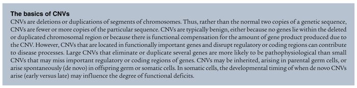
The aim of genetic studies of ASD should be to identify functional variants that contribute to ASD risk. A thorough recent review provides a detailed listing of up-to-date genetic findings (16). One approach with great promise for the identification of candidate genes and pathways is analysis of CNV (see The basics of CNVs) (18, 19). However, as with single gene mutations and common variants, CNV analyses need to be interpreted with extreme caution for a number of reasons. First, the presence of a de novo CNV in an individual with ASD does not necessarily imply it is associated with increased risk of developing the disorder. Further, CNVs typically are not fully penetrant, meaning that they may be present in individuals who do not have an ASD. CNVs were first described in healthy control individuals, with more than 11 CNVs per individual (20), indicating that having multiple CNVs is not pathologic. Only formal genetic association analyses involving large sample sizes should be used to imply a particular CNV is associated with disorder risk. Second, the presence of a CNV does not necessarily imply functional disruption. Analyses of CNVs in the human adult cerebral cortex indicate that more than 50% of mature neurons are aneuploid (21, 22), and experiments in mice indicate that CNVs in cortical neurons may have little impact on function (23). Further, germline deletion of both copies of certain genes in experimental animals can result in mutants without a detectable phenotype. This suggests that due to adaptive processes, gene dosage in the form of CNV does not lead necessarily to dramatic functional changes in vivo. Third, CNV in peripheral blood cells, the cells typically analyzed in humans, may not relate in a one-to-one fashion to CNVs in neurons. Indeed, the number of CNVs in the human cerebral cortex is approximately 7-fold higher than in peripheral blood cells (21), and thus, analysis of peripheral blood may identify some, but not necessarily all, of the CNVs occurring in neurons that contribute to ASD-related disturbances of brain architecture and circuitry. Fourth, de novo CNVs are observed in 7%–10% of cases from simplex families (families with only one child with ASD), 2%–3% of cases from multiplex families (families with more than one child with ASD), and 1% of controls (24, 25). The de novo CNVs that occur in a subset of individuals with ASD in multiplex families may influence the severity of the disorder, rather than contributing directly to the expression of the disorder. Despite these cautions, CNV analysis can be used to identify candidate genes that can be tested further for functional effects that may contribute to ASD susceptibility (18, 19).
We are beginning to recognize that inheritance of rare or common functional alleles is only one genetic mechanism that increases disorder risk. Private (de novo) functional mutations also impart genetic risk. Analyses suggest that, at the genetic and behavioral levels, multiplex families may be fundamentally different from simplex families (24, 26–29). Furthermore, multiple genes or even multiple mutations of the same gene may be involved in the etiology of the same clinically diagnosed disorder in different individuals. For example, there are over 50 genes that carry mutations known to cause nonsyndromic retinitis pigmentosa (30). Conversely, there are more than 130 distinct catalogued mutations of the 7-dehydrocholesterol reductase (DHCR7) gene in individuals with the monogenic disorder Smith-Lemli-Opitz syndrome (31). Heritable, high-risk mutations in breast cancer, such as those in the breast cancer 1, early onset (BRCA1) gene, are balanced by more common variants in multiple genes discovered through whole-genome association studies (WGASs). Given the range of possibilities for disorder etiology, sample numbers are clearly important. For example, WGASs have examined between 500,000 and 1,000,000 SNPs simultaneously in thousands of patient samples for diabetes and coronary artery disease (18, 32–34). These diseases, with arguably less complex pathophysiology than ASD, only recently have had the sample power to generate statistically reliable data that reveal common SNPs with disease-related heritability patterns in multiple genes. Similarly, rare functional mutations in ASD candidate risk genes initially may seem to be overrepresented in the clinical population compared with unrelated controls, but recent analysis demonstrates that even these types of studies more accurately reflect clinical findings when larger sample populations are assessed (35).
-
Syndromic disorders and rare mutations point the way
Although not understood from an etiological or pathophysiological perspective, it is now clear that rare neurodevelopmental disorders (<1 in 10,000) are becoming increasingly important to study in greater detail because of their relationship to ASD. Higher penetrance of ASD diagnosis (far greater than the 0.75% observed in the general population) is reported in children who have genetically diverse neurodevelopmental syndromic disorders, including Angelman syndrome, Fragile X syndrome (FraX), Rett syndrome, Smith-Lemli-Opitz syndrome, Timothy syndrome, neurofibromatosis, and tuberous sclerosis. It is important to emphasize that each syndrome is characterized by fundamentally different gene mutations, which presumably impart distinct molecular pathophysiologies (Table 1). However, there are few studies that examine closely the similarities and differences in phenotypic characteristics between single gene (syndromic) and multigenic (idiopathic) ASD (36). The neurodevelopmental syndromic disorders listed in Table 1 are characterized in part by intellectual disability (ID; formally termed mental retardation). A large minority (25%–40%) of individuals with ASD has ID, but ASD is not synonymous with ID. A recent structural MRI study suggests that individuals with FraX, with or without ASD diagnosis, are more closely related in the context of the size of brain structures than those with idiopathic ASD (37). Microarray analysis of lymphocytes from patients with FraX, chromosome 15q deletion, or idiopathic ASD reveal unique patterns of gene expression that may serve as a signature for each disorder, but with a potentially important small subset of overlapping changes in mRNA expression (38). Moreover, detailed neuropathological studies are lacking to compare these syndromes and idiopathic ASD. Although there is likely to be diversity in the pathological targets in each syndrome, there are suggestions of some commonalities. The triad of overlapping dysfunctions (social behavior, communication, and repetitive behavior) across ASD and the syndromes, together with the known brain neuropathology of some of the syndromes, suggests that later neurodevelopmental events, such as synapse formation and maturation, dendritic growth, and myelination, are probably most vulnerable. In addition to the evidence from syndromic disorders, the focus on later events in this Review is supported by the discovery of rare mutations in certain genes that regulate synaptogenesis. A substantial focus has been on the adhesive and structural elements needed for synapse formation, stability, and physiologic maturation. In ASD cases, rare mutations and CNVs have been identified in genes encoding neuroligins, neurexins, contactin-associated protein-2 (CNTNAP2), and SH3 and multiple ankyrin repeat domains 3 (SHANK3). The disruptions are likely to occur in shared forebrain and cerebral cortical circuits. Thus, while not identical to idiopathic ASD, biological and behavioral analyses of syndromic disorders and rare mutations provide a sound approach to discern potential overlapping molecular and brain targets (Table 1).

-
Intracellular kinase signaling in ASD-associated disorders
The potential contribution of defects in adhesion and structural proteins that build synapses to the etiology of ASD has been the subject of many reviews of ASD (16, 17, 39–41). In this Review, we suggest that some neurodevelopmental syndromic disorders and rare mutations point to an additional set of molecular targets. Thus, recognizing that we are attempting to resolve a spectrum of disorders that will not have a single, underlying etiology, findings from studies of certain syndromic disorders with high penetrance of ASD converge on the ERK and PI3K intracellular signaling pathways that we believe deserve increased scrutiny in all forms of ASD. ERK and PI3K activate mammalian target of rapamycin (mTOR), which through other kinases will increase mRNA translation to influence developmental functions as diverse as the cell cycle, cell survival, differentiation, and motility. Receptor tyrosine kinases (RTKs) can signal through either of these intracellular kinase pathways, with cell type and cellular milieu defining the intracellular response (Figure 2). Table 1 reports several syndromic disorders with high penetrance of ASD that involve a primary disruption in signaling through these pathways specifically and others that would disrupt RTK signaling, the primary membrane receptor class that transduces signals through ERK and PI3K. The most convincing connections between ERK/PI3K signaling disruption and ASD are evident in tuberous sclerosis and neurofibromatosis type 1, in which different elements of the ERK/PI3K pathway are disrupted genetically, leading to enhanced mTOR downstream activation (Figure 2). In addition, ERK and PI3K signaling is dependent in part on normal cholesterol biosynthesis, which is absent in Smith-Lemli-Opitz syndrome. For example, Ras signaling, a key upstream mediator of ERK activation, requires cholesterolization. Rare gene mutations of another element of the PI3K signaling pathway, phosphatase and tensin homolog (PTEN), are associated with high prevalence of ASD. Rett syndrome disrupts the X-linked methyl CpG binding protein 2 (MECP2) gene, which encodes a protein that binds to specific regulatory regions of certain genes (based on DNA methylation patterns) that control gene transcription. Methylation status and/or MECP2 binding directly regulates transcription of key genes involved in met proto-oncogene–RTK signaling (MET RTK signaling; MET is also known as HGFR), which our laboratory has implicated in ASD risk (see below). Those genes include those encoding MET, the MET coreceptor CD44, the MET transcriptional regulator SP1, and several proteins in the ERK/PI3K downstream signaling pathway (42).
 Figure 2
The MET RTK signaling pathway and genes implicated in ASD risk. Intracellular signaling of MET and other RTKs occurs via the PI3K or ERK1/2 pathways. Rare mutations and CNVs (which are both designated by ‡) or associated common alleles (which are designated by *) have been identified in individuals with ASD in seven genes encoding proteins involved in these signaling pathways. Of note, an association between common MET variants and ASD has been reported for five independent family cohorts. PLAUR and SERPINE1 associations with ASD have been determined in single, large family cohorts (>600 families). Ras disruption in Smith-Lemli-Opitz syndrome is due to alterations in cholesterol biosynthesis (which is designated by †). Also depicted are other proteins that interact with the MET signaling pathway, such as semaphorins, plexins, and other RTKs. MET can signal via the PI3K and the ERK pathway. RTKs, including MET, are involved in key neurodevelopmental processes, including axon guidance, synapse formation, and plasticity. Convergence of many different genetic etiologies suggests that risk via ERK/PI3K signaling may be common in ASD. Risk, severity of the pathophysiology (i.e., intellectual disability), and disorder heterogeneity may relate to differences in genetic and epigenetic points of entry to the pathways. Thus, the impact due to genetic risk, via regulators of ligand availability or RTKs such as MET, may be less severe than the more severe clinical impact (i.e., intellectual disability) from disruption downstream along the intracellular signaling pathways. c-cbl, E3 ubiquitin-protein ligase c-Cbl; rheb, Ras homolog enriched in brain; RSK, ribosomal S6 kinase; uPA, urokinase plasminogen activator.
The various neurodevelopmental syndromic disorders and rare mutations described thus far along the ERK/PI3K pathways result in an increased state of activation of mTOR (Figure 2). Additional evidence for involvement of these intracellular kinase pathways in ASD comes from recent treatment studies in genetically engineered mice that exhibit behavioral and neuropathologic phenotypes that are common in the human neurodevelopmental syndromic disorders. For example, systemic administration of drugs that reduce mTOR activation, such as rapamycin, wortmannin, and RAD001, can reverse behavioral and structural pathology in mice with Pten (43), tuberous sclerosis 1 (Tsc1) (44, 45), and neurofibromin 1 (Nf1) (46, 47) mutations, with no reported side effects.
ASD etiologies also are likely to include environmental factors that work together with genetic risk to drive neurodevelopment systems over the threshold for disorder expression (Figure 3). We therefore hypothesize that different genetic routes to altered RTK function, by way of modulation of ERK/PI3K signaling pathways, combine with environmental factors, such as biochemical stressors, that also modulate these signaling pathways. The G X E interactions either modulate the degree of dysfunction of the core clinical features of ASD or have an impact on neurobiological circuits that are at greater risk for dysfunction, because genetic vulnerability pushes the system closer to disorder threshold.
 Figure 3
Contributions of the PI3K pathway to ASD risk threshold. The degree of genetic risk is indicated by shading, with darker color indicating increased risk. The model presents common functional variants in the MET, PLAUR, and SERPINE1 genes that, along with other genetic risk alleles, contribute to risk of developing ASD. Adaptive processes may prevent presentation of ASD, but additional environmental factors or the presence of multiple risk alleles result in idiopathic (multiple genes, each having a small effect) ASD. Mutations further down the PI3K pathway result in syndromic disorders, with penetrance and phenotype severity determined by a decreasing availability of adaptive processes.
Given that ERK/PI3K signaling is widely distributed throughout multiple organ systems, where does disorder specificity arise? One way to think about the issue of specificity is to recognize that signaling through ERK/PI3K is highly influenced by cell type and timing of activation of the RTK signaling systems. For example, there is a potential dichotomy in the molecular mechanisms of ASD and cancer that would involve different genetic risk factors affecting ERK/PI3K signaling. Unequivocal evidence implicates hyperactivated PI3K signaling in a number of malignant cancer types (48–50). In contrast, decreased PI3K activation may contribute in some instances to ASD (26, 51, 52). We are unaware of any studies of cancer frequencies in individuals with ASD. However, disruption of PI3K signaling also has been implicated in other psychiatric disorders of neurodevelopmental origin, such as schizophrenia (53, 54). An altered incidence of various cancers in individuals with schizophrenia is debated (55, 56), but reduced cancer incidence is observed consistently in parents and siblings of individuals with schizophrenia compared with the general population (57–59). These data support the plausibility of a genetic impact, through different mutations or common variants, that increases risk for a neurodevelopmental disorder and decreases cancer risk, a hypothesis that can be tested by epidemiological studies and complete sequencing of candidate genes to identify mutations associated with specific disorders.
One testable facet of our hypothesis is that risk for more global neurodevelopmental disruptions increases when the genetic hits are downstream from the molecular components that are involved in initial RTK activation, which are the growth factors or receptors themselves. Consistent with this, mutations in NF1, AKT, TSC1, and TSC2 typically result in widespread and severe clinical problems such as mild to severe intellectual disabilities, seizure disorder, sensory-motor deficits, and medical dysfunctions (Figure 3). The corollary to this would be that mutations in upstream genes encoding RTKs or proteins that regulate growth factor availability would place signaling through this pathway at risk but require additional genetic and environmental insults to cause neurodevelopmental disruption. Disruption of the development of specific brain circuits would occur, because, unlike their intracellular mediators, upstream signaling elements are not distributed uniformly. Rather, RTKs and growth factors may be concentrated in developing circuits at key periods of development that mediate the maturation of connections underlying specific functions. This hypothesis is consistent with the identification of neuregulin 1 (the ligand for the RTK ERBB4) as a factor for schizophrenia susceptibility (60) and the RTK MET as a factor for ASD risk (26, 27, 52). Although both ERBB4 and MET activate PI3K signaling, the differential timing and patterns of expression of each of these RTKs in developing cerebral cortex (61) may account for the distinct neurodevelopmental disruptions characteristic of each disorder. We have shown that MET is enriched in neocortex, amygdala, septum, and cerebellum, regions implicated in ASD (62).


Article tools
- Download citation information
- Send a comment
- Terms of use
- Standard abbreviations
- Need help? Email the journal
Review Series
Rethinking Mental Disorders
-
Schizophrenia from a neural circuitry perspective: advancing toward rational pharmacological therapies
-
Children with obsessive-compulsive disorder: are they just “little adults”?
-
Bipolar disorder: from genes to behavior pathways
-
The genetic and neurobiologic compass points toward common signaling dysfunctions in autism spectrum disorders
-
Targeted electrode-based modulation of neural circuits for depression
-
Disruptive insights in psychiatry: transforming a clinical discipline
Metrics
Go to
- Top
- Abstract
- Distinct genetic mechanisms can result in ASD
- Distinct patterns of heritability of risk alleles in ASD
- Syndromic disorders and rare mutations point the way
- Intracellular kinase signaling in ASD-associated disorders
- MET in PI3K signaling and ASD
- G X E interactions in ASD etiology
- Final thoughts
- Footnotes
- References
- Version history


Copyright © 2025 American Society for Clinical Investigation
ISSN: 0021-9738 (print), 1558-8238 (online)