Go to JCI Insight
  • About
  • Editors
  • Consulting Editors
  • For authors
  • Publication ethics
  • Publication alerts by email
  • Advertising
  • Job board
  • Contact
  • Clinical Research and Public Health
  • Current issue
  • Past issues
  • By specialty
    • COVID-19
    • Cardiology
    • Gastroenterology
    • Immunology
    • Metabolism
    • Nephrology
    • Neuroscience
    • Oncology
    • Pulmonology
    • Vascular biology
    • All ...
  • Videos
    • Conversations with Giants in Medicine
    • Video Abstracts
  • Reviews
    • View all reviews ...
    • Pancreatic Cancer (Jul 2025)
    • Complement Biology and Therapeutics (May 2025)
    • Evolving insights into MASLD and MASH pathogenesis and treatment (Apr 2025)
    • Microbiome in Health and Disease (Feb 2025)
    • Substance Use Disorders (Oct 2024)
    • Clonal Hematopoiesis (Oct 2024)
    • Sex Differences in Medicine (Sep 2024)
    • View all review series ...
  • Viewpoint
  • Collections
    • In-Press Preview
    • Clinical Research and Public Health
    • Research Letters
    • Letters to the Editor
    • Editorials
    • Commentaries
    • Editor's notes
    • Reviews
    • Viewpoints
    • 100th anniversary
    • Top read articles

  • Current issue
  • Past issues
  • Specialties
  • Reviews
  • Review series
  • Conversations with Giants in Medicine
  • Video Abstracts
  • In-Press Preview
  • Clinical Research and Public Health
  • Research Letters
  • Letters to the Editor
  • Editorials
  • Commentaries
  • Editor's notes
  • Reviews
  • Viewpoints
  • 100th anniversary
  • Top read articles
  • About
  • Editors
  • Consulting Editors
  • For authors
  • Publication ethics
  • Publication alerts by email
  • Advertising
  • Job board
  • Contact
Developing DNA vaccines that call to dendritic cells
Michele A. Kutzler, David B. Weiner
Published November 1, 2004
Citation Information: J Clin Invest. 2004;114(9):1241-1244. https://doi.org/10.1172/JCI23467.
View: Text | PDF
Commentary

Developing DNA vaccines that call to dendritic cells

  • Text
  • PDF
Abstract

DNA vaccination is a novel immunization strategy that has great potential for the development of vaccines and immune therapeutics. This strategy has been highly effective in mice, while less immunogenic in nonhuman primates and humans. Enhancing DNA vaccine potency remains a challenge. It is likely that APCs, and especially DCs, play a paramount role in the presentation of vaccine antigen to the immune system. A new study reports the synergistic recruitment, expansion, and activation of DCs in vivo in a mouse model through covaccination with plasmids encoding macrophage inflammatory protein-1α (MIP-1α), fms-like tyrosine kinase 3 ligand (Flt3L), and the DNA vaccine. Such cooperative strategies delivering vaccine in a single, simple platform result in improved cellular immunity in vivo, including enhanced tetramer responses and IFN-γ secretion by antigen-specific cells.

Authors

Michele A. Kutzler, David B. Weiner

×

Figure 1

Options: View larger image (or click on image) Download as PowerPoint
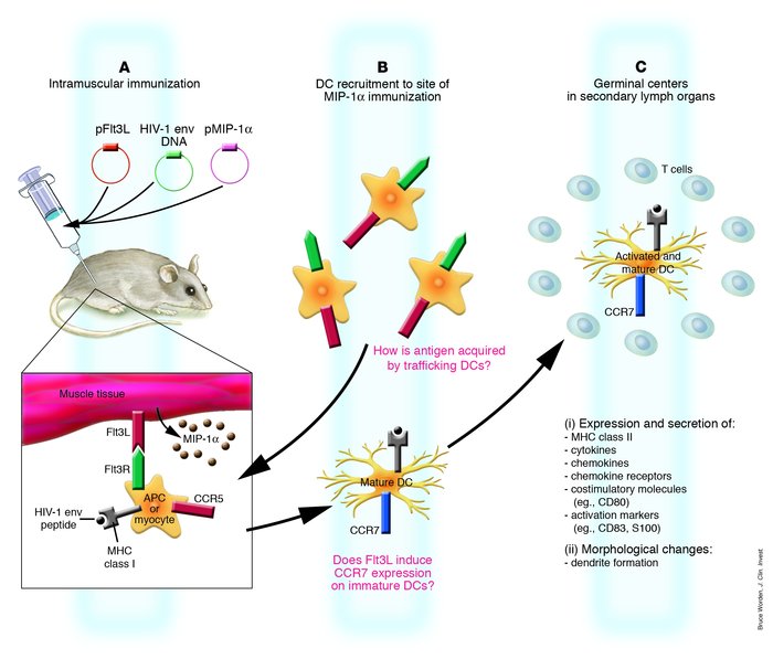
Proposed schematic of chemokine-induced traffic and activation of DCs fo...
Proposed schematic of chemokine-induced traffic and activation of DCs following DNA vaccination with plasmid-encoded Flt3L and MIP-1α. (A) Following intramuscular injection in mice of plasmid-encoded MIP-1α (pMIP-1α) and plasmid-encoded Flt3L (pFlt3L), in combination with HIV-1 env, muscle and resident APCs are transfected with plasmid, leading to protein production. MIP-1α protein is secreted, while Flt3L is expressed on muscle cells or on the surface of resident APCs. HIV-1 env is processed, and peptides are presented by MHC class I molecules; this peptide/MHC complex stimulates CD8+ T lymphocytes. Soluble protein released by transfected cells is taken up by APCs, and via the MHC Class II pathway, the peptide/MHC complex stimulates CD4+ T lymphocytes. Cross-priming occurs in which CD8+ T cell responses are primed by exogenous class I–restricted antigens that are not expressed in, but rather are acquired by, local APCs. (B) DCs that express CCR5 are recruited to the site of immunization in response to MIP-1α secretion. DCs bind Flt3L on the surface of local APCs or muscle cells, resulting in their expansion and maturation as they migrate to the draining lymph node. How antigen is acquired by the newly recruited DCs is an important question that remains poorly understood. These DCs acquire antigen, express CCR7, and are attracted by chemokines expressed in the draining lymph nodes. (C) The same DCs as in B traffic to the draining lymph nodes, where they prime naive T cells. Mature DCs express high levels of MHC class II, costimulatory molecules, activation markers, adhesion molecules, and chemokine receptors, secrete cytokines and chemokines, and form dendrites.

Copyright © 2025 American Society for Clinical Investigation
ISSN: 0021-9738 (print), 1558-8238 (online)

Sign up for email alerts