Development
Citation Information: J Clin Invest. 2005;115(2):268-281. https://doi.org/10.1172/JCI21848.
Abstract
B lymphocyte differentiation is coordinated with the induction of high-level Ig secretion and expansion of the secretory pathway. Upon accumulation of unfolded proteins in the lumen of the ER, cells activate an intracellular signaling pathway termed the unfolded protein response (UPR). Two major proximal sensors of the UPR are inositol-requiring enzyme 1α (IRE1α), an ER transmembrane protein kinase/endoribonuclease, and ER-resident eukaryotic translation initiation factor 2α (eIF2α) kinase (PERK). To elucidate whether the UPR plays an important role in lymphopoiesis, we carried out reconstitution of recombinase-activating gene 2–deficient (rag2–/–) mice with hematopoietic cells defective in either IRE1α- or PERK-mediated signaling. IRE1α-deficient (ire1α–/–) HSCs can proliferate and give rise to pro–B cells that home to bone marrow. However, IRE1α, but not its catalytic activities, is required for Ig gene rearrangement and production of B cell receptors (BCRs). Analysis of rag2–/– mice transplanted with IRE1α trans-dominant-negative bone marrow cells demonstrated an additional requirement for IRE1α in B lymphopoiesis: both the IRE1α kinase and RNase catalytic activities are required to splice the mRNA encoding X-box–binding protein 1 (XBP1) for terminal differentiation of mature B cells into antibody-secreting plasma cells. Furthermore, UPR-mediated translational control through eIF2α phosphorylation is not required for B lymphocyte maturation and/or plasma cell differentiation. These results suggest specific requirements of the IRE1α-mediated UPR subpathway in the early and late stages of B lymphopoiesis.
Authors
Kezhong Zhang, Hetty N. Wong, Benbo Song, Corey N. Miller, Donalyn Scheuner, Randal J. Kaufman
Citation Information: J Clin Invest. 2004;114(7):994-1001. https://doi.org/10.1172/JCI15925.
Abstract
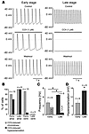
Parasympathetic slowing of the heart rate is predominantly mediated by acetylcholine-dependent activation of the G protein–gated potassium (K+) channel (IK,ACh). This channel is composed of 2 inward-rectifier K+ (Kir) channel subunits, Kir3.1 and Kir3.4, that display distinct functional properties. Here we show that subunit composition of IK,ACh changes during embryonic development. At early stages, IK,ACh is primarily formed by Kir3.1, while in late embryonic and adult cells, Kir3.4 is the predominant subunit. This change in subunit composition results in reduced rectification of IK,ACh, allowing for marked K+ currents over the whole physiological voltage range. As a consequence, IK,ACh is able to generate the membrane hyperpolarization that underlies the strong negative chronotropy occurring in late- but not early-stage atrial cardiomyocytes upon application of muscarinic agonists. Both strong negative chronotropy and membrane hyperpolarization can be induced in early-stage cardiomyocytes by viral overexpression of the mildly rectifying Kir3.4 subunit. Thus, a switch in subunit composition is used to adopt IK,ACh to its functional role in adult cardiomyocytes.
Authors
Bernd K. Fleischmann, Yaqi Duan, Yun Fan, Torsten Schoneberg, Andreas Ehlich, Nibedita Lenka, Serge Viatchenko-Karpinski, Lutz Pott, Juergen Hescheler, Bernd Fakler
Citation Information: J Clin Invest. 2004;114(4):485-494. https://doi.org/10.1172/JCI19596.
Abstract
One of the most perplexing questions in clinical genetics is why patients with identical gene mutations oftentimes exhibit radically different clinical features. This inconsistency between genotype and phenotype is illustrated in the malformation spectrum of holoprosencephaly (HPE). Family members carrying identical mutations in sonic hedgehog (SHH) can exhibit a variety of facial features ranging from cyclopia to subtle midline asymmetries. Such intrafamilial variability may arise from environmental factors acting in conjunction with gene mutations that collectively reduce SHH activity below a critical threshold. We undertook a series of experiments to test the hypothesis that modifying the activity of the SHH signaling pathway at discrete periods of embryonic development could account for the phenotypic spectrum of HPE. Exposing avian embryos to cyclopamine during critical periods of craniofacial development recreated a continuum of HPE-related defects. The craniofacial malformations included hypotelorism, midfacial hypoplasia, and facial clefting and were not the result of excessive crest cell apoptosis. Rather, they resulted from molecular reprogramming of an organizing center whose activity controls outgrowth and patterning of the mid and upper face. Collectively, these data reveal one mechanism by which the variable expressivity of a disorder such as HPE can be produced through temporal disruption of a single molecular pathway.
Authors
Dwight Cordero, Ralph Marcucio, Diane Hu, William Gaffield, Minal Tapadia, Jill A. Helms
Citation Information: J Clin Invest. 2004;113(12):1692-1700. https://doi.org/10.1172/JCI20384.
Abstract
Classical research has suggested that early palate formation develops via epithelial-mesenchymal interactions, and in this study we reveal which signals control this process. Using Fgf10–/–, FGF receptor 2b–/– (Fgfr2b–/–), and Sonic hedgehog (Shh) mutant mice, which all exhibit cleft palate, we show that Shh is a downstream target of Fgf10/Fgfr2b signaling. Our results demonstrate that mesenchymal Fgf10 regulates the epithelial expression of Shh, which in turn signals back to the mesenchyme. This was confirmed by demonstrating that cell proliferation is decreased not only in the palatal epithelium but also in the mesenchyme of Fgfr2b–/– mice. These results reveal a new role for Fgf signaling in mammalian palate development. We show that coordinated epithelial-mesenchymal interactions are essential during the initial stages of palate development and require an Fgf-Shh signaling network.
Authors
Ritva Rice, Bradley Spencer-Dene, Elaine C. Connor, Amel Gritli-Linde, Andrew P. McMahon, Clive Dickson, Irma Thesleff, David P.C. Rice
Citation Information: J Clin Invest. 2004;113(7):1051-1058. https://doi.org/10.1172/JCI20049.
Abstract
Congenital obstructive nephropathy is the principal cause of renal failure in infants and children. The underlying molecular and cellular mechanisms of this disease, however, remain largely undetermined. We generated a mouse model of congenital obstructive nephropathy that resembles ureteropelvic junction obstruction in humans. In these mice, calcineurin function is removed by the selective deletion of Cnb1 in the mesenchyme of the developing urinary tract using the Cre/lox system. This deletion results in reduced proliferation in the smooth muscle cells and other mesenchymal cells in the developing urinary tract. Compromised cell proliferation causes abnormal development of the renal pelvis and ureter, leading to defective pyeloureteral peristalsis, progressive renal obstruction, and, eventually, fatal renal failure. Our study demonstrates that calcineurin is an essential signaling molecule in urinary tract development and is required for normal proliferation of the urinary tract mesenchymal cells in a cell-autonomous manner. These studies also emphasize the importance of functional obstruction, resulting from developmental abnormality, in causing congenital obstructive nephropathy.
Authors
Ching-Pin Chang, Bradley W. McDill, Joel R. Neilson, Heidi E. Joist, Jonathan A. Epstein, Gerald R. Crabtree, Feng Chen
Citation Information: J Clin Invest. 2003;112(8):1152-1163. https://doi.org/10.1172/JCI17409.
Abstract
Embryo liver morphogenesis takes place after gastrulation and starts with a ventral foregut evagination that reacts to factor signaling from both cardiac mesoderm and septum transversum mesenchyme. Current knowledge of the progenitor stem cell populations involved in this early embryo liver development is scarce. We describe here a population of 11-day postcoitus c-Kitlow(CD45/TER119)– liver progenitors that selectively expressed hepatospecific genes and proteins in vivo, was self-maintained in vitro by long-term proliferation, and simultaneously differentiated into functional hepatocytes and bile duct cells. Purified c-Kitlow(CD45/TER119)– liver cells cocultured with cell-depleted fetal liver fragments engrafted and repopulated the hepatic cell compartments of the latter organoids, suggesting that they may include the embryonic stem cells responsible for liver development.
Authors
Susana Minguet, Isabel Cortegano, Pilar Gonzalo, José-Alberto Martínez-Marin, Belén de Andrés, Clara Salas, David Melero, Maria-Luisa Gaspar, Miguel A.R. Marcos
Citation Information: J Clin Invest. 2003;111(5):707-716. https://doi.org/10.1172/JCI17423.
Abstract
Kidney disease affects over 20 million people in the United States alone. Although the causes of renal failure are diverse, the glomerular filtration barrier is often the target of injury. Dysregulation of VEGF expression within the glomerulus has been demonstrated in a wide range of primary and acquired renal diseases, although the significance of these changes is unknown. In the glomerulus, VEGF-A is highly expressed in podocytes that make up a major portion of the barrier between the blood and urinary spaces. In this paper, we show that glomerular-selective deletion or overexpression of VEGF-A leads to glomerular disease in mice. Podocyte-specific heterozygosity for VEGF-A resulted in renal disease by 2.5 weeks of age, characterized by proteinuria and endotheliosis, the renal lesion seen in preeclampsia. Homozygous deletion of VEGF-A in glomeruli resulted in perinatal lethality. Mutant kidneys failed to develop a filtration barrier due to defects in endothelial cell migration, differentiation, and survival. In contrast, podocyte-specific overexpression of the VEGF-164 isoform led to a striking collapsing glomerulopathy, the lesion seen in HIV-associated nephropathy. Our data demonstrate that tight regulation of VEGF-A signaling is critical for establishment and maintenance of the glomerular filtration barrier and strongly supports a pivotal role for VEGF-A in renal disease.
Authors
Vera Eremina, Manish Sood, Jody Haigh, András Nagy, Ginette Lajoie, Napoleone Ferrara, Hans-Peter Gerber, Yamato Kikkawa, Jeffrey H. Miner, Susan E. Quaggin
Citation Information: J Clin Invest. 2003;111(4):453-461. https://doi.org/10.1172/JCI15924.
Abstract
Preadipocyte factor-1 (Pref-1) is a transmembrane protein highly expressed in preadipocytes. Pref-1 expression is, however, completely abolished in adipocytes. The extracellular domain of Pref-1 undergoes two proteolytic cleavage events that generate 50 and 25 kDa soluble products. To understand the function of Pref-1, we generated transgenic mice that express the full ectodomain corresponding to the large cleavage product of Pref-1 fused to human immunoglobulin-γ constant region. Mice expressing the Pref-1/hFc transgene in adipose tissue, driven by the adipocyte fatty acid–binding protein (aP2, also known as aFABP) promoter, showed a substantial decrease in total fat pad weight. Moreover, adipose tissue from transgenic mice showed reduced expression of adipocyte markers and adipocyte-secreted factors, including leptin and adiponectin, whereas the preadipocyte marker Pref-1 was increased. Pref-1 transgenic mice with a substantial, but not complete, loss of adipose tissue exhibited hypertriglyceridemia, impaired glucose tolerance, and decreased insulin sensitivity. Mice expressing the Pref-1/hFc transgene exclusively in liver under the control of the albumin promoter also showed a decrease in adipose mass and adipocyte marker expression, suggesting an endocrine mode of action of Pref-1. These findings demonstrate the inhibition of adipogenesis by Pref-1 in vivo and the resulting impairment of adipocyte function that leads to the development of metabolic abnormalities.
Authors
Kichoon Lee, Josep A. Villena, Yang Soo Moon, Kee-Hong Kim, Sunjoo Lee, Chulho Kang, Hei Sook Sul
Citation Information: J Clin Invest. 2002;110(11):1619-1628. https://doi.org/10.1172/JCI15621.
Abstract
Research Article
Authors
Akiyoshi Uemura, Minetaro Ogawa, Masanori Hirashima, Takashi Fujiwara, Shinji Koyama, Hitoshi Takagi, Yoshihito Honda, Stanley J. Wiegand, George D. Yancopoulos, Shin-Ichi Nishikawa
Citation Information: J Clin Invest. 2002;110(11):1629-1641. https://doi.org/10.1172/JCI13588.
Abstract
Research Article
Authors
Christine Fritsch, Elzbieta A. Swietlicki, Olivier Lefebvre, Michele Kedinger, Hristo Iordanov, Marc S. Levin, Deborah C. Rubin
No posts were found with this tag.


Copyright © 2025 American Society for Clinical Investigation
ISSN: 0021-9738 (print), 1558-8238 (online)


