Hiroshi Yamamoto, Charlotte E. Lee, Jacob N. Marcus, Todd D. Williams, J. Michael Overton, Marisol E. Lopez, Anthony N. Hollenberg, Laurie Baggio, Clifford B. Saper, Daniel J. Drucker, Joel K. Elmquist
Hiroshi Yamamoto, Charlotte E. Lee, Jacob N. Marcus, Todd D. Williams, J. Michael Overton, Marisol E. Lopez, Anthony N. Hollenberg, Laurie Baggio, Clifford B. Saper, Daniel J. Drucker, Joel K. Elmquist
Abstract
Research Article
Authors
Hiroshi Yamamoto, Charlotte E. Lee, Jacob N. Marcus, Todd D. Williams, J. Michael Overton, Marisol E. Lopez, Anthony N. Hollenberg, Laurie Baggio, Clifford B. Saper, Daniel J. Drucker, Joel K. Elmquist
Figure 2
Options:
View larger image
(or click on image)
Download as PowerPoint
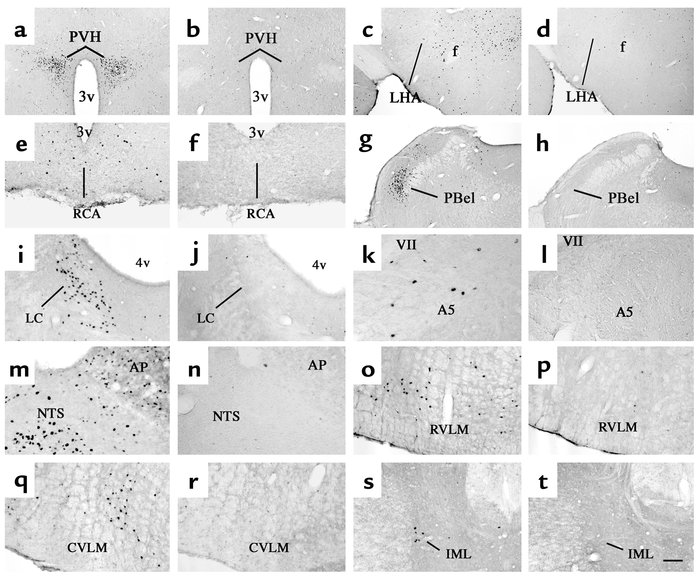
Distribution of i.v. EXN-4–induced Fos-IR in the brain. A series of photomicrographs demonstrates Fos-IR in neurons 2 hours after i.v. administration of EXN-4 (first and third columns) or PFS (second and fourth columns) in several brain regions. These regions include (a and b ) the paraventricular nucleus of the hypothalamus (PVH); (c and d ) the lateral hypothalamic area (LHA); (e and f ) the arcuate nucleus (Arc) and the retrochiasmatic area (RCA); (g and h ) the external lateral subdivision of the parabrachial nucleus (PBel); (i and j ) the locus coeruleus (LC); (k and l ) the A5 cell group (A5); (m and n ) the area postrema (AP) and the NTS; (o and p ) the rostral ventrolateral medulla (RVML); (q and r ) the caudal ventrolateral medulla (CVLM); and (s and t ) the IML in the spinal cord. 3v, third ventricle; f, fornix; 4v, fourth ventricle; VII, facial nerve. Scale bar = 250 μm in a –d , g , and h , and 100 μm in e , f , and i –t .


Copyright © 2025 American Society for Clinical Investigation
ISSN: 0021-9738 (print), 1558-8238 (online)

