
Oncology
Abstract
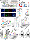
Glioblastoma (GBM) is a lethal brain tumor containing a subpopulation of GBM stem cells (GSCs) that interaction with surrounding cells, including infiltrating tumor-associated macrophages and microglia (TAMs). While GSCs and TAMs are in close proximity and likely interact to coordinate tumor growth, a limited number of mechanisms have been identified that support their communication. Here, we identified glycoprotein NMB (GPNMB) as a key factor mediating a unique bidirectional interaction between GSCs and TAMs in GBM. Specifically, GSCs educated macrophages and microglia to preferentially express GPNMB in the GBM tumor microenvironment. As a result, TAM-secreted GPNMB interacted with its receptor CD44 on GSCs to promote their glycolytic and self-renewal abilities via activating the PYK2/RSK2 signaling axis. Disrupting GPNMB-mediated GSC-TAM interplay suppressed tumor progression and self-renewal in GBM mouse models. Our study found a protumor function of GPNMB-mediated GSC-TAM bidirectional communication and supports GPNMB as a promising therapeutic target for GBM.
Authors
Yang Liu, Lizhi Pang, Fatima Khan, Junyan Wu, Fei Zhou, Craig Horbinski, Shideng Bao, Jennifer S. Yu, Justin D. Lathia, Peiwen Chen

Abstract
Immune checkpoint inhibitors (ICI) have revolutionized cancer therapy, but their use is limited by the development of autoimmunity in healthy tissues as a side effect of treatment. Such immune-related adverse events (IrAE) contribute to hospitalizations, cancer treatment interruption, and even premature death. ICI-induced autoimmune diabetes mellitus (ICI-T1DM) is a life-threatening IrAE that presents with rapid pancreatic β-islet cell destruction leading to hyperglycemia and life-long insulin dependence. While prior reports have focused on CD8+ T cells, the role for CD4+ T cells in ICI-T1DM is less understood. We identify expansion of CD4+ T follicular helper (Tfh) cells expressing IL-21 and IFN-γ as a hallmark of ICI-T1DM. Furthermore, we show that both IL-21 and IFN-γ are critical cytokines for autoimmune attack in ICI-T1DM. Because IL-21 and IFN-γ both signal through JAK/STAT pathways, we reasoned that JAK inhibitors (JAKi) may protect against ICI-T1DM. Indeed, JAKi provide robust in vivo protection against ICI-T1DM in a mouse model that is associated with decreased islet-infiltrating Tfh cells. Moreover, JAKi therapy impaired Tfh cell differentiation in patients with ICI-T1DM. These studies highlight CD4+ Tfh cells as underrecognized but critical mediators of ICI-T1DM that may be targeted with JAKi to prevent this grave IrAE.
Authors
Nicole L. Huang, Jessica G. Ortega, Kyleigh Kimbrell, Joah Lee, Lauren N. Scott, Esther M. Peluso, Sarah J. Wang, Ellie Y. Kao, Kristy Kim, Jarod Olay, Jaden N. Nguyen, Zoe Quandt, Trevor E. Angell, Maureen A. Su, Melissa G. Lechner
Abstract
The tumor microenvironment (TME) is highly heterogeneous and can dictate the success of therapeutic interventions. Identifying TMEs that are susceptible to specific therapeutic interventions paves the way for more personalized and effective treatments. In this study, using a spontaneous murine model of breast cancer, we characterize a TME that is responsive to inhibitors of the heme degradation pathway mediated by heme oxygenase (HO), resulting in CD8+ T- and NK-cell-dependent tumor control. A hallmark of this TME is a chronic type-I interferon (IFN) signal that is directly involved in orchestrating the anti-tumor immune response. Importantly, we identify that similar TMEs exist in human breast cancer which are associated with patient prognosis. Leveraging these observations, we demonstrate that combining a STING agonist, which induces type-I IFN responses, with an HO inhibitor produces a synergistic effect leading to superior tumor control. This study highlights HO activity as a potential resistance mechanism for type-I IFN responses in cancer offering a novel avenue for overcoming immune evasion in cancer therapy.
Authors
Dominika Sosnowska, Tik Shing Cheung, Jit Sarkar, James W. Opzoomer, Karen T. Feehan, Joanne E. Anstee, Chloé Amelia Woodman, Mohamed Reda Keddar, Kalum Clayton, Samira Ali, William Macmorland, Dorothy D. Yang, James Rosekilly, Cheryl E. Gillett, Francesca D. Ciccarelli, Richard Buus, James Spicer, Anita Grigoriadis, James N. Arnold
Abstract
Pancreatic ductal adenocarcinoma (PDAC) has a poor survival rate due to late detection. PDAC arises from precursor microscopic lesions, termed pancreatic intraepithelial neoplasia (PanIN), that develop at least a decade before overt disease––this provides an opportunity to intercept PanIN–to–PDAC progression. However, immune interception strategies require full understanding of PanIN and PDAC cellular architecture. Surgical specimens containing PanIN and PDAC lesions from a unique cohort of five treatment-naïve patients with PDAC were surveyed using spatial-omics (proteomic and transcriptomic). Findings were corroborated by spatial proteomics of PanIN and PDAC from tamoxifen-inducible KPC (tiKPC) mice. We uncovered the organization of lymphoid cells into tertiary lymphoid structures (TLSs) adjacent to PanIN lesions. These TLSs lacked CD21+CD23+ B cells compared to more mature TLSs near the PDAC border. PanINs harbored mostly CD4+ T cells with fewer Tregs and exhausted T cells than PDAC. Peri-tumoral space was enriched with naïve CD4+ and central memory T cells. These observations highlight the opportunity to modulate the immune microenvironment in PanINs before immune exclusion and immunosuppression emerge during progression into PDAC.
Authors
Melissa R. Lyman, Jacob T. Mitchell, Sidharth Raghavan, Luciane T. Kagohara, Amanda L. Huff, Saurav D. Haldar, Sarah M. Shin, Samantha Guinn, Benjamin Barrett, Gabriella Longway, Alexei Hernandez, Erin M. Coyne, Xuan Yuan, Lalitya Andaloori, Jiaying Lai, Yun Zhou Liu, Rachel Karchin, Anuj Gupta, Ashley L. Kiemen, André Forjaz, Denis Wirtz, Pei-Hsun Wu, Atul Deshpande, Jae W. Lee, Todd D. Armstrong, Nilofer S. Azad, Jacquelyn W. Zimmerman, Laura D. Wood, Robert A. Anders, Elizabeth D. Thompson, Elizabeth M. Jaffee, Elana J. Fertig, Won Jin Ho, Neeha Zaidi
Abstract
Androgen receptor positive prostate cancer (PC), castration resistant prostate cancer (CRPC) and neuroendocrine prostate cancer (NEPC) invariably become resistant to treatment with targeted and cytotoxic agents. Multiple pathways have been identified as being responsible for these pleotropic mechanisms of resistance. The MUC1 gene is aberrantly expressed in CRPC/NEPC in association with poor clinical outcomes; whereas, it is not known if the oncogenic MUC1-C/M1C protein drives treatment resistance. We demonstrated that MUC1-C is necessary for resistance of (i) PC cells to enzalutamide (ENZ), and (ii) CRPC and NEPC cells to docetaxel (DTX). Our results showed that MUC1-C-mediated resistance is conferred by upregulation of aerobic glycolysis and suppression of reactive oxygen species necessary for self-renewal. Dependence of these resistant phenotypes on MUC1-C for the cancer stem cell (CSC) state identified a potential target for treatment. In this regard, we further demonstrated that targeting MUC1-C with a M1C antibody-drug conjugate (ADC) is highly effective in suppressing (i) self-renewal of drug-resistant CRPC/NEPC CSCs and (ii) growth of t-NEPC tumor xenografts derived from drug-resistant cells and a patient with refractory disease. These findings uncovered a common MUC1-C-dependent pathway in treatment-resistant CRPC/NEPC progression and identified MUC1-C as a target for their treatment with a M1C ADC.
Authors
Keisuke Shigeta, Tatsuaki Daimon, Hiroshi Hongo, Sheng-Yu Ku, Hiroki Ozawa, Naoki Haratake, Atsushi Fushimi, Ayako Nakashoji, Atrayee Bhattacharya, Shinkichi Takamori, Michihisa Kono, Masahiro Rokugo, Yuto Baba, Takeo Kosaka, Mototsugu Oya, Justine Jacobi, Mark D. Long, Himisha Beltran, Donald W. Kufe
Abstract
Although obesity is a major risk factor for cancer, it may also improve the response to cancer therapy. Here we investigated the impact of obesity on the efficacy of immune checkpoint inhibitors (ICI). In male mice, obesity promoted tumor growth but enhanced the response to ICI. This was associated with higher expression of immune-related genes within the tumor and enhanced infiltration of tumor-specific CD8+ T cells. Further, obesity in mice was associated with higher estrogen levels and enrichment of estrogen response genes in the tumor, and anti-PD-1 efficacy was reduced upon administration of the aromatase inhibitor letrozole, which blocks the production of estrogens. Mechanistically, adipocyte-derived estrogens increased antigen presentation by dendritic cells and tumor-specific CD8+ T-cell cytotoxicity. Lastly, overweight and obese male melanoma patients responded better to ICI, high estrogen levels being associated with improved response and survival. Our results suggest that estrogens may serve as a predictive factor of response to ICI in men with melanoma.
Authors
Eloïse Dupuychaffray, Hélène Poinot, Aurélie Vuilleumier, Maxime Borgeaud, Montserrat Alvarez, Betül Taskoparan, Olivier Preynat-Seauve, Clarissa D. Voegel, Eliana Marinari, Denis Migliorini, Valérie Dutoit, Carole Bourquin, Aurélien Pommier
Abstract
The prognosis for colorectal cancer (CRC) patients with liver metastasis remains poor, and the molecular mechanisms driving CRC liver metastasis are still not fully understood. Hypoxia-induced extracellular vesicles (H-EVs) derived from tumors have emerged as key players in inducing angiogenesis by transferring non-coding RNAs. However, the specific role of CRC-derived hypoxic EVs (H-EVs) in regulating the formation of the pre-metastatic microenvironment (PMN) by inducing angiogenesis remains unclear. Our study demonstrates that H-EVs induce both angiogenesis and liver metastasis. Through microRNA microarray analysis, we identified a reduction in miR-6084 levels within H-EVs. We found that miR-6084 inhibits angiogenesis by being transferred to endothelial cells via EVs. In endothelial cells, miR-6084 directly targets ANGPTL4 mRNA, thereby suppressing angiogenesis through ANGPTL4-mediated JAK2/STAT3 pathway. Furthermore, we uncovered that SP1 acts as a transcription factor regulating miR-6084 transcription, while HIF1A decreases miR-6084 expression by promoting SP1 protein dephosphorylation and facilitating ubiquitin‒proteasome degradation in SW620 cells. In clinical samples, we observed low expression of miR-6084 in plasma-derived EVs from CRC patients with liver metastasis. In summary, our findings suggest that CRC-derived hypoxic EVs promote angiogenesis and liver metastasis through the HIF1A/SP1/miR-6084/ANGPTL4 axis. Additionally, miR-6084 holds promise as a potential diagnostic and prognostic biomarker for CRC liver metastasis.
Authors
Yang Zhang, Xuyang Yang, Su Zhang, Qing Huang, Sicheng Liu, Lei Qiu, Mingtian Wei, Xiangbing Deng, Wenjian Meng, Hai-Ning Chen, Yaguang Zhang, Junhong Han, Ziqiang Wang
Abstract
For over a century, scientists reported the disruption of normal nuclear shape and size in cancer. These changes have long been used as tools for diagnosis and staging of malignancies. However, to date, the mechanisms underlying these aberrant nuclear phenotypes and their biological significance remain poorly understood. Using a model of pancreatic ductal adenocarcinoma (PDAC), the major histological subtypes of pancreatic cancer, we found oncogenic mutant KRAS reduces nuclear size. Transcriptomic and protein expression analysis of mutant KRAS-expressing PDAC cells revealed differential levels of several nuclear envelope-associated genes. Further analysis demonstrated the nuclear lamina protein, Emerin (EMD), acted downstream of KRAS to mediate nuclear size reduction in PDAC. Analysis of human PDAC samples showed that increased EMD expression associates with reduced nuclear size. Finally, in vivo genetic depletion of EMD in a mutant KRAS-driven PDAC model resulted in an increased nuclear size and a reduced incidence of poorly differentiated PDAC. Thus, our data provides evidence of a novel mechanism underlying nuclear size regulation and its impact in PDAC carcinogenesis.
Authors
Luis F. Flores, David L. Marks, Renzo E. Vera, Ashley N. Sigafoos, Ezequiel J. Tolosa, Luciana L. Almada, David R. Pease, Merih D. Toruner, Brian Chang, Brooke R. Tader, Kayla C. LaRue-Nolan, Ryan M. Carr, Rondell P. Graham, Catherine E. Hagen, Matthew R. Brown, Aleksey V. Matveyenko, Katherine L. Wilson, David W. Dawson, Christopher L. Pin, Kyle J. Roux, Martin E. Fernandez-Zapico
Abstract
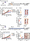
Despite aggressive chemoradiation treatment, the overall survival rate for patients with HPV– head and neck squamous cell carcinoma (HNSCC) remains poor, highlighting the urgent need for more effective drug-radiotherapy combinations to improve the therapeutic index of radiation therapy (RT). The fat mass and obesity-related gene (FTO) is emerging as a promising cancer therapeutic target; however, its role in the RT response has been underexplored. In our study, we found that both genetic and pharmacologic inhibition of FTO enhanced the efficacy of RT in human and mouse HNSCC tumor xenografts. Mechanistically, inhibition of FTO improved the RT response in HPV– HNSCC cells, which was associated with increased DNA damage, reduced efficiency of homology directed repair, and decreased formation of RAD51 homolog 1 (RAD51) foci. Importantly, pharmacologic inhibition of FTO did not exacerbate radiation-induced oral mucositis, a significant normal-tissue toxicity associated with HNSCC RT. In summary, our results indicate a role for FTO in regulating homologous recombination while identifying FTO as a potential therapeutic target to enhance the therapeutic index of RT in HPV– HNSCC treatment.
Authors
Lu Ji, Leighton Pu, Jinglong Wang, Hongbin Cao, Stavros Melemenidis, Subarna Sinha, Li Guan, Eyiwunmi E. Laseinde, Rie von Eyben, Sara A. Richter, Jin-Min Nam, Christina Kong, Kerriann M. Casey, Edward E. Graves, Richard L. Frock, Quynh Thu Le, Erinn B. Rankin
Abstract
Resistance to chemotherapy of pancreatic ductal adenocarcinoma (PDAC) is largely driven by intratumoral heterogeneity (ITH) due to tumor cell plasticity and clonal diversity. In order to develop novel strategies to overcome this defined mechanism of resistance, tools to monitor and quantify ITH in a rapid and scalable fashion are needed urgently. Here, we employed label-free digital holographic microscopy (DHM) to characterize ITH in PDAC. We established a robust experimental and machine learning analysis pipeline to perform single cell phenotyping based on DHM-derived phase images of PDAC cells in suspension. Importantly, we are able to detect dynamic changes in tumor cell differentiation and heterogeneity of distinct PDAC subtypes upon induction of epithelial-to-mesenchymal transition and under treatment-imposed pressure in murine and patient-derived model systems. This platform allows us to assess phenotypic ITH in PDAC on a single cell level in real-time. Implementing this technology into the clinical workflow has the potential to fundamentally increase our understanding of tumor heterogeneity during evolution and treatment response.
Authors
Katja Wittenzellner, Manuel Lengl, Stefan Röhrl, Carlo Maurer, Christian Klenk, Aristeidis Papargyriou, Laura Schmidleitner, Nicole Kabella, Akul Shastri, David E. Fresacher, Farid Harb, Nawal Hafez, Stefanie Bärthel, Daniele Lucarelli, Carmen Escorial-Iriarte, Felix Orben, Rupert Öllinger, Ellen Emken, Lisa Fricke, Joanna Madej, Patrick Wustrow, I. Ekin Demir, Helmut Friess, Tobias Lahmer, Roland M. Schmid, Roland Rad, Günter Schneider, Bernhard Kuster, Dieter Saur, Oliver Hayden, Klaus Diepold, Maximilian Reichert
No posts were found with this tag.


