Issue published November 15, 2004 Previous issue | Next issue

- Volume 114, Issue 10
Go to section:

-
Science and Society
×
Abstract
Human embryonic stem cells offer the promise of a new regenerative medicine in which damaged adult cells can be replaced with new cells. Research is needed to determine the most viable stem cell lines and reliable ways to promote the differentiation of pluripotent stem cells into specific cell types (neurons, muscle cells, etc.). To create new cell lines, it is necessary to destroy preimplantation blastocysts. This has led to an intense debate that threatens to limit embryonic stem cell research. The profound ethical issues raised call for informed, dispassionate debate.
Authors
Gerald D. Fischbach, Ruth L. Fischbach


-
Review Series
×
Abstract
Effective immune responses against pathogens are sometimes accompanied by strong inflammatory reactions. To minimize damage to self, the activation of the immune system also triggers anti-inflammatory circuits. Both inflammatory and anti-inflammatory reactions are normal components of the same immune response, which coordinately fight infections while preventing immune pathology. IL-10 is an important suppressive cytokine, produced by a large number of immune cells in addition to the antigen-driven IL-10–producing regulatory and the naturally occurring suppressor CD4+ T cells, which is a key player in anti-inflammatory immune responses. However, additional mechanisms have evolved to ensure that pathogen eradication is achieved with minimum damage to the host. Here we discuss those mechanisms that operate to regulate effector immune responses.
Authors
Anne O’Garra, Pedro L. Vieira, Paulo Vieira, Anne E. Goldfeld
×Abstract
NKT cells are a unique T lymphocyte sublineage that has been implicated in the regulation of immune responses associated with a broad range of diseases, including autoimmunity, infectious diseases, and cancer. In stark contrast to both conventional T lymphocytes and other types of Tregs, NKT cells are reactive to the nonclassical class I antigen–presenting molecule CD1d, and they recognize glycolipid antigens rather than peptides. Moreover, they can either up- or downregulate immune responses by promoting the secretion of Th1, Th2, or immune regulatory cytokines. This review will explore the diverse influences of these cells in various disease models, their ability to suppress or enhance immunity, and the potential for manipulating these cells as a novel form of immunotherapy.
Authors
Dale I. Godfrey, Mitchell Kronenberg
×Abstract
Allergic diseases such as asthma, rhinitis, and eczema are increasing in prevalence and affect up to 15% of populations in Westernized countries. The description of Tregs as T cells that prevent development of autoimmune disease led to considerable interest in whether these Tregs were also normally involved in prevention of sensitization to allergens and whether it might be possible to manipulate Tregs for the therapy of allergic disease. Current data suggest that Th2 responses to allergens are normally suppressed by both CD4+CD25+ Tregs and IL-10 Tregs. Furthermore, suppression by these subsets is decreased in allergic individuals. In animal models, Tregs could be induced by high- or low-dose inhaled antigen, and prior induction of such Tregs prevented subsequent development of allergen sensitization and airway inflammation in inhaled challenge models. For many years, allergen-injection immunotherapy has been used for the therapy of allergic disease, and this treatment may induce IL-10 Tregs, leading to both suppression of Th2 responses and a switch from IgE to IgG4 antibody production. Improvements in allergen immunotherapy, such as peptide therapy, and greater understanding of the biology of Tregs hold great promise for the treatment and prevention of allergic disease.
Authors
Douglas S. Robinson, Mark Larché, Stephen R. Durham
×Abstract
The induction and maintenance of immune tolerance to transplanted tissues constitute an active process involving multiple mechanisms that work cooperatively to prevent graft rejection. These mechanisms are similar to inherent tolerance toward self antigens and have a requirement for active immunoregulation, largely T cell mediated, that promotes specific unresponsiveness to donor alloantigens. This review outlines our current understanding of the Treg subsets that contribute to allotolerance and the mechanisms by which these cells exert their effects as well as their potential for therapy.
Authors
Patrick T. Walsh, Devon K. Taylor, Laurence A. Turka


-
Commentaries
×
Abstract
Polycystin-1, the protein encoded by the principal gene involved in autosomal dominant polycystic kidney disease, has been implicated in extracellular sensing as well as in cell-cell and cell-matrix interactions. However, the precise mechanisms involved in polycystin-1 signaling are not well defined. A report in this issue of the JCI demonstrates that the C-terminal tail of polycystin-1 is cleaved from the membrane through regulated intramembrane proteolysis (RIP) and that this domain translocates to the nucleus, where it activates the AP-1 transcription pathway. This translocation appears to be modulated by polycystin-2, with which polycystin-1 is thought to interact. These findings provide what we believe to be the first evidence that polycystin-1 can signal directly to the nucleus and that polycystin-1–polycystin-2 interactions do not require colocalization of these proteins in the same membrane compartment.
Authors
Lisa M. Guay-Woodford
×Abstract
Autosomal dominant disorders of the skin may present in a pattern following the lines of embryologic development of the ectoderm. In these cases, the surrounding skin is normal, and molecular studies have shown that the causative mutation is confined to the affected ectodermal tissue (type 1 mosaicism). Rarely, an individual shows skin lesions that follow the pattern of type 1 mosaicism, but the rest of the skin shows a milder form of the disorder (type 2 mosaicism). A new study provides the molecular basis for type 2 mosaicism .
Authors
Amy S. Paller
×Abstract
SCID, a syndrome characterized by the absence of T cells and adaptive immunity, can result from mutations in multiple genes that encode components of the immune system. Three such components are cytokine receptor chains or signaling molecules, five are needed for antigen receptor development, one is adenosine deaminase — a purine salvage pathway enzyme, and the last is a phosphatase, CD45. In this issue of the JCI, a report describes how complete deficiency of the CD3ε chain of the T cell antigen receptor/CD3 complex causes human SCID.
Authors
Rebecca H. Buckley
×Abstract
There have been exciting recent advances in our understanding of the structural and molecular biology of the glomerular slit diaphragm, as described in a report in this issue of the JCI. These findings, combined with data on the permeability of the basement membrane and evidence that the endothelium may be a more important barrier than often supposed, are allowing a clearer understanding to emerge of how the 3 parts of the glomerular capillary wall jointly determine its functional properties.
Authors
William M. Deen
×Abstract
The Randle cycle, which has been invoked to explain the reciprocal relationship between fatty acid oxidation and glucose oxidation, has long been implicated as a potential mechanism for hyperglycemia and type 2 diabetes mellitus (T2DM). Now genetic, functional genomic, and transgenic approaches have identified PPARγ coactivators (PGC-1α and PGC-1β) as key regulators of mitochondrial number and function. They regulate adaptive thermogenesis as well as glucose and fat oxidation in muscle and fat tissue, gluconeogenesis in liver, and even glucose-regulated insulin secretion in β cells. PGC-1α and PGC-1β mRNA levels and the mitochondrial genes they regulate are decreased in muscle of people with prediabetes and T2DM. A new report indicates that PGC-1α and PGC-1β mRNA levels decrease with age in individuals with a genetic variant in PGC-1α, and these decreases correlate with alterations in whole-body glucose and fatty acid oxidation. These findings provide insights into how aging modifies genetic susceptibility to alterations in oxidative phosphorylation and T2DM.
Authors
Alan R. Shuldiner, John C. McLenithan


-
Research Articles
×
Abstract
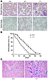
Scatter factor (SF), also known as hepatocyte growth factor, is ubiquitously present in the extracellular matrix of tissues in the form of an inactive precursor (pro-SF). In order to acquire biological activity, pro-SF must be cleaved by specific proteases present on the cell surface. The mature form of SF controls invasive cues in both physiological and pathological processes through activation of its receptor, the Met tyrosine kinase. By substituting a single amino acid in the proteolytic site, we engineered an unprocessable form of pro-SF (uncleavable SF). Using lentivirus vector technology, we achieved local or systemic delivery of uncleavable SF in mice. We provide evidence that (a) uncleavable SF inhibits both protease-mediated pro-SF conversion and active SF–induced Met activation; (b) local expression of uncleavable SF in tumors suppresses tumor growth, impairs tumor angiogenesis, and prevents metastatic dissemination; and (c) systemic expression of uncleavable SF dramatically inhibits the growth of transplanted tumors and abolishes the formation of spontaneous metastases without perturbing vital physiological functions. These data show that proteolytic activation of pro-SF is a limiting step in tumor progression, thus suggesting a new strategy for the treatment or prevention of the malignant conversion of neoplastic lesions.
Authors
Massimiliano Mazzone, Cristina Basilico, Silvia Cavassa, Selma Pennacchietti, Mauro Risio, Luigi Naldini, Paolo M. Comoglio, Paolo Michieli
×Abstract
Polycystin-1, which is encoded by a gene that is mutated in autosomal dominant polycystic kidney disease (ADPKD), is involved in cell-matrix interactions as well as in ciliary signaling. The precise mechanisms by which it functions, however, remain unclear. Here we find that polycystin-1 undergoes a proteolytic cleavage that releases its C-terminal tail (CTT), which enters the nucleus and initiates signaling processes. The cleavage occurs in vivo in association with alterations in mechanical stimuli. Polycystin-2, the product of the second gene mutated in ADPKD, modulates the signaling properties of the polycystin-1 CTT. These data reveal a novel pathway by which polycystin-1 transmits messages directly to the nucleus.
Authors
Veronique Chauvet, Xin Tian, Herve Husson, David H. Grimm, Tong Wang, Thomas Hieseberger, Peter Igarashi, Anton M. Bennett, Oxana Ibraghimov-Beskrovnaya, Stefan Somlo, Michael J. Caplan
×Abstract
Proteinase-activated receptor–1 (PAR1), a G protein–coupled receptor activated by thrombin, is highly expressed in different cell types of the gastrointestinal tract. The activity of thrombin and of other proteinases is significantly increased in the colon of inflammatory bowel disease (IBD) patients. Since PAR1 activation in tissues other than the gut provoked inflammation, we hypothesized that PAR1 activation in the colon is involved in the pathogenesis of IBD. Here, we demonstrate that PAR1 is overexpressed in the colon of IBD patients. In mice, intracolonic administration of PAR1 agonists led to an inflammatory reaction characterized by edema and granulocyte infiltration. This PAR1 activation–induced inflammation was dependent on B and T lymphocytes. Moreover, PAR1 activation exacerbated and prolonged inflammation in a mouse model of IBD induced by the intracolonic administration of trinitrobenzene sulfonic acid (TNBS), while PAR1 antagonism significantly decreased the mortality and severity of colonic inflammation induced by TNBS and dextran sodium sulfate. In these 2 models, colitis development was strongly attenuated by PAR1 deficiency. Taken together, these results imply an important role for PAR1 in the pathogenesis of experimental colitis, supporting the notion that PAR1 inhibition may be beneficial in the context of IBD and possibly in other chronic intestinal inflammatory disorders.
Authors
Nathalie Vergnolle, Laurie Cellars, Andrea Mencarelli, Giovanni Rizzo, Sunita Swaminathan, Paul Beck, Martin Steinhoff, Patricia Andrade-Gordon, Nigel W. Bunnett, Morley D. Hollenberg, John L. Wallace, Giuseppe Cirino, Stefano Fiorucci
×Abstract
Hemoglobin (Hb) A production during red blood cell development is coordinated to minimize the deleterious effects of free α- and β-Hb subunits, which are unstable and cytotoxic. The α-Hb–stabilizing protein (AHSP) is an erythroid protein that specifically binds α-Hb and prevents its precipitation in vitro, which suggests that it may function to limit free α-Hb toxicities in vivo. We investigated this possibility through gene ablation and biochemical studies. AHSP–/– erythrocytes contained hemoglobin precipitates and were short-lived. In hematopoietic tissues, erythroid precursors were elevated in number but exhibited increased apoptosis. Consistent with unstable α-Hb, AHSP–/– erythrocytes contained increased ROS and evidence of oxidative damage. Moreover, purified recombinant AHSP inhibited ROS production by α-Hb in solution. Finally, loss of AHSP worsened the phenotype of β-thalassemia, a common inherited anemia characterized by excess free α-Hb. Together, the data support a model in which AHSP binds α-Hb transiently to stabilize its conformation and render it biochemically inert prior to Hb A assembly. This function is essential for normal erythropoiesis and, to a greater extent, in β-thalassemia. Our findings raise the possibility that altered AHSP expression levels could modulate the severity of β-thalassemia in humans.
Authors
Yi Kong, Suiping Zhou, Anthony J. Kihm, Anne M. Katein, Xiang Yu, David A. Gell, Joel P. Mackay, Kazuhiko Adachi, Linda Foster-Brown, Calvert S. Louden, Andrew J. Gow, Mitchell J. Weiss
×Abstract
Hailey-Hailey disease (HHD) is an autosomal dominant trait characterized by erythematous and oozing skin lesions preponderantly involving the body folds. In the present unusual case, however, unilateral segmental areas along the lines of Blaschko showing a rather severe involvement were superimposed on the ordinary symmetrical phenotype. Based on this observation and similar forms of mosaicism as reported in other autosomal dominant skin disorders, we postulated that in such cases, 2 different types of segmental involvement can be distinguished. Accordingly, the linear lesions as noted in the present case would exemplify type 2 segmental HHD. In the heterozygous embryo, loss of heterozygosity occurring at an early developmental stage would have given rise to pronounced linear lesions reflecting homozygosity or hemizygosity for the mutation. By analyzing DNA and RNA derived from blood and skin samples as well as keratinocytes of the index patient with various molecular techniques including RT-PCR, real-time PCR, and microsatellite analysis, we found a consistent loss of the paternal wild-type allele in more severely affected segmental skin regions, confirming this hypothesis for the first time, to our knowledge, at the molecular and cellular level.
Authors
Pamela Poblete-Gutiérrez, Tonio Wiederholt, Arne König, Frank K. Jugert, Yvonne Marquardt, Albert Rübben, Hans F. Merk, Rudolf Happle, Jorge Frank
×Abstract
Nephrin is a key functional component of the slit diaphragm, the structurally unresolved molecular filter in renal glomerular capillaries. Abnormal nephrin or its absence results in severe proteinuria and loss of the slit diaphragm. The diaphragm is a thin extracellular membrane spanning the approximately 40-nm-wide filtration slit between podocyte foot processes covering the capillary surface. Using electron tomography, we show that the slit diaphragm comprises a network of winding molecular strands with pores the same size as or smaller than albumin molecules, as demonstrated in humans, rats, and mice. In the network, which is occasionally stratified, immunogold-nephrin antibodies labeled individually detectable globular cross strands, about 35 nm in length, lining the lateral elongated pores. The cross strands, emanating from both sides of the slit, contacted at the slit center but had free distal endings. Shorter strands associated with the cross strands were observed at their base. Immunolabeling of recombinant nephrin molecules on transfected cells and in vitrified solution corroborated the findings in kidney. Nephrin-deficient proteinuric patients with Finnish-type congenital nephrosis and nephrin-knockout mice had only narrow filtration slits that lacked the slit diaphragm network and the 35-nm-long strands but contained shorter molecular structures. The results suggest the direct involvement of nephrin molecules in constituting the macromolecule-retaining slit diaphragm and its pores.
Authors
Jorma Wartiovaara, Lars-Göran Öfverstedt, Jamshid Khoshnoodi, Jingjing Zhang, Eetu Mäkelä, Sara Sandin, Vesa Ruotsalainen, R. Holland Cheng, Hannu Jalanko, Ulf Skoglund, Karl Tryggvason
×Abstract
Desmogleins (Dsgs), cadherin-type cell adhesion molecules, are targeted in skin-blistering diseases such as pemphigus and staphylococcal scalded skin syndrome (SSSS). The role of Dsg4, a new isoform, was investigated in these diseases. Dsg4 was recognized by 30 (77%) of 39 pemphigus sera containing anti-Dsg1 IgG but not by 16 pemphigus sera containing no anti-Dsg1 IgG or by 34 normal control sera. The Dsg4 immunoreactivity of these sera was abolished by removal of anti-Dsg1 IgG. Conversely, the removal of anti-Dsg4 IgG from pemphigus sera reduced the immunoreactivity against Dsg1 only 13.8% ± 8.8% (n = 23) and did not affect its ability to induce blisters in neonatal mice. IgG that was affinity-purified on Dsg4 recognized Dsg1 but failed to induce blisters, while IgG purified on Dsg1 from the same pemphigus foliaceus sera induced blisters. Thus, pemphigus sera show Dsg4 reactivity due to cross-reactivity of a subset of anti-Dsg1 IgG, and the Dsg4/Dsg1–cross-reacting IgG has no demonstrable pathogenic effect. In addition, Dsg4 was not cleaved by exfoliative toxins that induce blisters in SSSS. These findings suggest that Dsg4 may play a role other than adhesion and that the cross-reactivity of desmoglein autoantibodies should be factored into the framework of future studies of autoimmune mechanisms in pemphigus.
Authors
Takeshi Nagasaka, Koji Nishifuji, Takayuki Ota, Neil V. Whittock, Masayuki Amagai
×Abstract
The apolipoprotein apoC-III plays an important role in plasma triglyceride metabolism. It is predominantly produced in liver, and its hepatic expression is inhibited by insulin. To elucidate the inhibitory mechanism of insulin in apoC-III expression, we delivered forkhead box O1 (Foxo1) cDNA to hepatocytes by adenovirus-mediated gene transfer. Foxo1 stimulated hepatic apoC-III expression and correlated with the ability of Foxo1 to bind to its consensus site in the apoC-III promoter. Deletion or mutation of the Foxo1 binding site abolished insulin response and Foxo1-mediated stimulation. Likewise, Foxo1 also mediated insulin action on intestinal apoC-III expression in enterocytes. Furthermore, elevated Foxo1 production in liver augmented hepatic apoC-III expression, resulting in increased plasma triglyceride levels and impaired fat tolerance in mice. Transgenic mice expressing a constitutively active Foxo1 allele exhibited hypertriglyceridemia. Moreover, we show that hepatic Foxo1 expression becomes deregulated as a result of insulin deficiency or insulin resistance, culminating in significantly elevated Foxo1 production, along with its skewed nuclear distribution, in livers of diabetic NOD or db/db mice. While loss of insulin response is associated with unrestrained apoC-III production and impaired triglyceride metabolism, these data suggest that Foxo1 provides a molecular link between insulin deficiency or resistance and aberrant apoC-III production in the pathogenesis of diabetic hypertriglyceridemia.
Authors
Jennifer Altomonte, Lin Cong, Sonal Harbaran, Anja Richter, Jing Xu, Marcia Meseck, Hengjiang Henry Dong
×Abstract
The intracellular signals that mediate skeletal muscle protein loss and functional deficits due to muscular disuse are just beginning to be elucidated. Previously we showed that the activity of an NF-κB–dependent reporter gene was markedly increased in unloaded muscles, and p50 and Bcl-3 proteins were implicated in this induction. In the present study, mice with a knockout of the p105/p50 (Nfkb1) gene are shown to be resistant to the decrease in soleus fiber cross-sectional area that results from 10 days of hindlimb unloading. Furthermore, the marked unloading-induced activation of the NF-κB reporter gene in soleus muscles from WT mice was completely abolished in soleus muscles from Nfkb1 knockout mice. Knockout of the B cell lymphoma 3 (Bcl3) gene also showed an inhibition of fiber atrophy and an abolition of NF-κB reporter activity. With unloading, fast fibers from WT mice atrophied to a greater extent than slow fibers. Resistance to atrophy in both strains of knockout mice was demonstrated clearly in fast fibers, while slow fibers from only the Bcl3–/– mice showed atrophy inhibition. The slow-to-fast shift in myosin isoform expression due to unloading was also abolished in both Nfkb1 and Bcl3 knockout mice. Like the soleus muscles, plantaris muscles from Nfkb1–/– and Bcl3–/– mice also showed inhibition of atrophy with unloading. Thus both the Nfkb1 and the Bcl3 genes are necessary for unloading-induced atrophy and the associated phenotype transition.
Authors
R. Bridge Hunter, Susan C. Kandarian
×Abstract
We investigated the molecular mechanism underlying a severe combined immunodeficiency characterized by the selective and complete absence of T cells. The condition was found in 5 patients and 2 fetuses from 3 consanguineous families. Linkage analysis performed on the 3 families revealed that the patients were carrying homozygous haplotypes within the 11q23 region, in which the genes encoding the γ, δ, and ε subunits of CD3 are located. Patients and affected fetuses from 2 families were homozygous for a mutation in the CD3D gene, and patients from the third family were homozygous for a mutation in the CD3E gene. The thymus from a CD3δ-deficient fetus was analyzed and revealed that T cell differentiation was blocked at entry into the double positive (CD4+CD8+) stage with the accumulation of intermediate CD4–single positive cells. This indicates that CD3δ plays an essential role in promoting progression of early thymocytes toward double-positive stage. Altogether, these findings extend the known molecular mechanisms underlying severe combined immunodeficiency to a new deficiency, i.e., CD3ε deficiency, and emphasize the essential roles played by the CD3ε and CD3δ subunits in human thymocyte development, since these subunits associate with both the pre-TCR and the TCR.
Authors
Geneviève de Saint Basile, Frédéric Geissmann, Elisabeth Flori, Béatrice Uring-Lambert, Claire Soudais, Marina Cavazzana-Calvo, Anne Durandy, Nada Jabado, Alain Fischer, Françoise Le Deist
×Abstract
Genetic and environmental factors contribute to age-dependent susceptibility to type 2 diabetes. Recent studies have reported reduced expression of PPARγ coactivator 1α (PGC-1α) and PGC-1β genes in skeletal muscle from type 2 diabetic patients, but it is not known whether this is an inherited or acquired defect. To address this question we studied expression of these genes in muscle biopsies obtained from young and elderly dizygotic and monozygotic twins without known diabetes before and after insulin stimulation and related the expression to a Gly482Ser variant in the PGC-1α gene. Insulin increased and aging reduced skeletal muscle PGC-1α and PGC-1β mRNA levels. This age-dependent decrease in muscle gene expression was partially heritable and influenced by the PGC-1α Gly482Ser polymorphism. In addition, sex, birth weight, and aerobic capacity influenced expression of PGC-1α in a complex fashion. Whereas expression of PGC-1α in muscle was positively related to insulin-stimulated glucose uptake and oxidation, PGC-1β expression was positively related to fat oxidation and nonoxidative glucose metabolism. We conclude that skeletal muscle PGC-1α and PGC-1β expression are stimulated by insulin and reduced by aging. The data also suggest different regulatory functions for PGC-1α and PGC-1β on glucose and fat oxidation in muscle cells. The finding that the age-dependent decrease in the expression of these key genes regulating oxidative phosphorylation is under genetic control could provide an explanation by which an environmental trigger (age) modifies genetic susceptibility to type 2 diabetes.
Authors
Charlotte Ling, Pernille Poulsen, Emma Carlsson, Martin Ridderstråle, Peter Almgren, Jørgen Wojtaszewski, Henning Beck-Nielsen, Leif Groop, Allan Vaag


Copyright © 2026 American Society for Clinical Investigation
ISSN: 0021-9738 (print), 1558-8238 (online)